Apical 4C Testout Protocol / knowing the different waveforms
1/61
There's no tags or description
Looks like no tags are added yet.
Name | Mastery | Learn | Test | Matching | Spaced | Call with Kai |
|---|
No study sessions yet.
62 Terms
1 step
Obtain and hold apical 4 chamber view
Step 2
Obtain zoomed LA view 2D
Step 3
Obtain Pulmonary veins with color (optimization 2-5 sample volume)
Step 4
Obtain PW of pulmonary veins
Step 5
Demonstrate CFI over MV to the LA and over IAS for MR
MR?
Step 6
correctly obtain CW doppler for MS (scale at 6) show above the baseline (compression at 12)
Step 7
Obtain a PW doppler for MV inflow
Step 8
Obtain a septal TDI (w/proper sample volume gate) gate 5-10mm
step 9
Obtain a Lateral TDI (w/proper sample volume gate) gate 5-10mm
Step 10
Zoom on AV and demonstrate 2D+ color in A5C
Step 11
Perform, CW doppler through AV for stenosis
step 12
perform CW doppler through AV regurgitation
step 13
perform LVOT PW doppler for SV with proper gate size (2-5)
step 14
performs CW between AV and AMVL for IVRT
step 15
obtain a IVS with color
step 16
Obtain an on axis images for the right heart
step 17
obtain an on axis RV focused view
step 18
Perform a TAPSE mmode at the TV annulus
step 19
Perform S’ TDI doppler at the TV annulus
Step 20
Zoom 2D + CF on the RA (Color should color the valve to the roof)
step 21
Perform a CW for TR
Step 22
obtain a parapical view 2D, color, and doppler for TR
step 23
Obtain a “mayo” view 2D, color, and doppler for TR
Step 24
obtain, hold, and image an apex shot in A4C
MEASUREMENTS
Correctly measure LA area and length in A4C
step 1
Correctly measure LA area and length in A4C
step 2
Correctly measure a CW doppler for MS
step 3
correctly measure a PW doppler for Mitral inflow (E wave, A wave, and decel time)
step 4
correctly measure septal and lateral TDI (e’ and a’)
step 5
correctly measure and Ar-A wave duration
step 6
correctly measure IVRT (AVO-MVC)
step 7
correctly measure a PW LVOT TVI for SV
step 8
measure a CW AV VTI for AS
Step 9
measure a TAPSE and S’ TDI
Step 10
If full TR envelope is present, measure the peak velocity
step 11
Correctly measure RA area and length at end-systole
Step 12
Correctly measure a RV (Base, mid and length)
Step 13
Correctly measure a RV end diastolic for FAC% (area)
step 14
Correctly measure RV end systolic for FAC% (area)
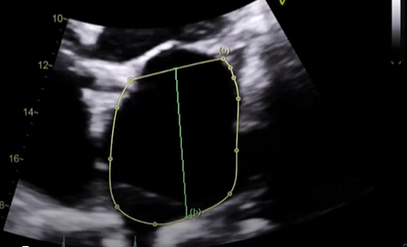
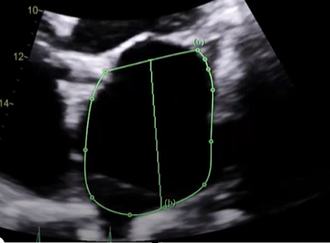
what measure measurement is this, explain how would want to measure this and what View,
Measured the LA area and length in A4C, at its largest dimension before the leaflet tips (MV) open at end-systole, want to exclude the Pulmonary vein and mitral annulus, this is when you need to trace the chamber and length. the normal LA area is going to be less than or equal to 20 or 24? (THIS IS NOT OUT LAV INDEX)
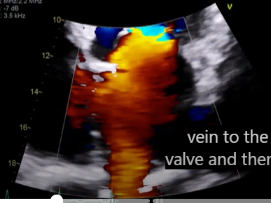
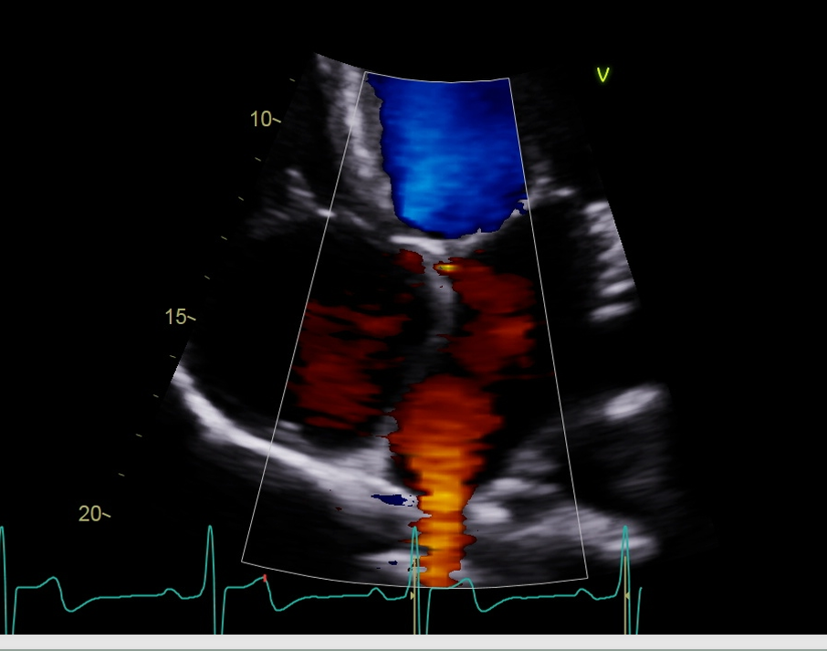
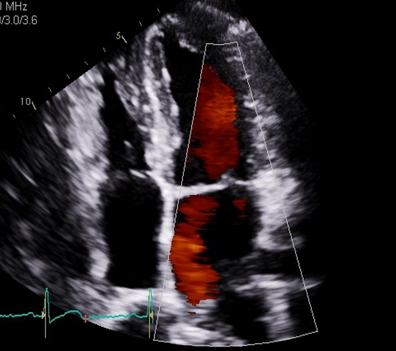
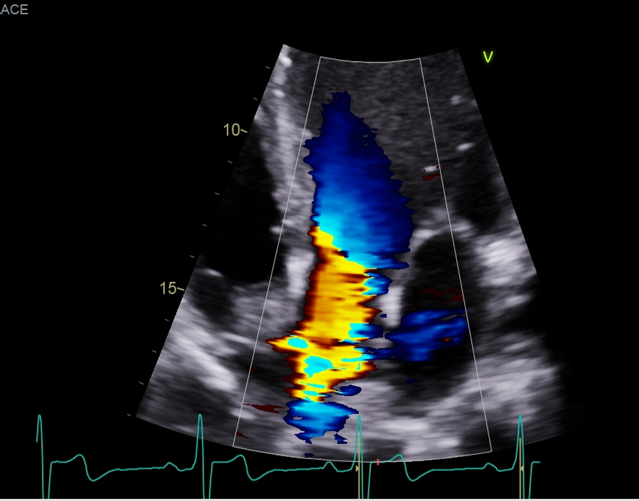
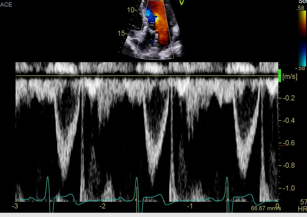
when we do our color flow what are we looing for the step we look at the pulmonary vein with color?
So, we look at the color of the pulmonary veins and for any MR, the normal flow pulmonary vein to LA to MV and the to LV, so any blue flow is going to be that MR reaching back and this red flow will be the pulmonary vein
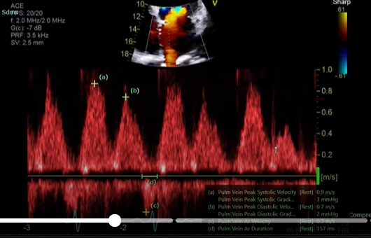
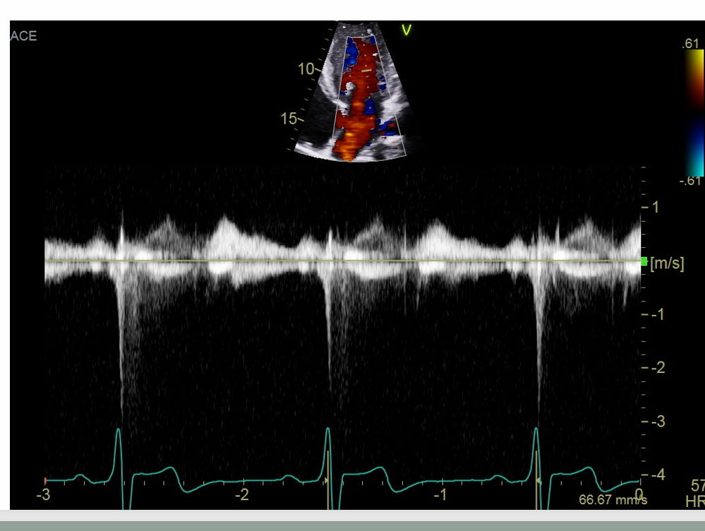
what single is this measurement for what are you going to peak, why would this doppler be important
this measure is for the pulmonary vein
Pulmonary vein peak systolic velocity (peak the S wave)
Peak D wave
peak the velocity of the atrial reversal or atrial duration
This will important when interrogate for MR, If the S wave is shorter than ourt D that is blunting so this coulf point to moderate or severe regurgitation if you see a reversal in these two
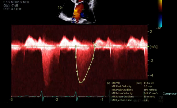
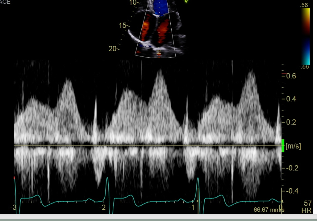
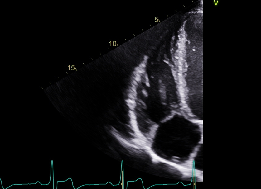
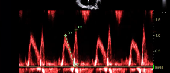
what measurement would this be
trace this measure when you get a really nice envelope, this is she MVR PISA method and measure MVR VTI (the jet is u-shaped)
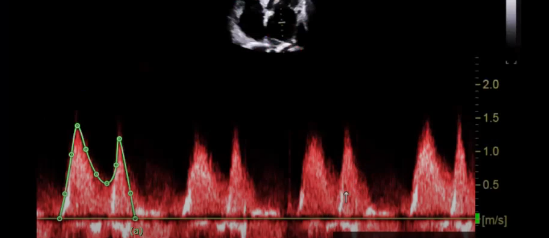
what is this measurement for
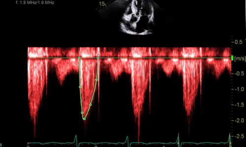
This measuring above the baseline for stenosis, this is our MV VTI
you want to Trace the E and A, a normal MV mean gradient is going to be less than five (if we see a very heavy calcified vale that is not opening well the CW is going to assessing for)
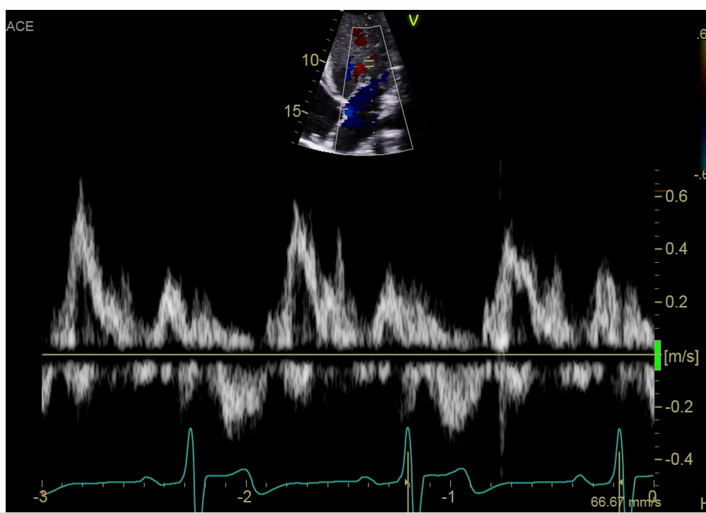
what measurement is this showing
PW doppler will be for diastology, placing the gate at the MV Leaflet tips and we have our E and A wave and will peak them and the MV decel time
The E wave is showing the early rapid LV fiiling it usally sits around the end of the T wave and then in early diastole
The A wave is our late LV filling an atrial contraction this will be right before our r wave
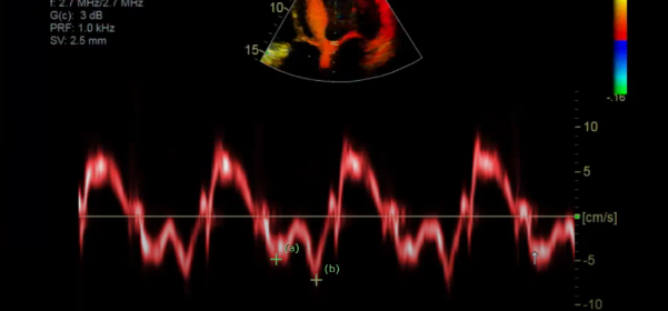
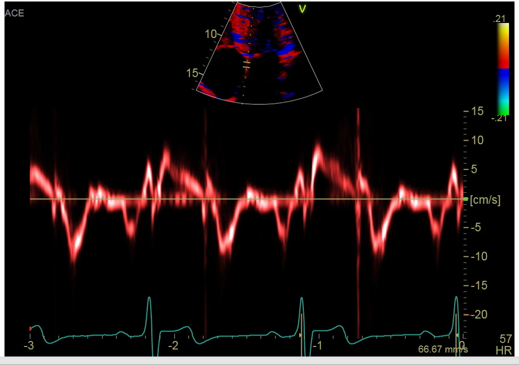
what is this measurement for
Septal side
measure the MV septal E
and then MV septal A velocity
gate should be 5-10
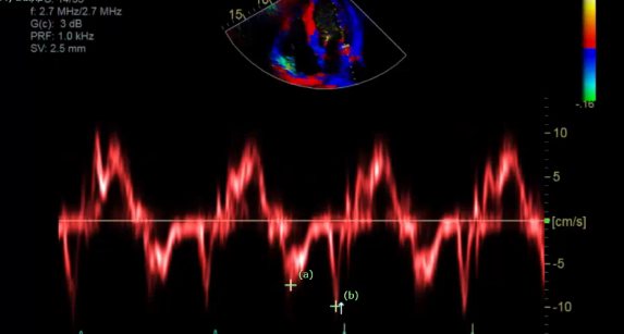
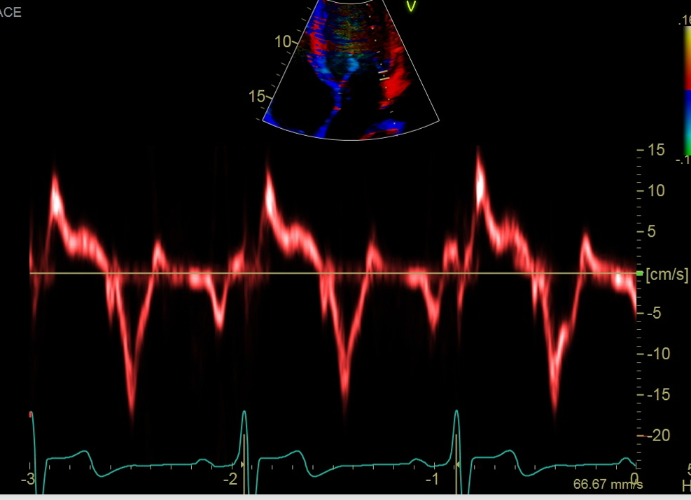
what measurement is this for
Lateral side
Peak lateral e’ and a’
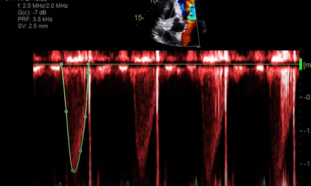
what measurement is this
Strove volume measurement
PW signal of the LVOT VTI
this will be a trace, trace the oustide
what measurement is this
now for assessing for stenosis AV VTI using CW
this will be a trace
what measurement is this for
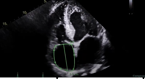
RA volume, measure when the chamber is at its largest, just right before the TV open, at end-systole
Trace the chamber excluding the TV annulus
and we also do a length
a normal RA size is going to be less than 19 centimeters squared
what is this measurement for
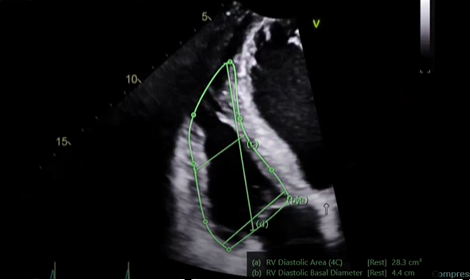
RV focused view this will be a lateral tilt
do a base, mid, and length measure along with the fractional area of change measurement (FAC%)
measure at the end of the QRS / end-diastole
RV diastolic area, trace it
when doing the base measurement do not bring it down past the annulus you will be right at the base of the actual ventricle
mid length, in the middle of the ventricle
and then perform the diastolic length, so RV apex all the way down
the area and the diastolic area and the systolic area give us fractional area of change this is showing our radio function, how the msucle is moving inwards that is our radial function that is our FAC %
what measurement is this
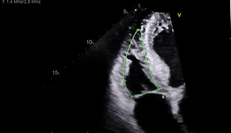
this will be for FAC% but of the RV but at end-systole you want it at the smallest dimension but before the leaflets pop open
this will be for our RV systolic area, make sure when you trace bringing it all the way up to the RV apex this will be RV systolic area
what is the TAPSE and S’ measurements
TAPSE and S’ is the longitudinal function measure how well the muscle is moving up and down
our TAPSE and S’ should match normal numbers b/c they are measuring the same thing so it should match normal
what is the measurement for
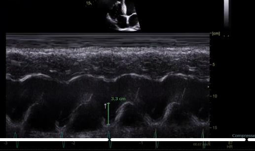
this is going to be the RV systolic function, this is the TAPSE measurement from the bottom of the hill to the top of the hill the normal values for TAPSE is >1.7- <2.4
what is the measuring
This is our S’, should be >9.5
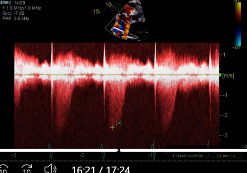
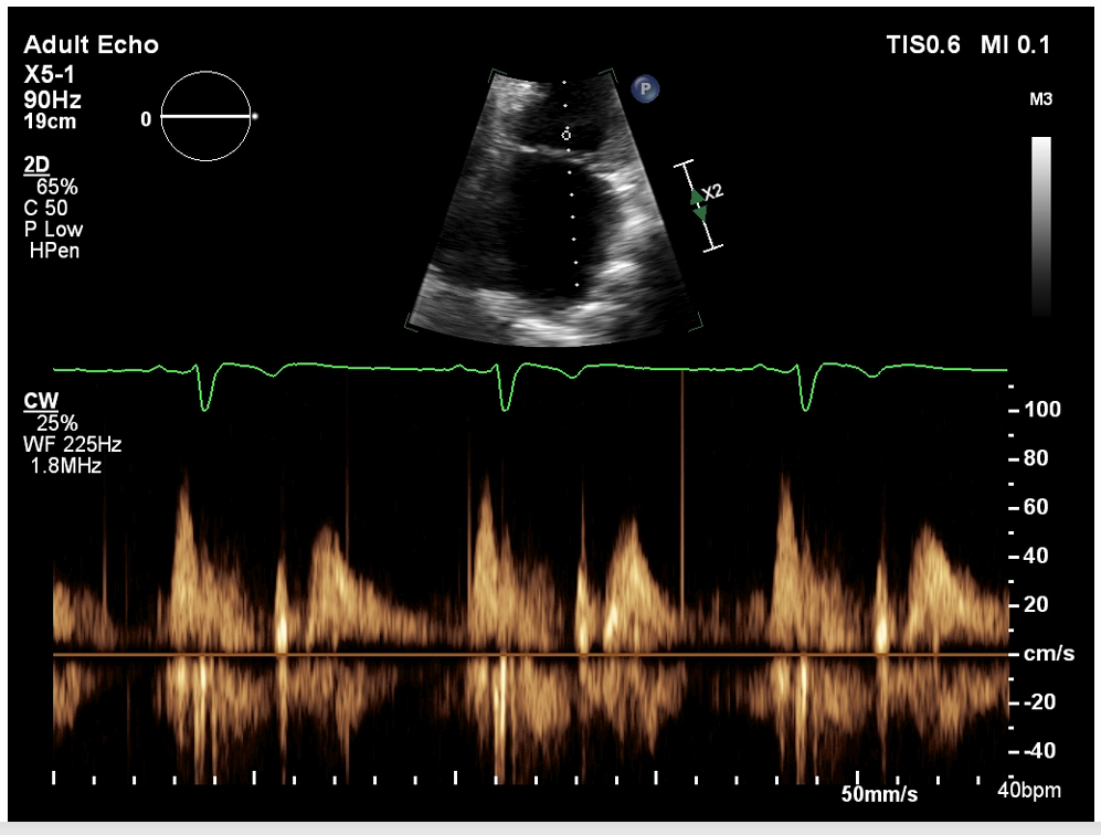
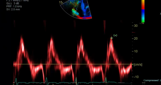
what measurement is this for
TR, peak the TR
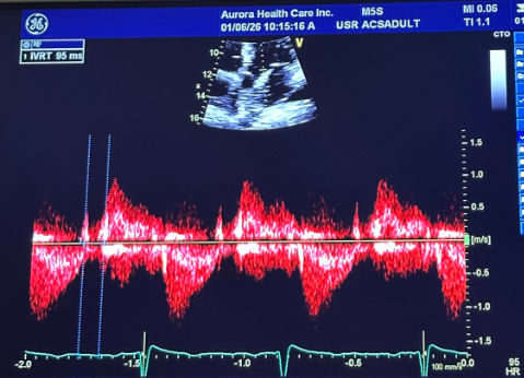
what measurement is this for
IVRT, perform CW between aortic valve and AMVL for IVRT (measure AVC-MVO)
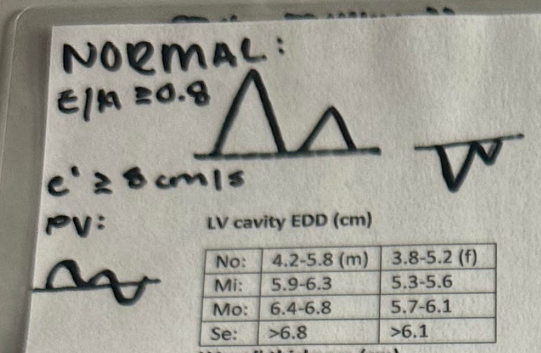
what is the normal mitral inflow waveform look like and the pulmonary vein flow and TDI look like what’s its e’ and E/A ratio?
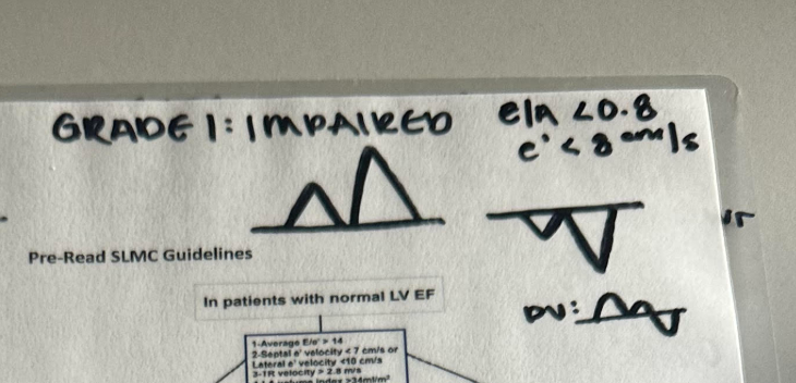
what does the impaired mitral inflow waveform look like and the pulmonary vein flow and TDI look like what’s its e’ and E/A ratio?
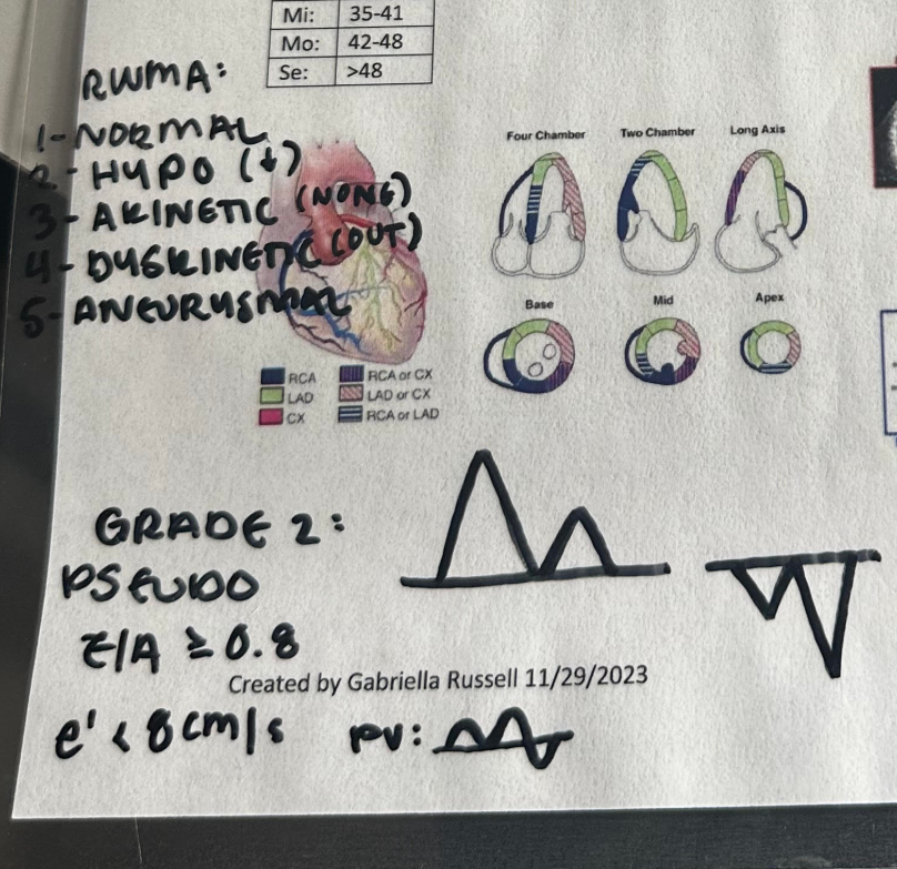
what does the pseudonormal mitral inflow waveform look like and the pulmonary vein flow and TDI look like what’s its e’ and E/A ratio?
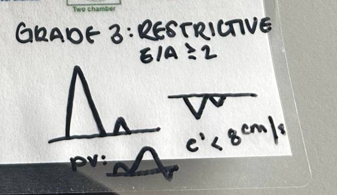
what does the restrictive mitral inflow waveform look like and the pulmonary vein flow and TDI look like what’s its e’ and E/A ratio?