AnaPhy: Ch.9 Incomplete
1/103
There's no tags or description
Looks like no tags are added yet.
Name | Mastery | Learn | Test | Matching | Spaced | Call with Kai |
|---|
No analytics yet
Send a link to your students to track their progress
104 Terms
skeletal muscle
voluntary muscle and constitutes about 40%of the body’s weight; attached to bone;
functions as body movement; locomotion, facial expressions, posture, respiratory functions, speech
skeletal muscle
type of muscles attached to bone and with striations; functions as body movement
cell shape of skeletal muscle
very long and cylindircal
smooth muscle
involuntary muscle most widely distributed in the body; found in walls of hollow organs, blood vessels, eyes, glands, and skin
smooth muscle function
propels urine through the urinary tract, mixes food in the stomach and the small intestines, regulates blood flow in blood vesseld
cell shape of smooth muscle
spindle shaped; single centrally located
skeletal muscle
type of muscle not capable of spontaneous contraction
cardiac muscle
involuntary muscle found in the heart; with striations
cardiac muscle
functions: pumps blood; contraction provide the major force for propelling blood through blood vessels
functions of the three types of muscle
movement of the body, maintenance of posture, respiration, production of body heat, communication, constriction of organs and blood vessels, contraction of the heart
movement of the body
most skeletal muscles are attached to bones and are responsible for the majority of body movement; walking, running, chewing
maintenance of posture
skeletal muscles constantly maintain tone, which keeps us sitting or standing erect
respiration
contraction of the skeletal muscles of the thorax and diaphragm help us breathe
production of body heat
when skeletal muscles contract, heat is given off as a by-product
communication
skeletal muscles are involved in all aspects of communication; speaking, writing, typing, gesturing, smiling or frowning
constriction of organs and vessels
contraction of smooth muscles within walls of internal organs causes the structures to constrict which help propel and mix food in the digestive tract, remove materials in organs
constriction due to smooth muscle contraction
help propel and mix food and water in the digestive tract; remove materials from organs, and regulate blood flow
contraction of the heart
contraction of the cardiac muscle causes heart to beat, propelling blood to all parts of the body
four major functional/general properties of muscle tissue
contractility, excitability, extensibility, elasticity
contractility
the ability of muscles to shorten forcefully and contract; lifting books
when muscles contract, it can cause
structures to which it is attached to move or increase pressure inside a blood vessel' or hollow organ
forces that oppose contraction
cause muscle to lengthen passively; gravity pulling on a limb
excitability
the capacity of muscle to respond to an electrical stimulus
smooth muscle and cardiac muscle respond to stimulation by
nerves and hormones
extensibility
a muscle can be stretched beyond its normal resting length and still be able to contract; retrieving a fallen pencil on the ground
elasticity
the ability of muscle to spring back to its original resting length after its been stretched; taking a deep breath
skeletal muscles are composed of
skeletal muscle tissues, nervous tissue, connective tissue, and adipose tissue
each muscle cell is called
muscle fiber
connective tissue coverings
each skeletal muscle is surrounded by these tissue layers that support the muscle during contraction
three layers of connective tissue in a skeletal muscle
epimysium, perimysium, endomysium
epimysium
forms a connective tissue sheath that surrounds each skeletal muscle; a layer of dense irregular connective tissue
epimysium protein fibers merge with
a connective tissue whose protein fibers gradually merge with muscular fascia between adjacent muscles and between muscles and skin
muscular fascia
outer layers of connective tissue that keep muscles separate from surrounding tissues and organs
perimysium
subdivides each whole muscle into numerous, visible bundles of muscle fibers called fascicles
fascicles
visible bundles of muscle fibers
perimysium
a loose connective tissue serving as passageways for blood vessels and nerves that supply each fascicle
endomysium
a delicate layer of connective tissue that covers and separates the individual muscle fibers within each fascicle
endomysium
serves as passageways for nerve fibers and blood vessels that supply each separate muscle fiber
epimysium
outer most layer of connective tissue covering that surrounds muscle organs
perimysium
middle layer of connective tissue covering that surrounds each fascicle
endomysium
innermost layer of connective tissue covering that surrounds the muscle fibers
collagen fibers of the connective tissue coverings
converge at the ends of muscle and together form tendons and aponeuroses; they are interwoven and blend into one another
epimysium of one muscle directly attaching to tendons or fascia of another muscle
attachments that serve to move bones or skin for locomotion, facial expressions, and other types of movement
skeletal muscle fibers
very unique cells that develop from the fusion of several hundred embryonic cells called myoblast
myoblast
embryonic cells that contain its own nucleus
sarcoplasm
muscle cell cytoplasm; contains high amounts of myoglobin and glycogen
myoglobin
conjugated protein which is the oxygen-transporting pigment of muscle
sarcolemma
muscle fiber cell membrane; contain transverse tubules (T-tubules); transmits electrical impulses to the interior of the muscle fiber
t-tubules
tube-like inward folds of the sarcolemma; carry electrical impulses into the center of the muscle fiber
sarcoplasmic reticulum
highly specialized smooth endoplasmic reticulum in skeletal muscle fibers that stores high levels of Ca; release of Ca from the sarcoplasmic reticulum is a “switch” for muscle contraction
sarcoplasm
muscle fiber containing organelles such as mitochondria and energy-storing glycogen granules that constitute a kind of cytoplasm
terminal cisternae
t-tubules that lie next to enlarged portions of the sarcoplasmic reticulum
t-tubules
allow action potentials to quickly spread to the myofibrils
two channel types contributing to the electrical properties of both resting and stimulated cell
ligand-gated channels, voltage-gated channelsl
ligand-gated channels
open when a specific ligand, a chemical signal such as neurotransmitter binds to a receptor
voltage-gated ion channel
are gated membrane channels that open and close in response to a specific membrane potential
voltage-gated channels that play major roles in an action potential
voltage-gated Na, K, Ca channels
electrically excitable cells
are polar; specialized to respond to electrical stimuli
intercalated disk
helping muscles contract
hypertrophy
increase in size of each muscle fiber
mechanical component structures
myofibrils, myofilaments (actin, myosin)
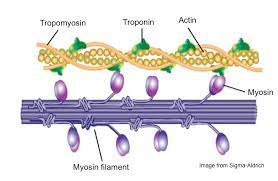
actin myofilaments
thin filaments made up of 2 long-coiling protein strands; connected to Z disks of sarcomeres; located between each myosin filament

myosin myofilaments
thick filaments arranged in parallel; have a main shaft and a globular head on each end
sarcomeres
join end to end forming myofibrils; smallest portion that can contract; contractile structures formed by overlapping actin and myosin
3 regulatory proteins of actin
globular actin, tropomyosin, troponin
G actin
molecules that are globular subunits that form a long chain of 200 G actins subunits; active site for myosin head binding during contraction
F actin (fibrous)
chain of 200 G actin subunits

tropomyosin
a long fibrous protein that lies in the groove along the fibrous actin strand

troponin
consist of three subunits: TnC- binds to Ca, TnT- binds to tropomyosin (prevents the tropomyosin from uncovering G actin active site) TnI- binds to G actin (inhibits actin to myosin binding)
myosin components
2 myosin heavy chains (forms the rod portion), 2 myosin heads
neuromuscular junctions
also called an end plate; point of contact of motor neuron axon branches with the muscle fiber
action potential
electrical signals
motor neuorons
carries action potential signals which stimulate muscle fiber action potentials followed by muscle cotraction
presynaptic terminal
axon terminals
synaptic cleft
space between the presynaptic terminal and the muscle fiber
motor-end plate/ postsynaptic membrane
muscle plasma membrane/ sarcolemma in the area of the junction; depression in the sarcolemma of the adjacent muscle fiber, n close association with the synaptic knob
synaptic vesicles
numerous mitochondria and spherical sacs in the presynaptic terminals; contains neurotransmitters called acetylcholine (ACh)
neurotransmitter
a molecule that allows a neuron to communicate with its target
neurotransmitters (ACh)
inhibits the production of action potential in the motor-end plate by binding to ligand-gated ion channels

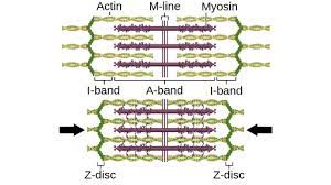
sarcomere
region between two Z lines
Z discs
filamentous networks of proteins form a stationary anchor for actin myosin filaments
I bands (isotropic)
2 two lighter straining regions; consisting only of thin filaments
A bands
darker straining band in the center of each sarcomere; formed by entire length of thick myosin filaments
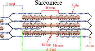
H zone
smaller band at the center of each A band
M line
dark line at the middle of each H zone; consists of delicate filaments that hold the myosin myofilaments in place
threshold
is the membrane potential at which voltage-gated ion channels open
depolarization phase
the action potential is a brief period during which further depolarization occurs and the inside of the cell becomes even more positively charged
repolarization phase
return of the membrane potential to its resting value
triad
a skeletal muscle substructure responsible for the regulation of excitation-contraction coupling
excitation-contraction coupling
link between an action potential on the sarcolemma and the sarcomere shortening
power stroke
movement of myosin head in cross-bridging
muscle twitch
response of a muscle fiber to a single AP along its motor neurons
myograph
recording produced from a single isolated twitch
phases of a twitch
lag phase, contraction phase, relaxation phase
lag phase/ latent phase
time during which action potential is traveling along the axon, the events at neuromuscular junction occurs, and the AP travels along the sarcolemma
contraction phase
Ca is released from SR and cross-bridge cycling occurs
relaxation phase
concentration of Ca in the sarcoplasm decreases slowly and Ca is actively transported back into the SR
action potential
electrochemical event
muscle contraction
mechanical event
tension
muscle contraction measured as a force