PHYS 335 Week 3 Flashcards on Neurobiology Concepts
1/95
There's no tags or description
Looks like no tags are added yet.
Name | Mastery | Learn | Test | Matching | Spaced | Call with Kai |
|---|
No analytics yet
Send a link to your students to track their progress
96 Terms
electrogenic pump
An active transport protein that creates a net flow of charge across a membrane while pumping ions.
- The sodium-potassium pump works by moving three sodium ions out of the cell for every two potassium ions pumped in. This process creates a net outward current, making the inside of the cell more negative
leak channels (ungated channels)
specialized proteins in the plasma membrane that allow ions to pass through and contribute to the resting membrane potential of cells
- allow ions to move along their gradient
ligand-gated ion channels
integral membrane proteins/transmembrane proteins with a pore that allow selected ions to pass through a cell membrane in response to the binding of chemical signaling molecules, such as a neurotransmitter
- also known as ionotropic receptors
- ion flux is passive and driven by the electrochemical gradient for the permeant ions
mechanically gated ion channels
proteins in cell membranes that open in response to mechanical stress, such as pressure, touch, sound, and temperature
- allow ions to pass through, which changes the electrical properties of the cell membrane and can trigger an action potential, which is sent to the CNS, which can cause a response.
resting membrane potential (RMP)
The electrical potential difference across a cell's plasma membrane when the cell is at rest and not actively transmitting signals
- RMP is the basis for how neurons communicate and send and receive signals. The charge of the membrane can change in response to neurotransmitter molecules and environmental stimuli.
voltage-gated ion channels
proteins that allow certain ions to pass through cell membranes, responding to changes in the cell's electrical potential.
- vital for electrical signaling, muscle contraction, cell secretion, and hormone regulation
- give a membrane the ability to undergo action potentials
decremental current
the flow of charge decreases as the distance from the site of origin of the graded potential increases
depolarized
the process by which a cell's membrane potential becomes less negative, or more positive, relative to its environment
excitability
the ability of a cell to respond to a stimulus by generating an electrical signal, or action potential
- present in neurons, muscle cells, and cardiac cells
excitable membranes
neurons are capable of generating action potentials that propagate rapidly along the length of their axons
graded potentials
local changes in membrane potential that can be depolarizing or hyperpolarizing
- proportional to the stimulus size, decrease in strength over time and space, and are conducted over short distances
hyperpolarized
phase of an action potential when the membrane potential becomes more negative at a specific location on the neuron's membrane.
overshoot
when an action potential causes a neuron's membrane potential to exceed its resting potential (over 0mV)
- happens when ion channels in the neuron's membrane open and close during the depolarization and repolarization stages of the action potential
pacemaker potential
spontaneous gradual depolarization to threshold of some neurons and muscle cells' plasma membrane
receptor potential
An initial response of a receptor cell to a stimulus, consisting of a change in voltage across the receptor membrane proportional to the stimulus strength.
- generally a depolarizing event resulting from inward current flow
- generated in the sensory receptors at the peripheral ends of the neurons, which are at the ends farthest from the CNS
repolarized
the process that returns a neuron's membrane potential to its resting negative state after depolarization
- occurs when potassium ions (K+) flow out of the neuron's membrane through channels
summation
the process that determines whether or not an action potential will be generated by the combined effects of excitatory and inhibitory signals, both from multiple simultaneous inputs (spatial), and from repeated inputs (temporal)
synaptic potential
the potential difference across the postsynaptic membrane that results from the action of neurotransmitters at a neuronal synapse
- two forms of synaptic potential: excitatory and inhibitory
absolute refractory period
the period immediately following the firing of a nerve fiber when it cannot be stimulated no matter how great a stimulus is applied
action potentials
short periods of electrical activity at the membrane of a neuron, responsible for the transmission of signals within the neuron
action potential propagation
the process by which a nerve impulse, or action potential, travels along a neuron
- differs between myelinated and unmyelinated axons
myelinated axons
Action potentials propagate through these axons via saltatory conduction, which is a node-to-node propagation pattern. Depolarizing current travels rapidly through the myelin-insulated cytoplasm until it reaches a node of Ranvier, where the membrane depolarizes and sodium ions flow in. This initiates the action potential again, and the process repeats.
unmyelinated axons
Action potentials propagate through these axons via continuous conduction. Depolarization of the cell membrane spreads to the adjacent region, raising the potential until it reaches the threshold voltage. This process is slower than saltatory conduction because of the constant influx of sodium
afterhyperpolarization
phase of an action potential when the membrane potential falls below the resting potential, also known as the undershoot phase.
- a slow hyperpolarization that often occurs after an action potential
all-or-none
referring to the fact that an action potential/neuron either fires completely or does not fire at all
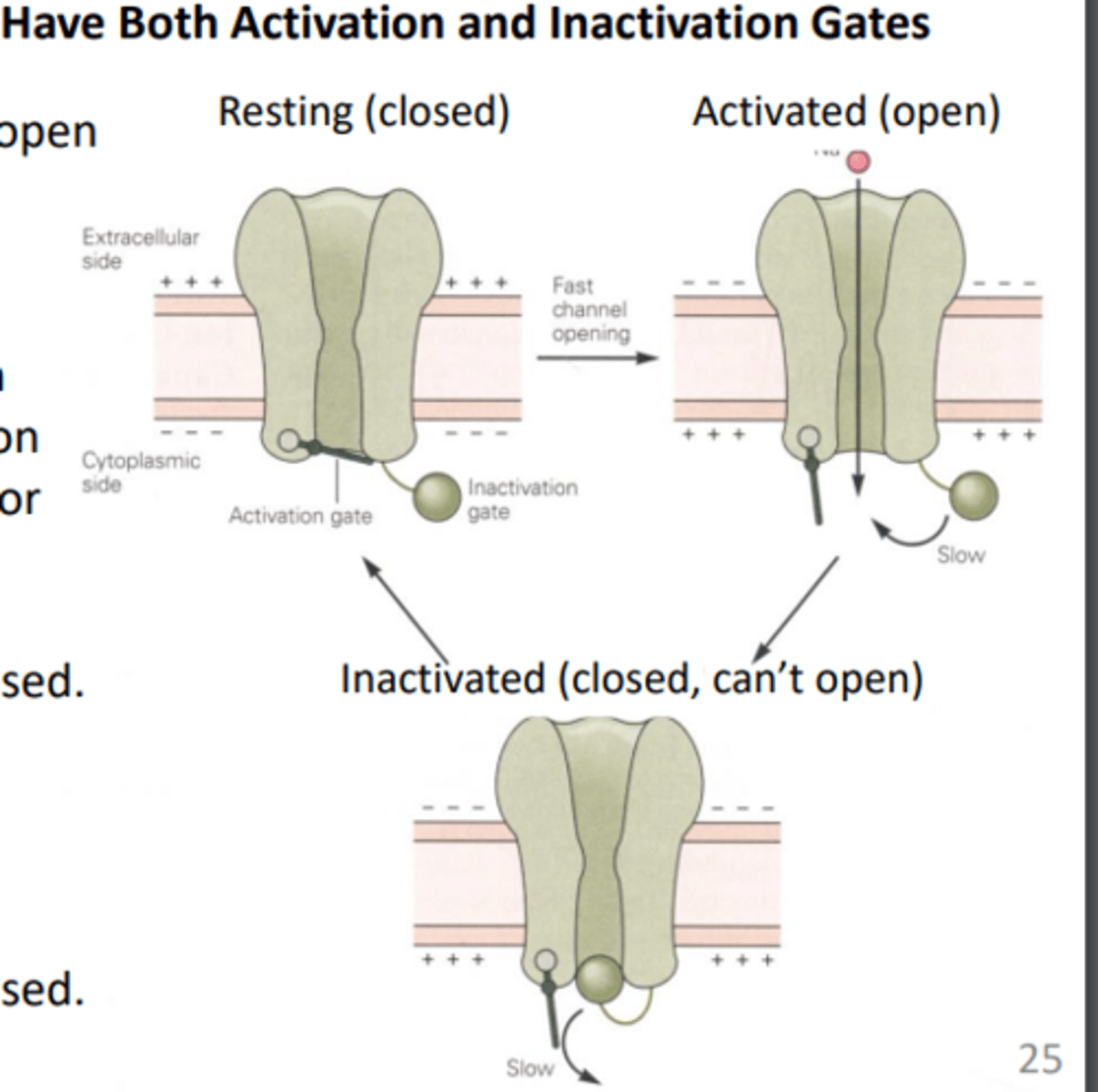
inactivation gates of voltage-gated Na+ channels
describes how a ball domain blocks the pore of a voltage-gated ion channel, preventing ions from passing through
relative refractory period
the period of time following an action potential, when it is possible, but difficult, for the neuron to fire a second action potential, due to the fact that the membrane is further from threshold potential (hyperpolarized)
- need stimulus to be greater than the previous to create AP
saltatory conduction
the propagation of action potentials along myelinated axons from one node of Ranvier to the next node, increasing the conduction velocity of action potentials.
subthreshold stimuli
produce no observable contractions, stimuli that aren't strong enough to reach threshold to cause action potential
threshold potential
the minimum membrane potential that must be reached in order for an action potential to be generated.
- usually (-55mV)
threshold stimuli
the weakest stimuli that results in an action potential
active zones
region within an axon terminal where neurotransmitter vesicles are clustered prior to secretion
chemical synapses
specialized for release and reception of chemical neurotransmitters.
convergence
multiple neurons can send signals to a single postsynaptic neuron, effectively combining inputs from various sources into one output, allowing a single neuron to integrate information from a large network of cells
divergence
a single neuron sending its signal to multiple other neurons, essentially branching out its connections to influence a wide range of downstream targets, allowing one neuron to communicate with many other neurons within a neural network
electrical synapses
synapses that transmit information via the direct flow of electrical current at gap junctions.
excitatory postsynaptic potential (EPSP)
a slight depolarization of a postsynaptic cell, bringing the membrane potential of that cell closer to the threshold for an action potential
excitatory synapse
a synapse in which an action potential in a presynaptic neuron increases the probability of an action potential occurring in a postsynaptic cell
ionotropic receptors
receptors that are coupled to ion channels and affect the neuron by causing those channels to open
inhibitory postsynaptic potential (IPSP)
a temporary hyperpolarization of postsynaptic membrane caused by the flow of negatively charged ions into the postsynaptic cell
inhibitory synapse
synapse at which a neurotransmitter causes the receiving cell to stop firing
metabotropic receptors
a type of membrane receptor that ithat act through a second messenger system to modulate cell activity
- associated with signal proteins and G proteins
SNARE proteins
molecular motors that mediate membrane fusion between organelles and the plasma membrane
- synaptotagmin
synaptic cleft
a gap into which neurotransmitters are released from the axon terminal
synaptic vesicles
Membrane-bounded compartments in which synthesized neurotransmitters are kept.
synaptotagmins
proteins present in wall of synaptic vesicle that bind calcium and help stimulate the process of exocytosis
- closely regulates the SNARE zipping
acetylcholine (ACh)
A neurotransmitter that enables learning and memory and also triggers muscle contraction
acetylcholinesterase
the enzyme that breaks down acetylcholine in the synaptic cleft
- located on the presynaptic and postsynaptic membranes
- releases choline and acetate.
adrenergic neurons
nerve cells that release neurotransmitters like adrenaline, noradrenaline, or dopamine
agonists
drugs that increase the action of a neurotransmitter
alpha-adrenergic receptors
Receptors to norepinephrine that generally produce an excitatory response
- when stimulated, can cause constriction of blood vessels.
AMPA receptors
an ionotropic glutamate receptor that controls a sodium channel; when open, it produces EPSPs
antagonists
chemical substances that block or reduce a cell's response to the action of other chemicals or neurotransmitters.
autoreceptors
signal the presynaptic neuron to stop releasing the neurotransmitter
axo-axonic synapse
a type of synapse that occurs when one neuron's axon terminals connect with another neuron's axon
beta-adrenergic receptors
Receptors located on postsynaptic cells that are stimulated by specific autonomic nerve fibers.
- Beta1-adrenergic receptors are located primarily in the heart
- Beta2-adrenergic receptors are located in the smooth muscle fibers of the bronchioles, arterioles, and visceral organs.
catecholamines
hormones secreted by the adrenal medulla that affect the sympathetic nervous system in stress response
- dopamine, norepinephrine, epinephrine (adrenaline)
cholinergic neurons
nerve cells that produce and release acetylcholine (ACh) to send messages throughout the nervous system
dopamine
a neurotransmitter associated with movement, attention and learning and the brain's pleasure and reward system.
endogenous opioids
Chemicals produced by the body that reduce pain, enhance positive mood, and suppress appetite.
- endorphins, enkephalins, dynorphins
epinephrine
Neurotransmitter secreted by the adrenal medulla in response to stress.
- known as adrenaline.
excitotoxicity
the property by which neurons die when overstimulated, as with large amounts of glutamate
Gamma-aminobutyric acid (GABA)
an amino acid that acts as the main inhibitory neurotransmitter in the brain and spinal cord. - responsible for reducing neuronal excitability by inhibiting nerve transmission.
- known for its calming effect and is thought to play a role in controlling anxiety, stress, and fear.
glutamate
A major excitatory neurotransmitter that plays a vital role in brain function
- essential for learning, memory, and mood regulation.
glutamatergic neurons
Neurons that use glutamate as a transmitter.
glycine
an amino acid that helps the body build proteins for tissue and hormone maintenance
long-term potentiation
an increase in a synapse's firing potential after brief, rapid stimulation.
- believed to be a neural basis for learning and memory.
muscarinic ACh receptors
a general type of cholinergic receptor stimulated not only by acetylcholine but by muscarine (a poison contained in some mushrooms)
- receptors are metabotropic and couple with G proteins, which then alter the activity of a number of different enzymes and ion channels.
- prevalent at some cholinergic synapses in the brain and at junctions where a major division of the PNS innervates peripheral glands, tissues, and organs
neuromodulators
chemicals released in the nervous system that influence the sensitivity of the receiving neuron to neurotransmitters
neuropeptides
Small protein-signaling molecules released by neurons that act as neurotransmitters or neuromodulators to coordinate long-lasting biological responses.
neurotransmitters
Chemical messengers released by neurons at synapses to transmit signals to other neurons, muscle cells, or glands.
nicotinic ACh receptors
Ionotropic receptors that function as ligand-gated cation channels, primarily allowing Na^{+} and K^{+} flux to depolarize the postsynaptic membrane.
NMDA receptors
Ionotropic glutamate receptors permeable to Na^{+}, K^{+}, and Ca^{2+}; critical for long-term potentiation (LTP).
norepinephrine
A catecholamine neurotransmitter
peptidergic neurons
use neuropeptides as their main signaling molecules (or as important co-transmitters), instead of—or in addition to—classic small-molecule neurotransmitters like glutamate, GABA, or dopamine.
presynaptic facilitation
is when a neuron releases more neurotransmitter than usual because something at the presynaptic terminal boosts release probability.
presynaptic inhibition
when neurotransmitter release is reduced at the presynaptic terminal, even though the action potential still arrives.
receptor desensitization
when a receptor becomes less responsive despite the ligand still being present.
serotonin
monoamine neurotransmitter best known for mood regulation—but it’s really a global neuromodulator with fingers in everything.
spatial summation
how a neuron adds up inputs coming from different locations on its membrane at the same time.
temporal summation
how a neuron adds up inputs over time from the same synapse when they arrive close together.
adequate stimulus
is the specific type of energy a sensory receptor is most sensitive to—the stimulus it detects with the lowest threshold
chemoreceptors
are sensory receptors that detect chemical substances—either in the external environment or inside the body—and convert that information into neural signals.
mechanoreceptors
specialized sensory receptors in the body that detect mechanical stimuli, such as pressure, stretch, vibration, and touch. They convert physical forces into electrical signals (nerve impulses) that the nervous system can interpret. These receptors are essential for sensing the environment and for proprioception (awareness of body position and movement).
nociceptors
are sensory receptors that detect painful or potentially harmful stimuli. Unlike mechanoreceptors, which respond to mechanical forces, nociceptors are specialized to alert the body to tissue damage or the threat of injury.
osmoreceptors
are specialized sensory receptors that detect changes in the osmotic pressure (osmolality) of body fluids, primarily in the blood. They are key players in maintaining fluid balance and homeostasis.
perception
the process by which the brain interprets sensory information received from the environment and the body, turning raw sensory signals into meaningful experiences. It’s essentially how we “make sense” of the world around us.
photoreceptors
are specialized sensory cells in the retina of the eye that detect light and convert it into electrical signals for the brain, enabling vision.
proprioception
body’s ability to sense its own position, movement, and orientation in space without relying on vision. In simple terms, it’s your body’s “internal GPS” that tells you where your limbs are and how they’re moving
rapidly adapting (phasic) receptors
are a type of sensory receptor that respond quickly at the onset of a stimulus but stop firing if the stimulus remains constant. They are specialized for detecting changes in the environment rather than continuous stimulation.
sensory information
data collected by the body’s sensory receptors about the internal and external environment. This information is sent to the nervous system, where it is processed and interpreted to guide perception, behavior, and bodily responses.
sensory receptors
are specialized cells or nerve endings that detect stimuli from the environment or the body and convert them into electrical signals (nerve impulses) that the nervous system can process. They are the starting point for all sensation and perception.
sensory system
the collection of organs, receptors, and neural pathways that allow the body to detect, transmit, and interpret information from the internal and external environment. It’s essentially how we sense and respond to the world.
sensory transduction
the process by which sensory receptors convert a stimulus from the environment into an electrical signal (nerve impulse) that the nervous system can interpret. It is the first and essential step in sensation.
slowly adapting (tonic) receptors
sensory receptors that continue to respond to a stimulus as long as it is present, though the firing rate may gradually decrease over time. They are specialized for monitoring sustained or constant stimuli.
thermoreceptors
are sensory receptors that detect temperature changes—both heat and cold—in the environment or the body. They allow the nervous system to maintain thermal homeostasis and respond to potentially harmful temperature extremes.