602 Auditory system, Vestibular system, Autonomic Nervous System 3/17
1/105
There's no tags or description
Looks like no tags are added yet.
Name | Mastery | Learn | Test | Matching | Spaced | Call with Kai |
|---|
No analytics yet
Send a link to your students to track their progress
106 Terms
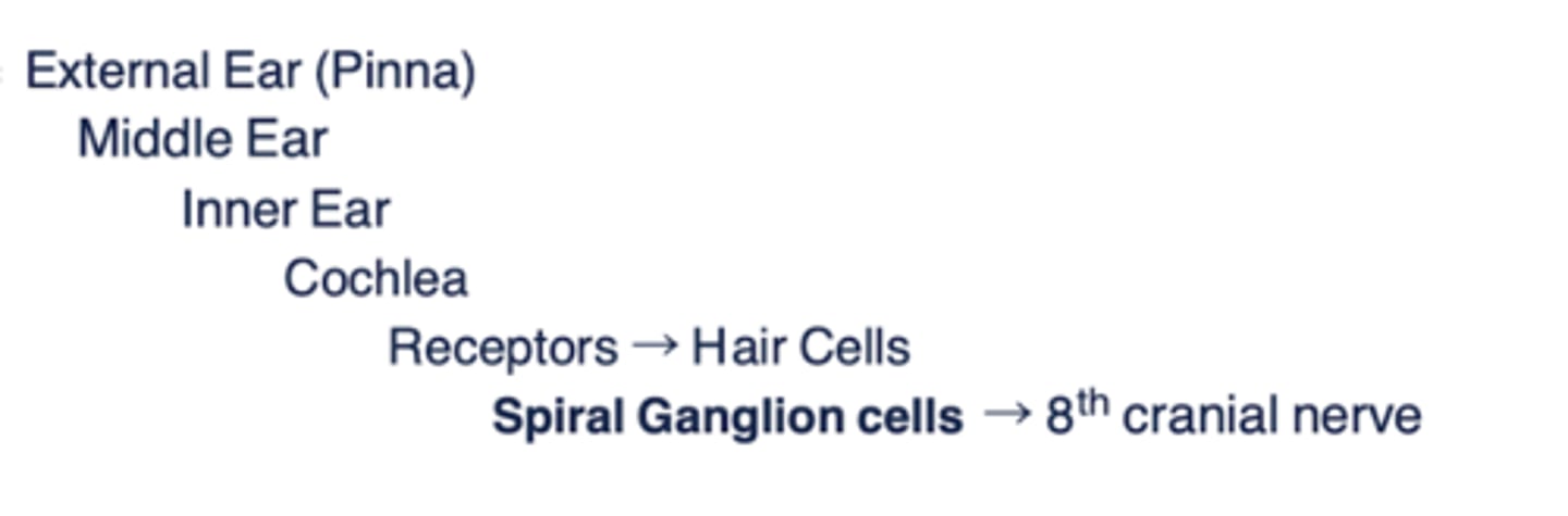
Peripheral structures auditory system
- External ear (pinna)
- Middle ear
- Inner ear
- Cochlea
- Receptors (hair cells)
- Spiral ganglion cells --> 8th cranial nerve
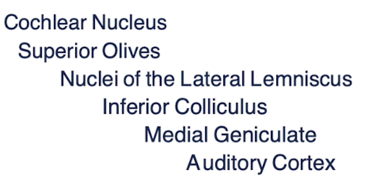
Central structures auditory system
- Cochlear nucleus
- Superior olives
- Nuclei of the lateral lemniscus
- Inferior colliculus
- Medial geniculate
- Audotry cortex
Hearing function (2)
- Sound identification
- Localization
Define:
- Frequency
- Amplitude
- Complexity
- Frequency: (pitch; hertz = diff musical notes)
- Amplitude (loudness; decibels = volume)
- Complexity (uniqueness = same not diff instruction) --> ex: guitar vs piano playing same key
Sound waves = ________________ in auditory system
physical energy
= wave of compressions and expansions of molecules of air
- increasing/decreasing air pressure
The auditory system has _____________ (unilateral/bilateral) processing
bilateral
Young individuals can hear from _____ Hz to ___________ Hz
20 to 20,000
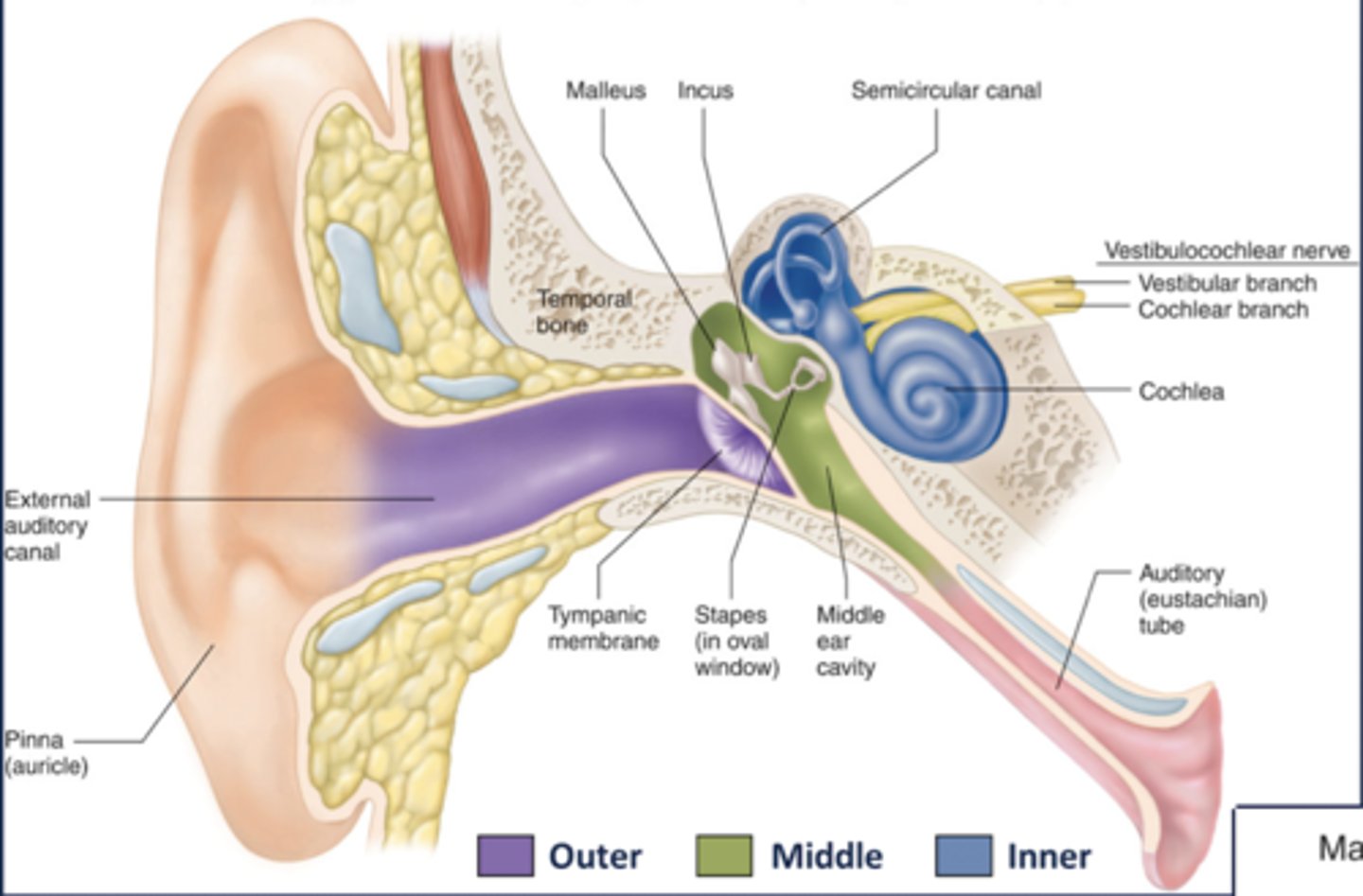
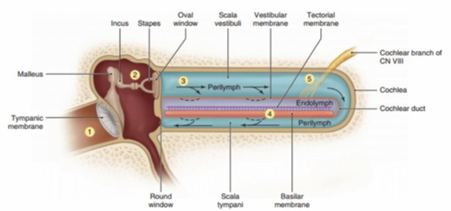
3 sections of the ear and structures within
Outer ear
- Pinna (auricle)
- External meatus
- Tympanic membrane
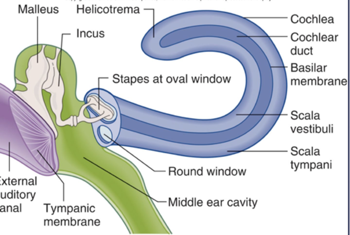
Middle ear (ossicles = bones)
- Malleus
- Incus
- Stapes
Inner ear (fluid filled)
- Oval window
- Round window
- Scala vestibuli
- Scala tympani
- Scala media = Cochlear duct
-- organ of corti
3 compartments of the cochlea
- what kind of fluid does each hold?
- Scala vestibuli (perilymph)
- Scala tympani (perilymph)
- Scala media (endolymph)
The cochlea transduces physical energy into ________________ that are routed to the _________
Action potentials
CNS
Movement within the ear - steps (5)
1. Tympanic membrane vibrates ossicles
2. Ossicles amplify vibration
3. Stapes vibrates against oval window, membrane moves back and forth, and creates pressure waves in cochlea
-- sound waves converted to fluid waves
4. Waves travel through scala vestibuli and scala tympani --> round window compensates for pressure changes
5. Waves cause scala media to vibrate
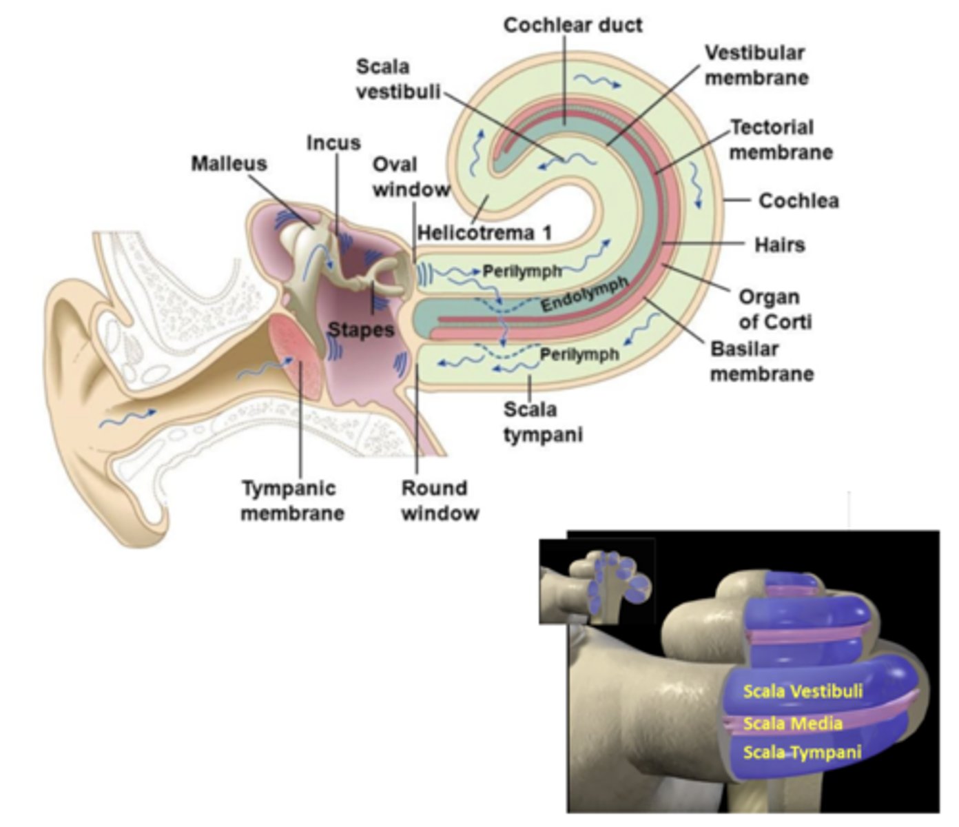
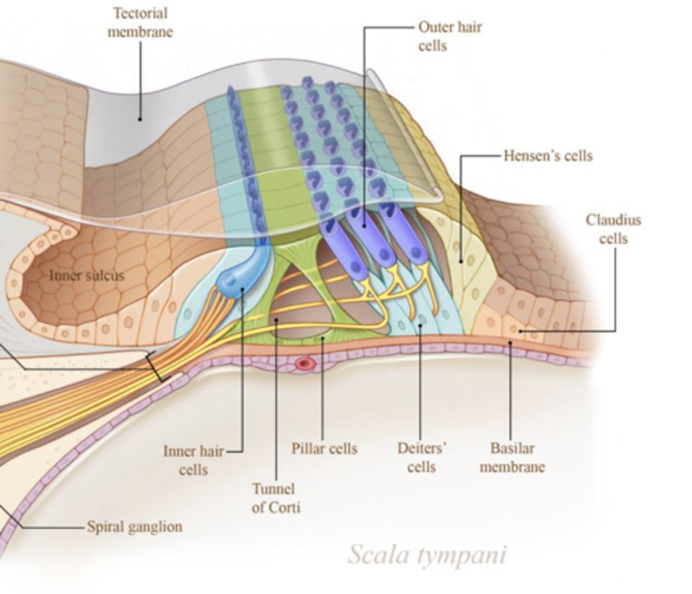
What type of epithelium on organ of corti?
Specialized neuroepithelium on basilar membrane
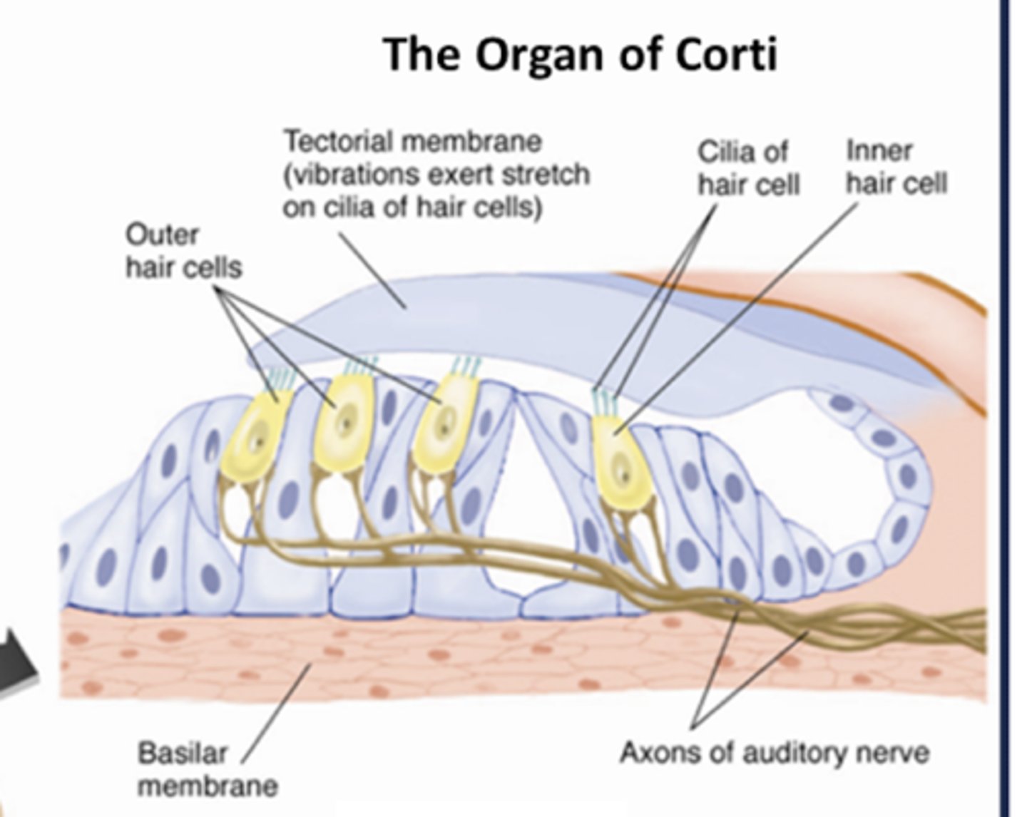
What does the organ of corti contain?
Hair cells (~15k) & variety of other supporting cells
The stereocilia of hair cells attach to the:
tectorial membrane
- moves relative to basilar membrane
- causes shearing of the hair cells = depolarization
Hair cells in the organ of corti are innervated by which CN?
The cochlear portion of CN VIII (vestibulocochlear)
Describe the transduction of sound
- Movement of the stapes against the oval window creates pressure waves within the cochlear perilymph
- Perilymph pressure waves vibrate the endolymph and the basilar membrane
- This vibration is detected and transduced by hair cells
Within the organ of corti, inner hair cells are responsible for ____________, while outer hair cells are responsible for __________
- Inner: transduction
- Outer: amplification
Endolymph and Perilymph - high or low in K+ and Na+?
- which resembles ICF and ECF?
- where is each found?
Endolymph: high in K+ and low in Na+ (resembles intracellular fluid)
- within scala media/cochlear duct
Perilymph: low in K+ and high in Na+ (resembles extracellular fluid)
- within scala vestibuli and tympani
How does cochlear fluid play a role in transduction in hair cells?
- Stereocilia connect to tectorial membrane (connect to kinocilium via tip links)
- Pressure wave-induced swaying toward the kinocilium opens mechanically gated K+ ion channels in sterocilia --> K+ ions enter hair cell = depolarization
- Depolarization opens Ca2+ ion channels
- Ca2+ influx triggers release of glutamate onto axons of CN VIII
- If depolarization is large enough, will fire APs to auditory cortex
TOWARD kinocilum = DEPOLARIZATION
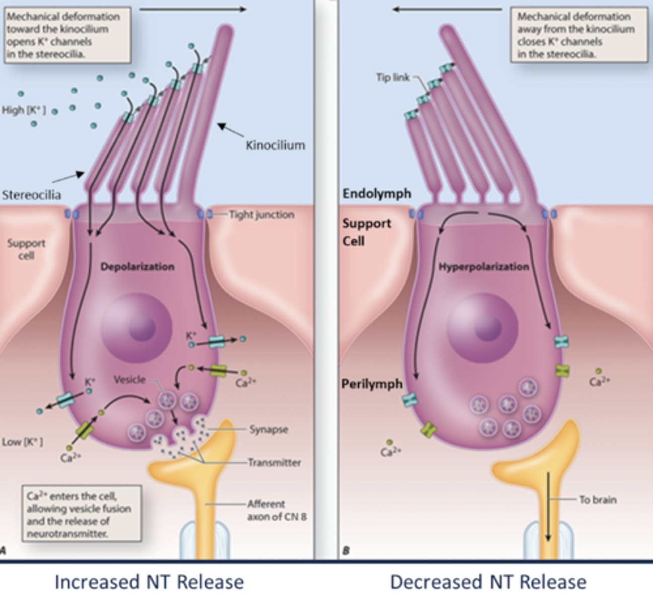
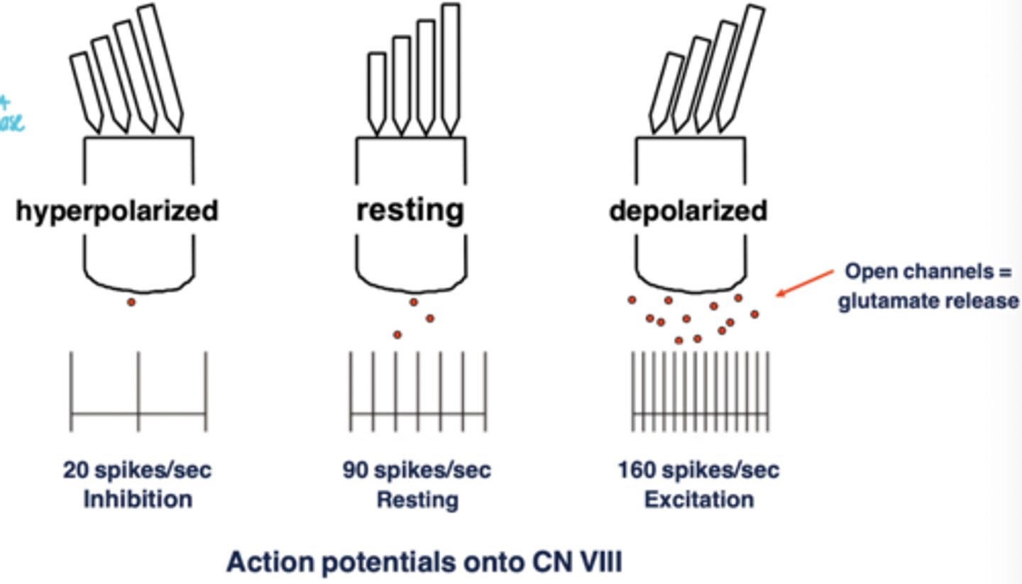
3 possible scenarios for hair cell transduction
- Hyperpolarization (sways away; slack)
- Resting
- Depolarized (sways toward; taught) = open channels --> glutamate release
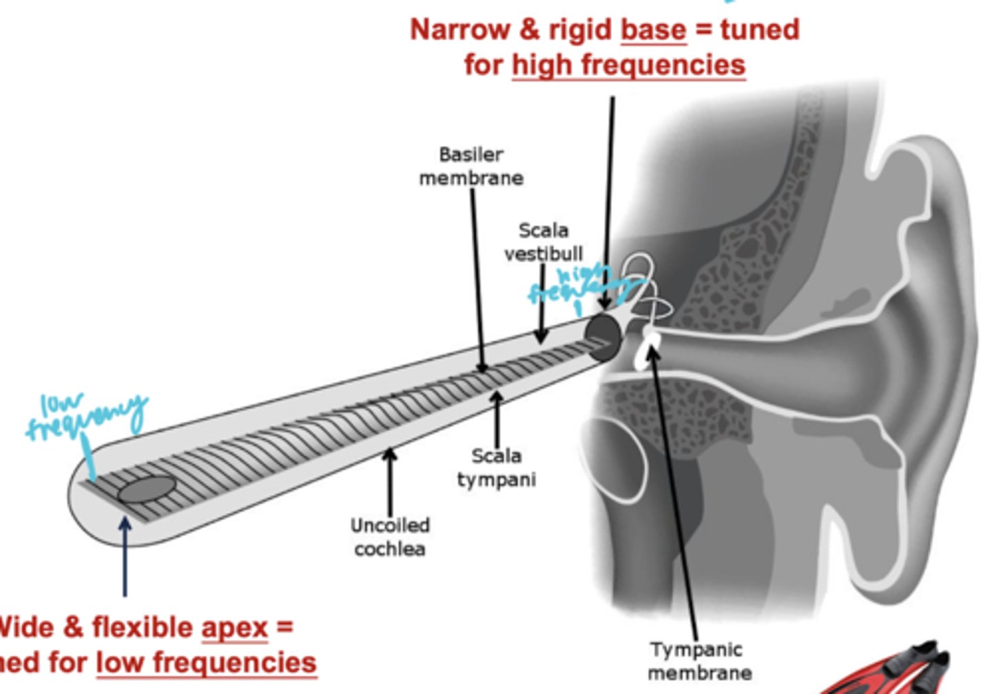
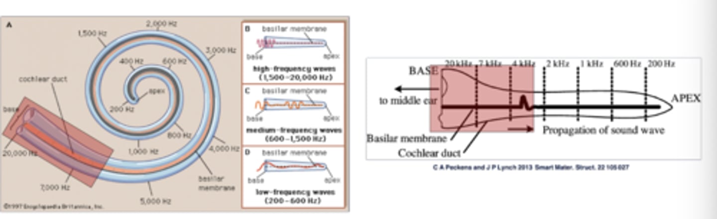
Describe the tonotopic organization on the basilar membrane
- Base = high tones (frequencies) --> near the tympanic membrane
- Apex = low tones (frequencies)--> tip of the uncoiled snail
Role of the CNS in the auditory pathway
- Interprets the pattern of hair cell stimulation as a sound of a particular frequency
- Detects small timing differences between sound arriving in each ear --> important for sound localization
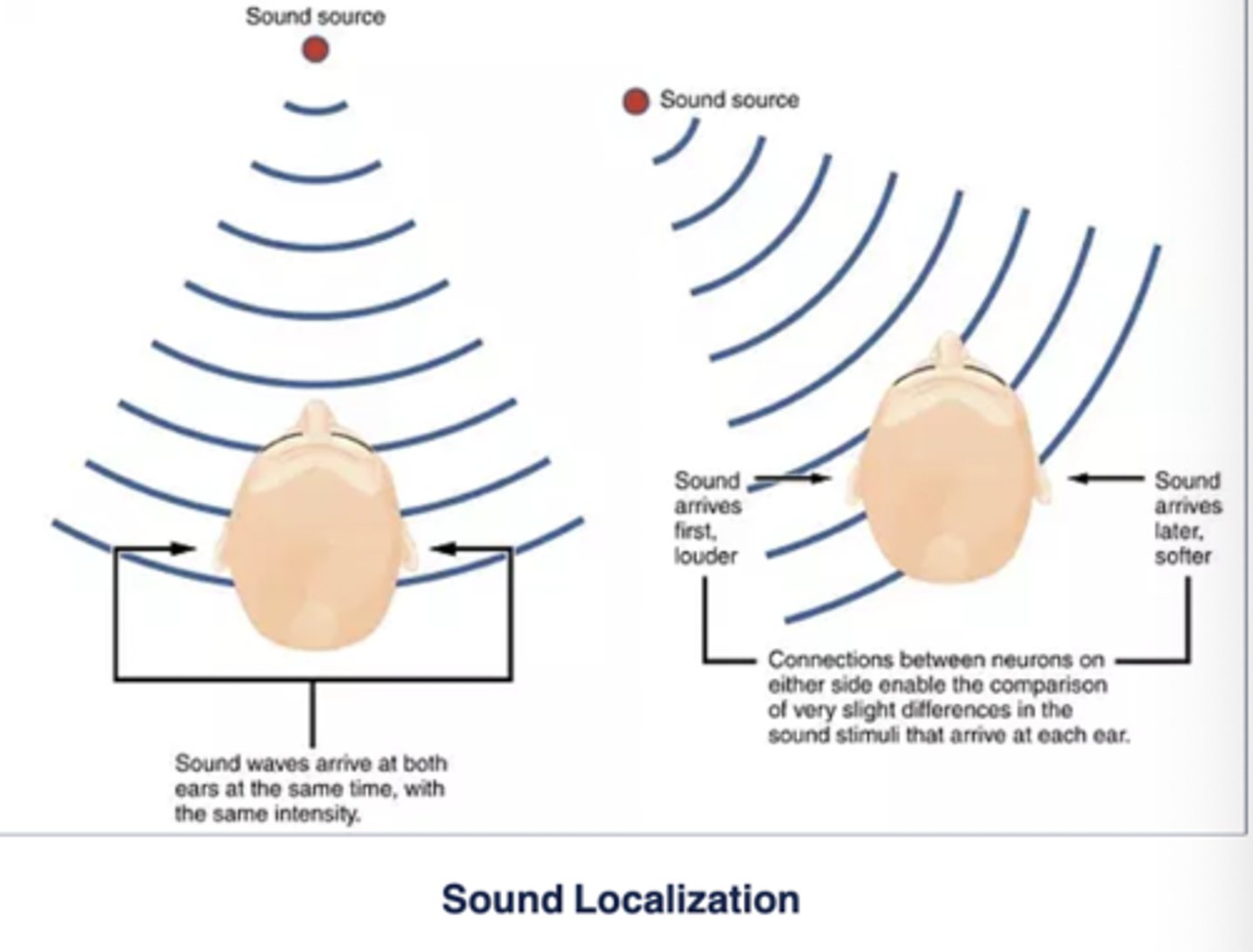
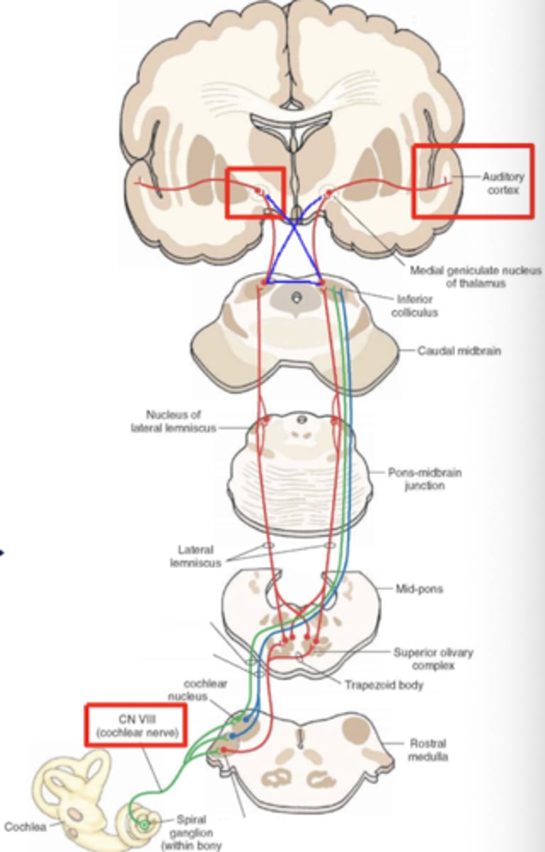
Information in the auditory pathway arrives ____________ (contralaterally/ipsilaterally)
Ipsilaterally
- quickly becomes bilateral after entering the CNS at the medulla
Where does the auditory pathway terminate?
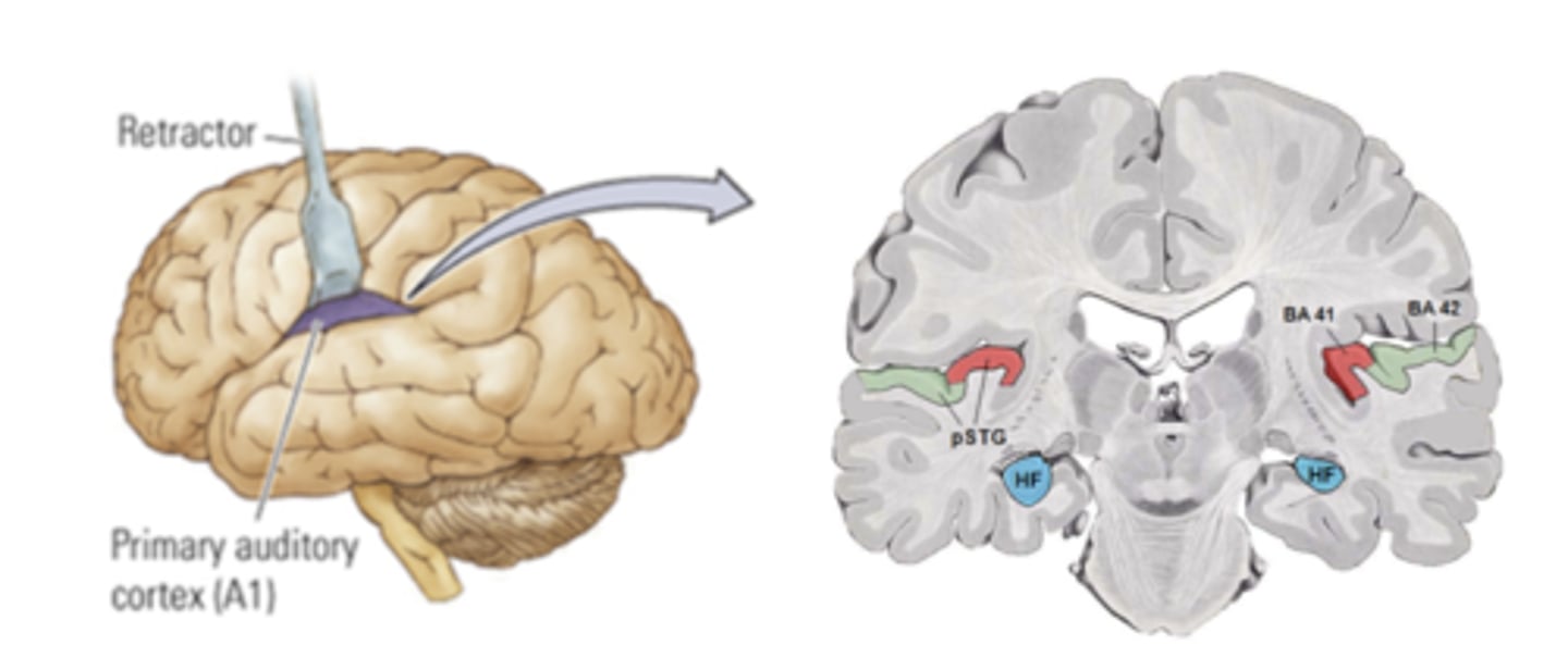
Primary auditory cortex (temporal lobe)
- Superior temporal gyrus = Heschl's Gyrus
- medial face of the lateral fissure in temporal lobe
T/F: Tonotopic organization begins in the cochlea and is maintained throughout the entire pathway to the primary auditory cortex
True
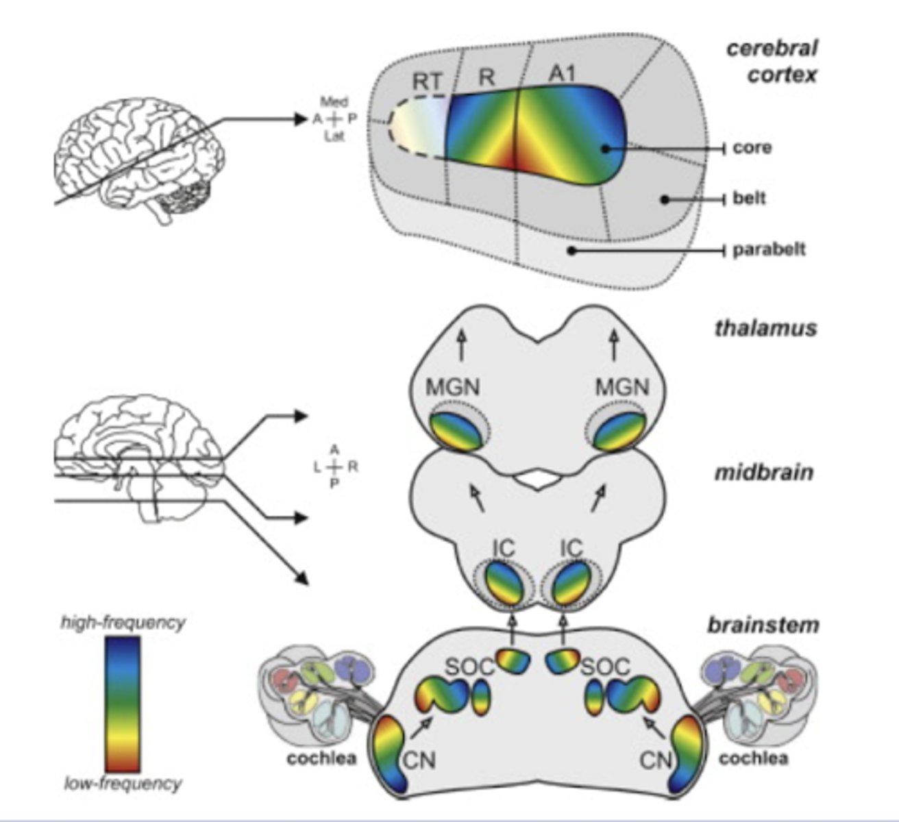
Auditory pathway - Steps
- Cochlea: hair cells --> cochlear/spiral ganglion --> cochlear (VIII nerve) -->
- Medulla: Cochlear nuclei --> ipsilateral and contralateral (trapezoid body) projections -->
- Pons: Superior olivary nuclei --> Nucleus of the Lateral leminiscus
- Midbrain: Inferior colliculi -->
- Thalamus: Medial geniculate nucleus -->
- Temporal lobe: Primary auditory cortex
2 types of acquired deafness:
- Conductive hearing loss
- Sensorineural hearing loss
What is conductive hearing loss?
= Mechanical issue
- sound waves are impeded
- sudden, extreme noise = rupture of tympanic membrane
What is sensorineural hearing loss?
- Possible causes? (4)
= Damage/loss of hair cells
Causes:
- Noise-induced: damage to sterocilia in Organ of Corti (loss of high-frequency first)
- Certain medications (e.g. gentamycin, streptomycin, neomycin)
- Infection of inner ear
- Presbycusis
What is Presbycusis?
Aging-related progressive sensorineural hearing loss
- Due to destruction of hair cells at the cochlear base
- often loss of higher frequencies and preserved low-frequency hearing at apex
As we age, we begin to lose the functioning of the hair cells in the ___________ of the basilar membrane, where the __________ tones are encoded
base; high
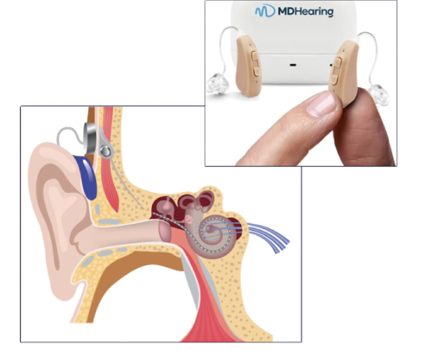
Difference between hearing aids and cochlear implants
- Hearing aids: amplify sound and are suitable for mild to moderate loss
- Cochlear implants: directly stimulate the auditory nerve and are used for severe to profound loss where hearing aids are ineffective
Function:
- Outer ear
- Middle ear
- Inner ear
- Outer ear: captures sound
- Middle ear: amplifies the pressure changes
- Inner ear: transduces sound energy into neural signals
Auditory pathway includes a synapse in the:
thalamus (MGN)
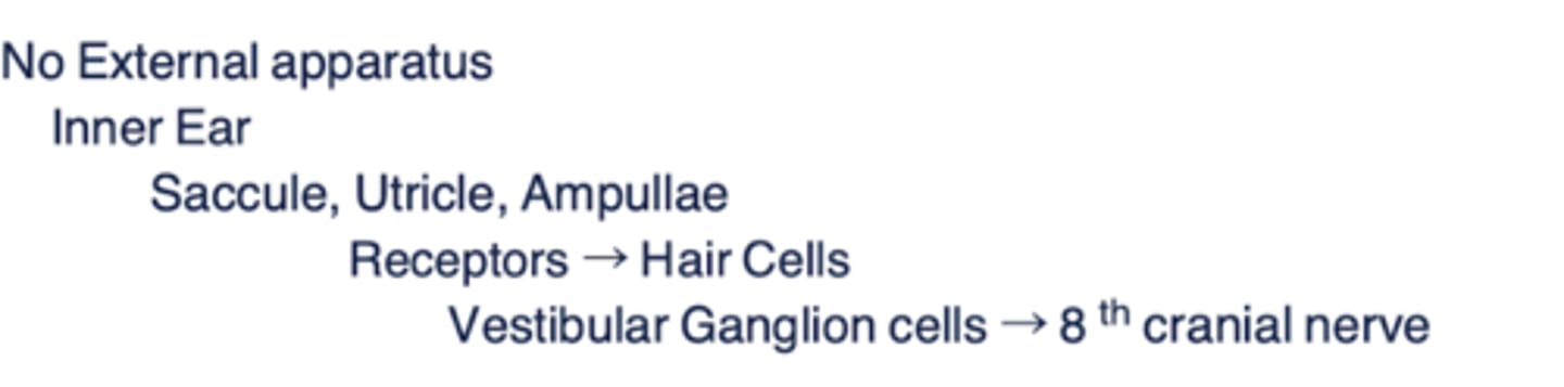
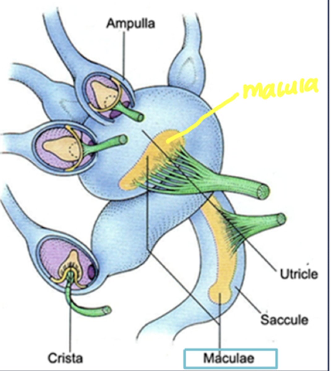
Vestibular system - Peripheral structures
- No external apparatus
- Inner ear
- Saccule, utricle, ampullae
- Receptors --> Hair cells
- Vestibular ganglion cells --> 8th CN
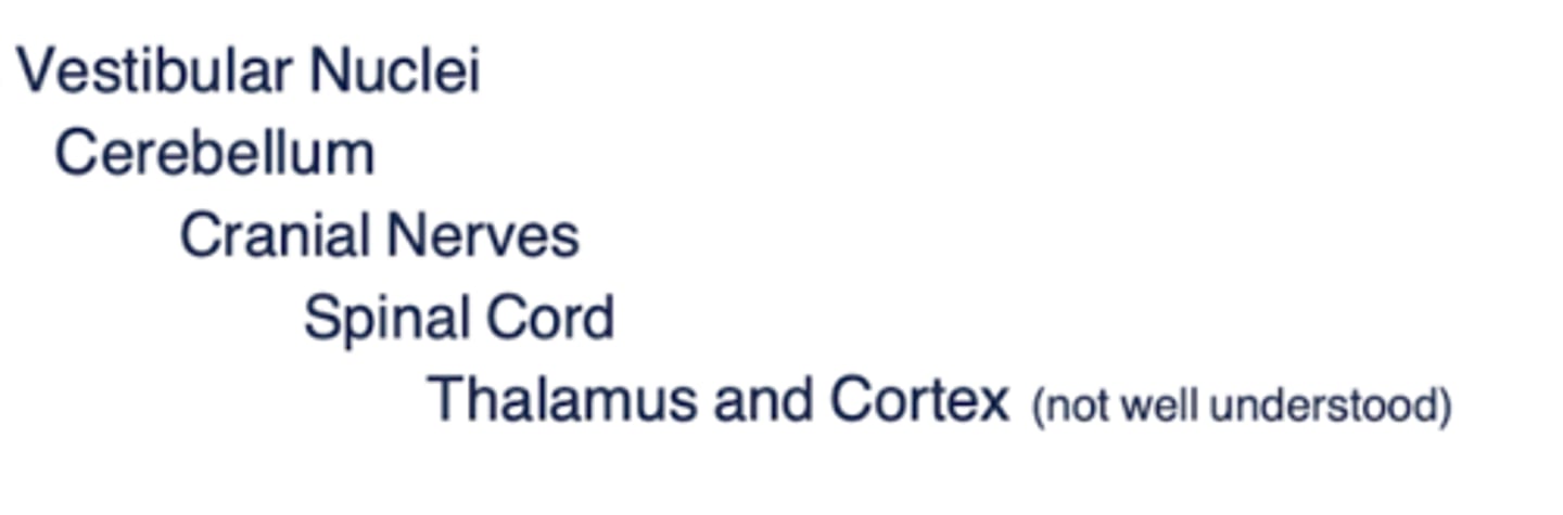
Vestibular system - Central structures
- Vestibular nuclei
- Cerebellum
- Cranial nerves
- Spinal cord
- Thalamus and cortex
Vestibular sense - Functions (6)
- Monitors the position and movement of the head
- Balance and equilibrium
- Posture and adjustments
- Coordinates movements of the head and eyes
- Focus on an object as we move our heads in space
- Vestibulo-autonomic reflexes
3 types of mechanical energy
= changes in head movement
- Linear motion (acceleration/deceleration)
- Angular motion (acceleration/deceleration)
- Tilt
What is linear motion?
Position of head/body relative to gravity
- Acceleration/deceleration
- Motion along each axis with static head positions
-- x: front or back
-- y: left or right
-- z: up or down
What is angular motion?
Rotating head positions, turning about each axis
- x: side-to-side; "maybe"
- y: up and down; "yes"
- z: left and right: "no"
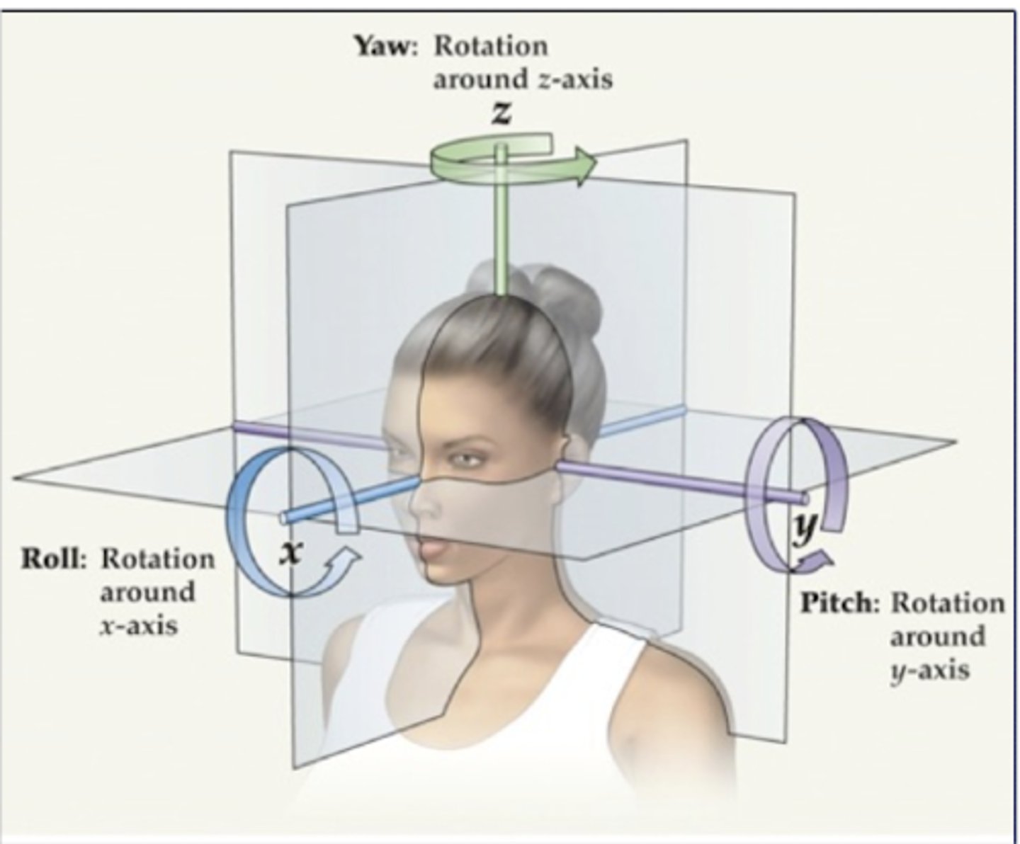
What is tilt?
Head is right-side up or tilted
- static position
Which organs are responsible for detection in the vestibular system?
- what kind of motion do they detect?
- Utricle and saccule (otolith organs) --> detect linear motion (static head); tilt
- Semicircular canals --> detect angular motion (head rotation)
Vestibular sensory organs are routed through the:
Vestibulococchlear nerve (CN VIII) --> CNS
- through sensory afferents
- Utricle detects ____________ in the ____________ plane
- Saccule detects _______________ in the ___________ plane
Utricle detects linear acceleration in the horizontal plane (oriented horizontally)
- x and y axis --> forward/backward, side to side
Saccule detects linear acceleration in the vertical plane (oriented vertically)
- z axis --> up and down , gravitational force
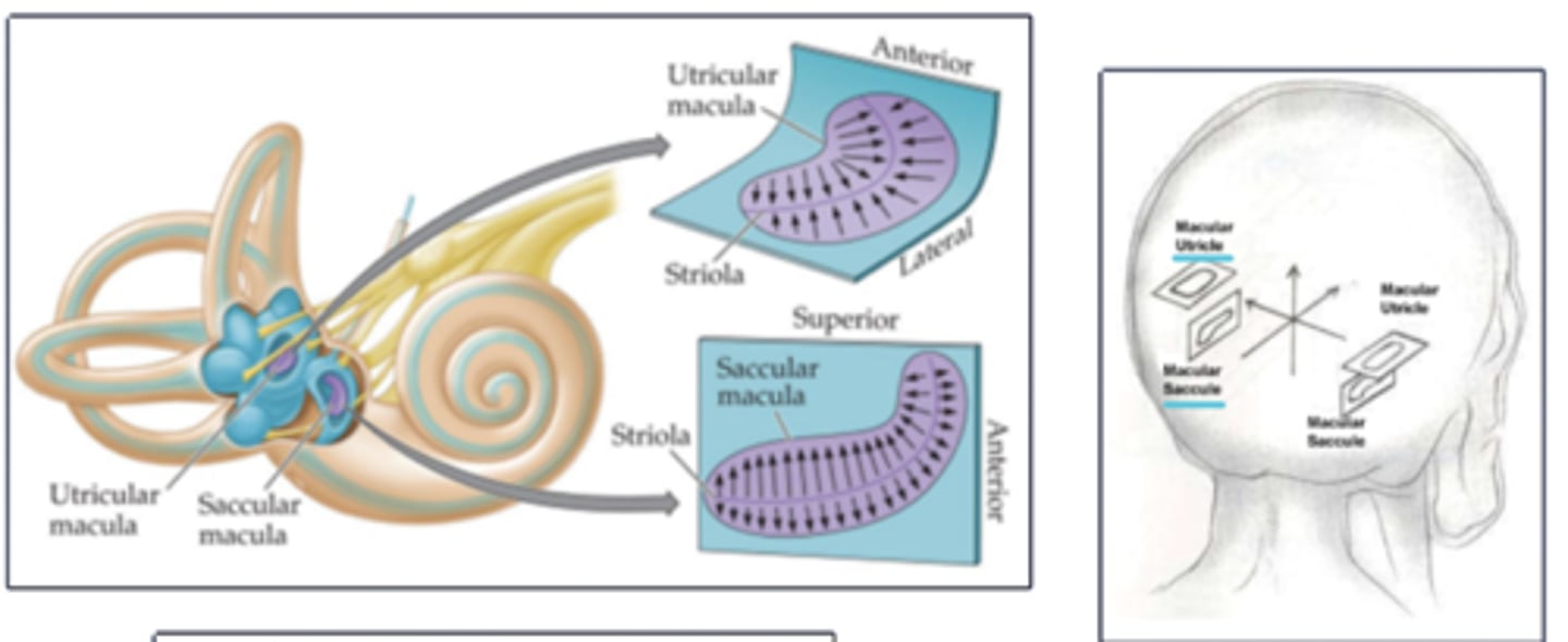
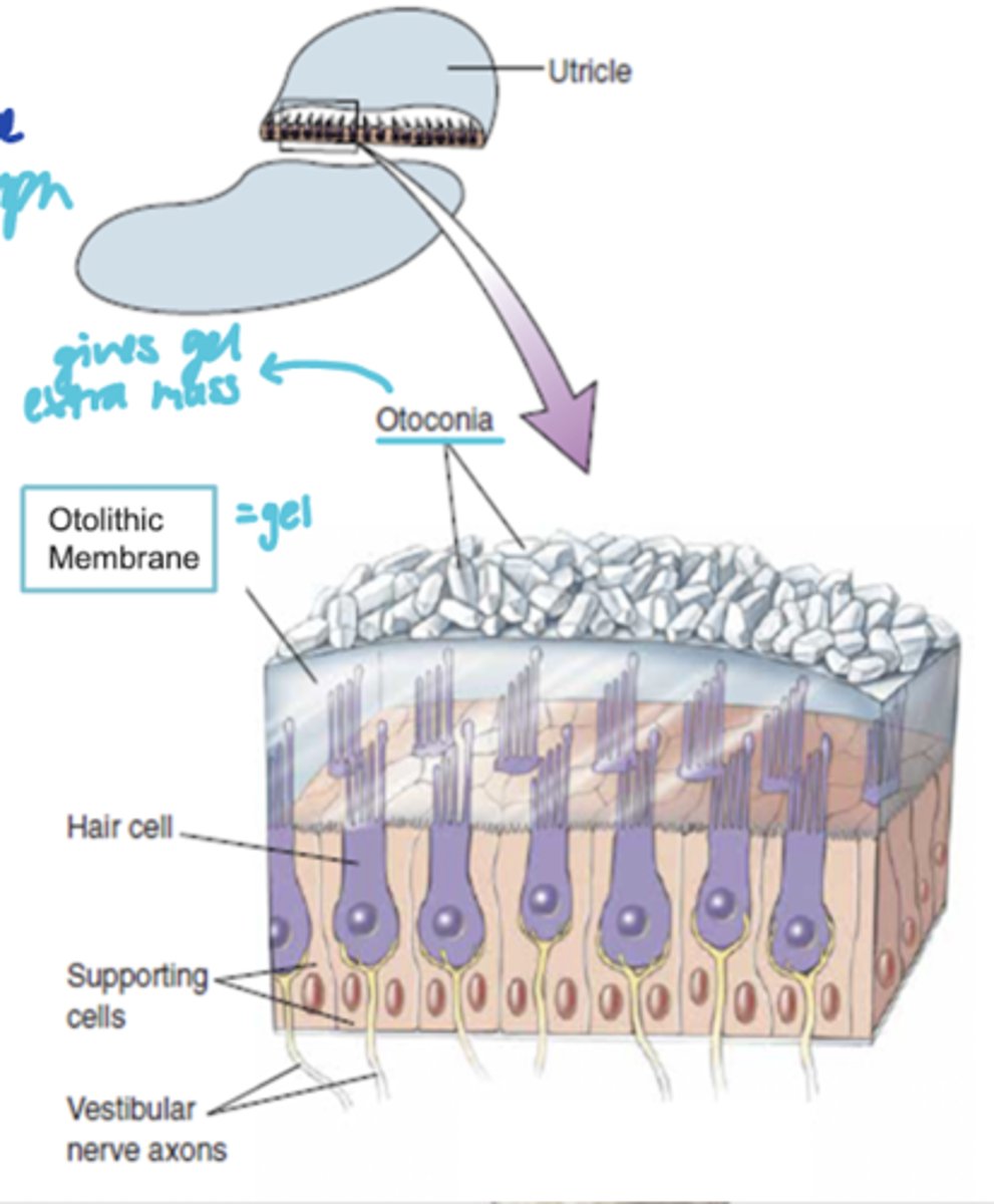
What is the Macula?
- what does it contain?
= Sensory organ within the utricle and saccule
- Contains hair cells and supporting cells
Describe the organization of the macula
- what structures (2)?
Otolithic membrane
- gelatinous substance
- surrounded by endolymph
- hair cell cilia project into this gel
Otoconia
- calcium carbonate crystals on surface of otolithic membrane
- gives extra mass
When occurs in the maccula when the head accelerates or tilts?
- Force exerted on otoconia and otolithic membrane
--> This bends the sterocilia
- Bending toward the kinocilium = depolarization
- Hair cells fire APs in CN VIII
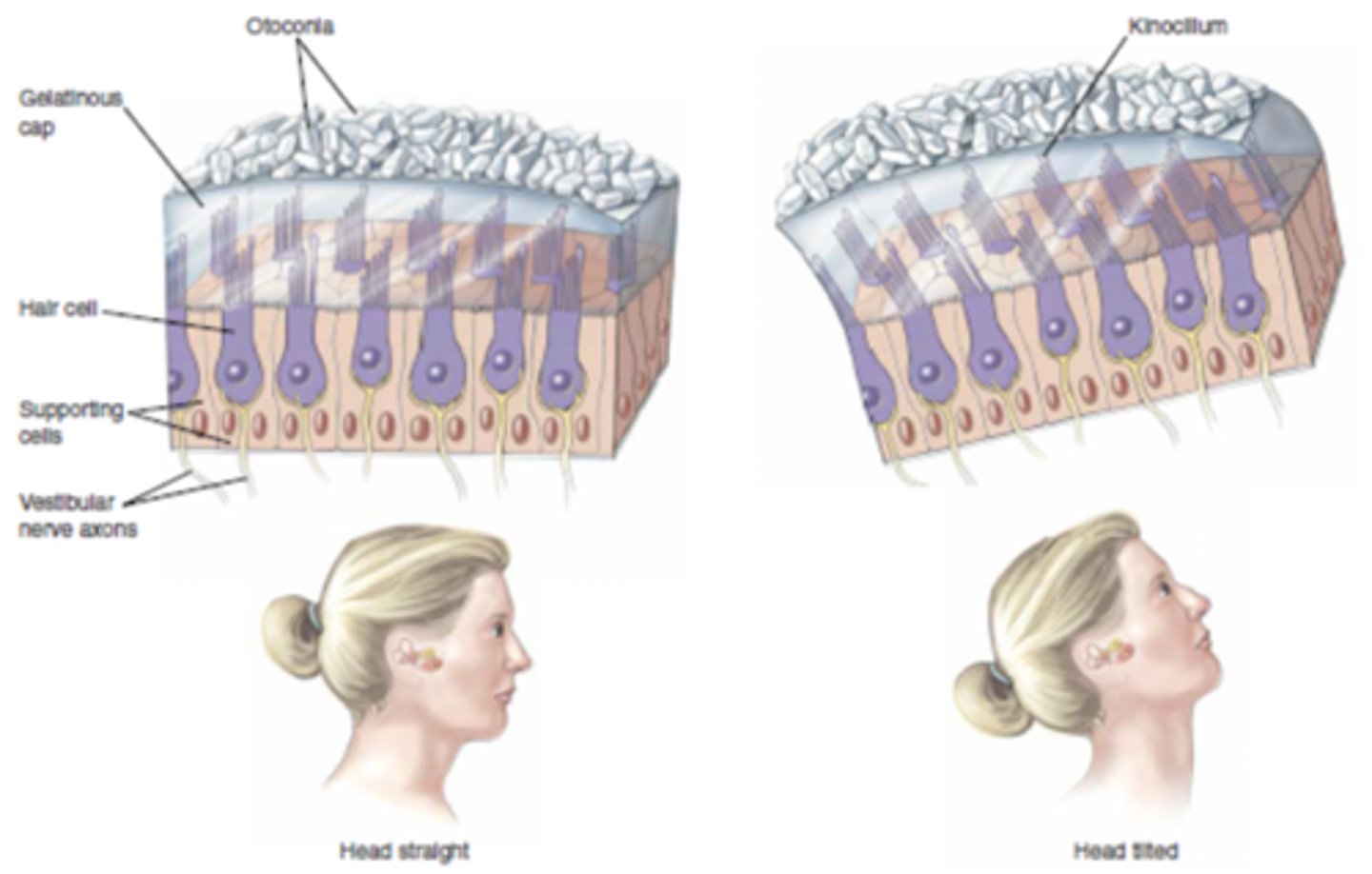
Hair cells have a _______________ orientation of the saccule and utricle on either side
bilateral, mirror-image
- excitation on one side of head = inhibition on opposite side
There are ______ semicircular canals
- oriented ______ degrees from each other
3
90
- detect angular accelerations in all 3 planes
What is the crista?
- where is it?
- what does it contain?
= Sensory organ of semicircular canals, housed within the ampulla (bulbous expansion at base of each semicircular canal
- contains hair cells
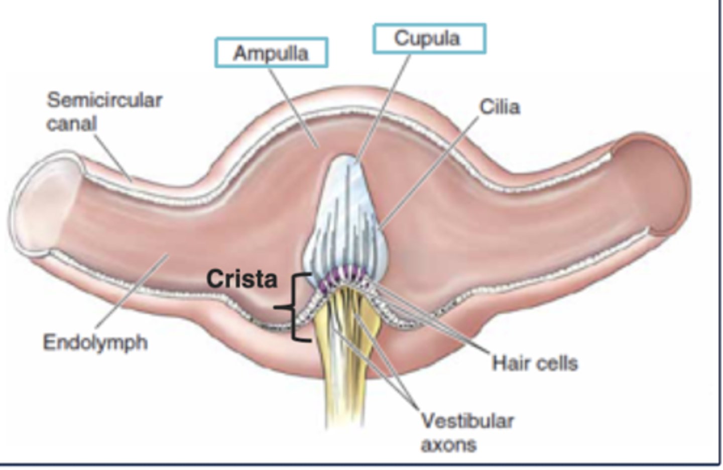
The hair cell stereocilia and kinocilium in the crista of the semicircular canals project into the gelatinous mass called the _______________ which is surrounded by ______________
Cupula
Endolymph
When occurs in the crista when the head rotates in one plane?
- Movement of endolymph produces a force across the cupula
- Cupula (and endolymph) is displaced (in opposite direction of head turn)
- Bends the stereoceilia --> bending toward kinocilium = depolarization
- Hair cells fire APs in CN VIII
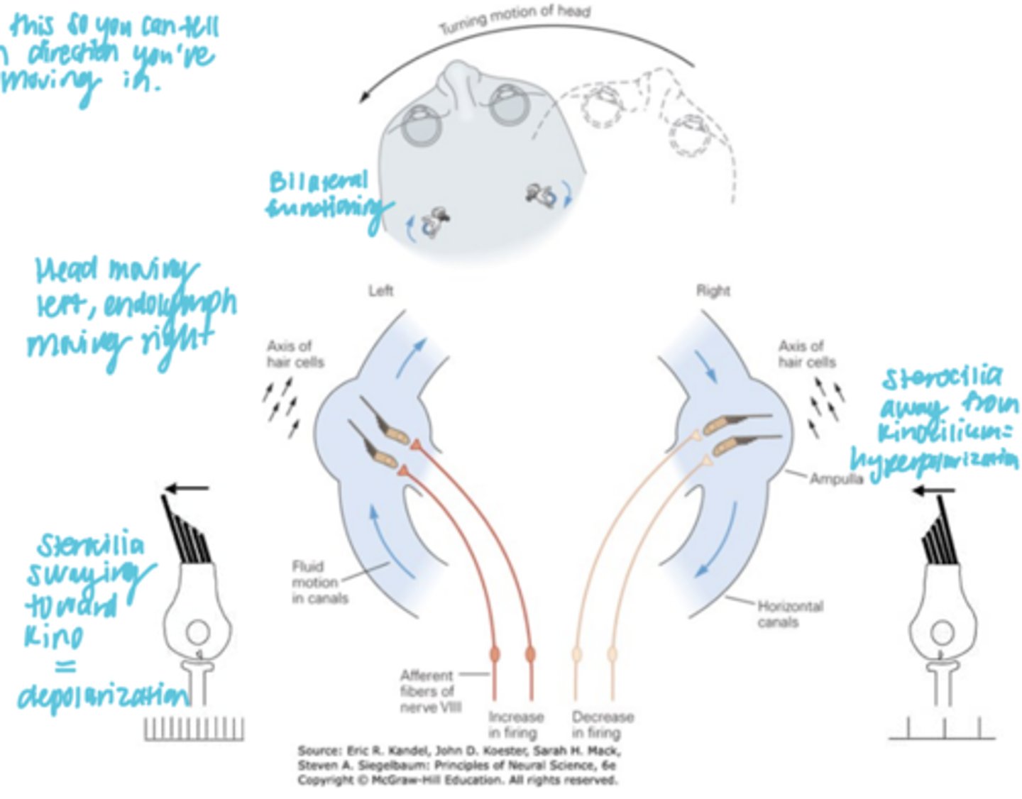
*Bilateral, mirror-image oreintation of the semicircular canal*
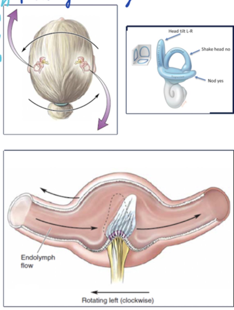
Describe the bilateral function in the semicircular canals
- Each semicircular canal works in concert with a partner on the opposite side of the head
- Head rotation bends the cupula in opposing directions for the two partners, resulting in opposite changes in firing rates
--> When you turn your head to the left, the cupula and endolymph move to the right. The LEFT side of your head semicircular canals are stimulated, resulting in depolarization. The RIGHT side of your head semicircular canals are inhibited (bc sterocilia away from kinocilium), resulting in hyperpolarization.
Hair cells in the vestibular system exist in____________ ganglion
Scarpa's

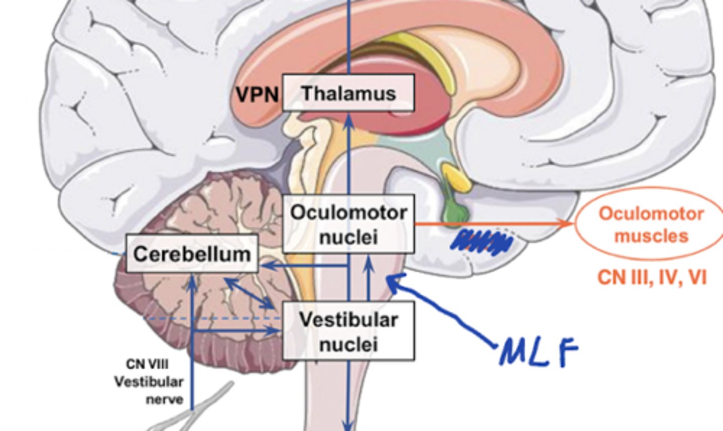
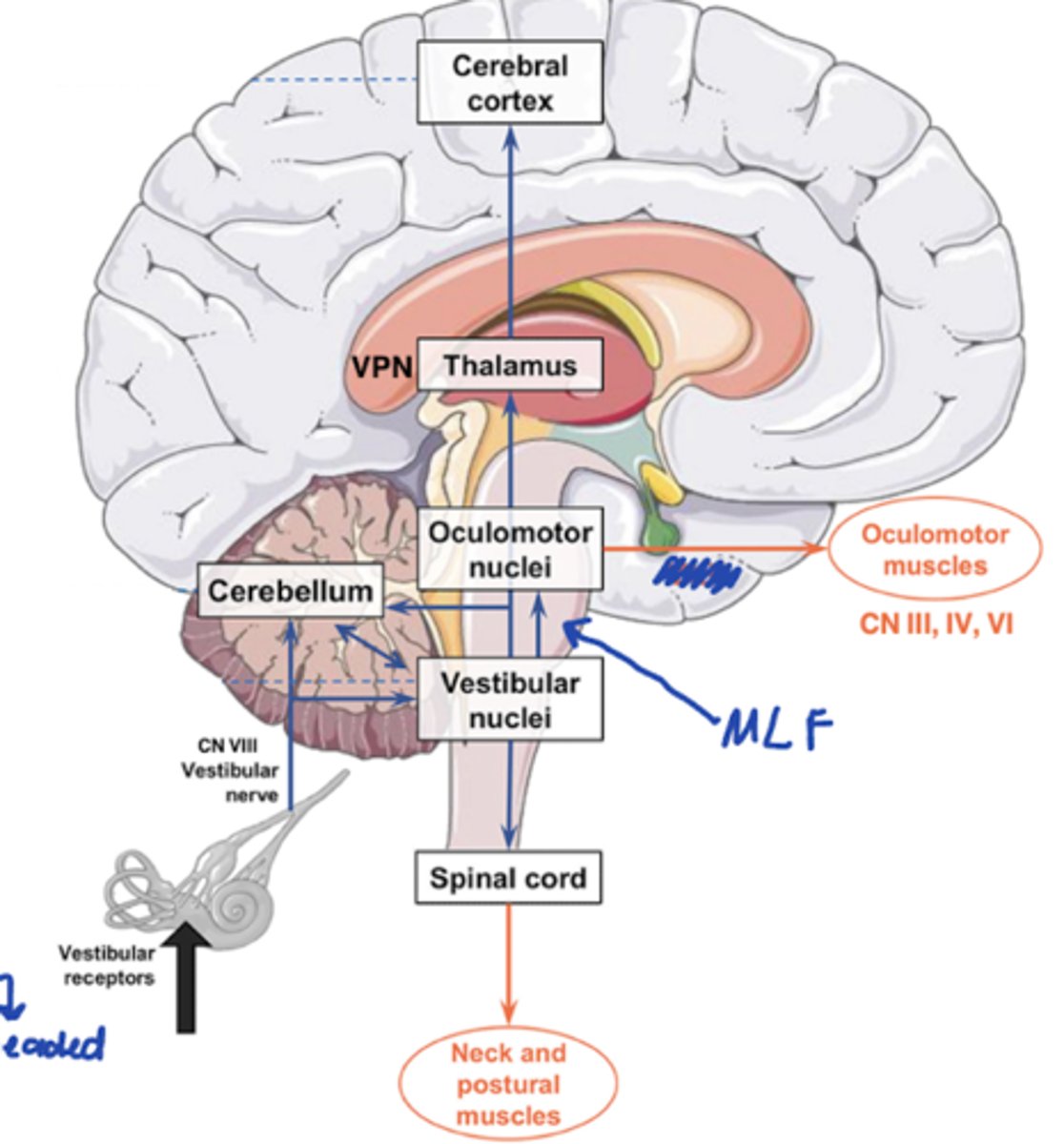
CN VIII afferents route to: (2)
1. Vestibular nuclei
-- caudal pons and rostral medulla
2. Cerebellum directly
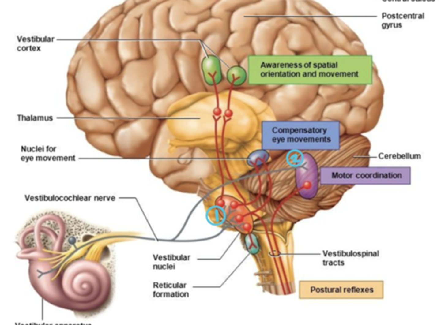
Vestibular nuclei receives input from (3)
- Cortex
- Cerebellum
- Brainstem (e.g. visual and proprioceptive)
5 Vestibular pathways
- Vestibulospinal tract
- Vestibuloccular tract
- Vestibulocerebellar tract
- Vestibulo-thalamo-cortico tract
- Vestibuloautonomic tract
Vestibulospinal tract
- pathway?
- function?
- VN --> spinal cord
- Modulate neck and trunk muscles for posture and balance

Vestibuloccular tract
- pathway?
- function?
- VN --> Cranial nuclei that control eye movements (III, IV, VI) via the Medial Longitudinal Fasciculus (MLF)
- Eye movements: VOR = maintain steady gaze as we move our head
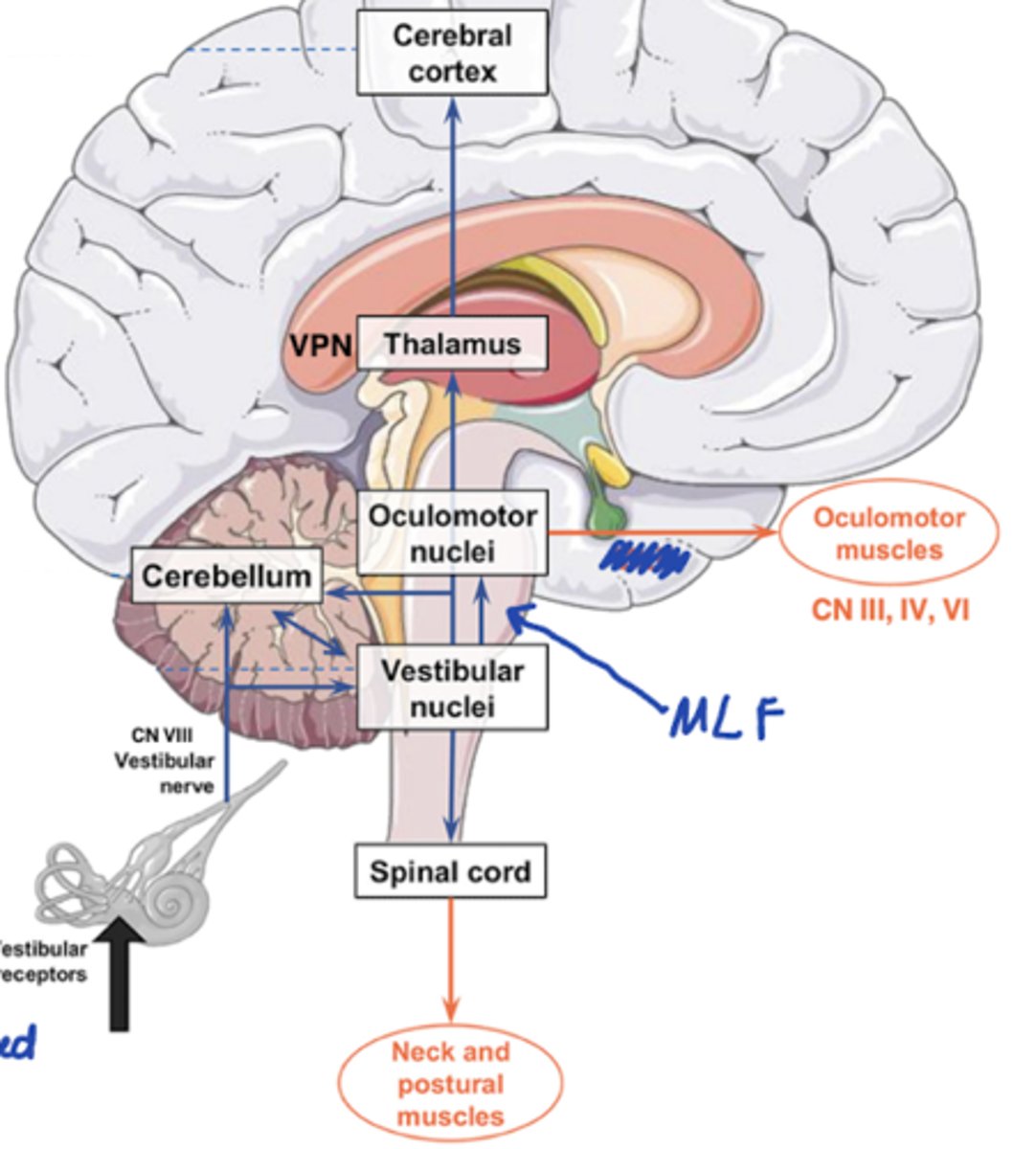
Vestibulocerebellar tract
- pathway?
- function?
- VN <--> Cerebellum; feedback loop
- Posture, balance, eye movements, and motor control
Vestibulo-thalamo-cortico tract
- pathway?
- function?
- VN --> Thalamus to cortex
- Conscious perception and control of balance
Vestibuloautonomic tract
- pathway?
- function?
- VN --> Reticular formation and other brainstem areas
- Stabilizes autonomic processes during motion and changes relative to gravity
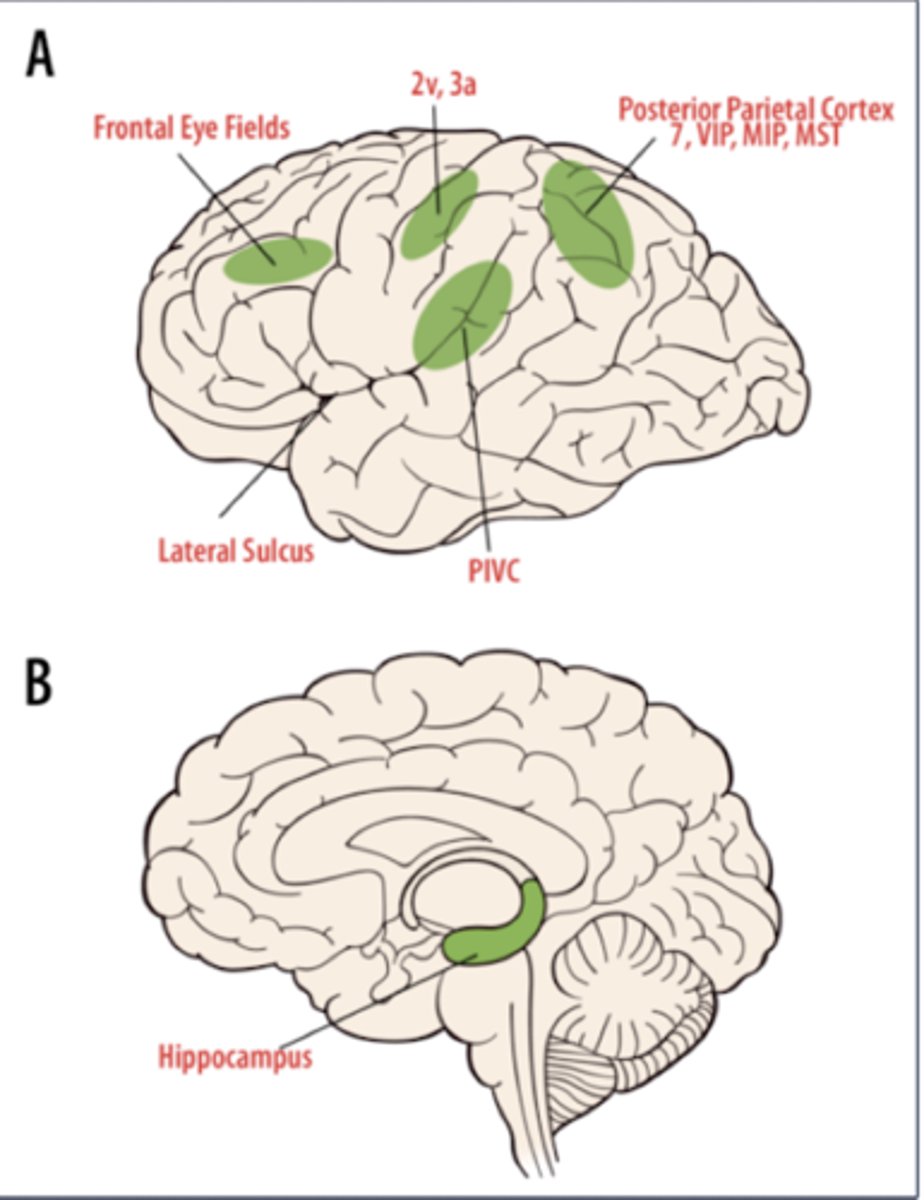
What do these cortical projections control in the vestibular system
- Frontal eye fields
- Areas 2v and 3a
- Area PIVC
- Posterior parietal cortex
- Hippocampus
- Frontal eye fields: control voluntary eye movements
- Areas 2v and 3a: somatosensory areas that map body location and movement signals
- Area PIVC: responds to body and head motion information (parieto-insular vestibular cortex)
- Posterior parietal cortex: motion perception and responds to both visual and vestibular motion cues
- Hippocampus: spatial orientation and navigation
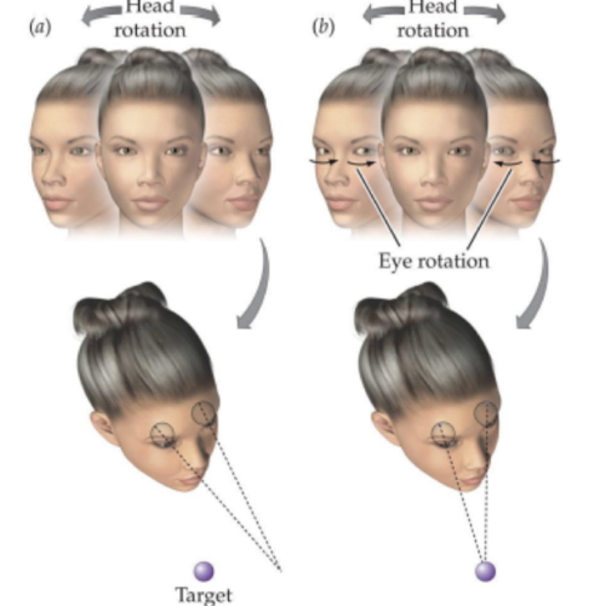
What is the Vestibulo-ocular reflex (VOR)?
Eyes move to opposite direction of head to maintain gaze on target
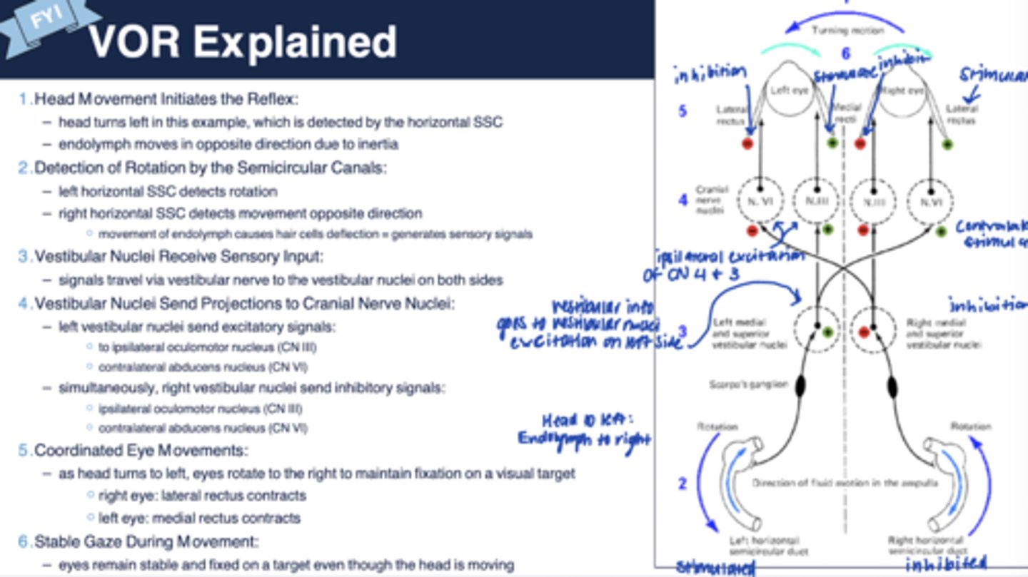
VOR explained
she said not rlly testable, just FYI
Vestibular impairment - Symptoms (7)
- Spatial disorientation
- Dizziness
- Vertigo
- Imbalance
- Blurred vision
- Illusory self-motion
- Motion sickness
What is Mal de debaruqement syndrome?
= "Getting your sea legs"
- when you get off a boat and still feels the waves; perception that you are still moving
What is Meniere's Syndrome?
- Sudden dizziness, imbalance, and spatial disorientation
- Sudden falling down
- Vomiting from severe motion sickness
What is Nystagmus?
= Abnormal eye reflex causing nodding of the eyes
--> Impairment of the VOR
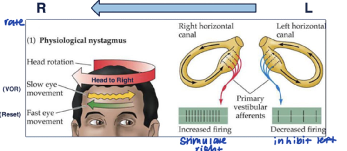
Describe physiologic (normal) nystagmus
Scenario: move head to right
- As the head turns, ipsilateral vestibular afferents increase firing rates while, contralateral vestibular afferents decrease firing rates (stimulate right side and inhibit left)
- Net difference in firing rates between two sides leads to a:
--- SLOW movement of the eyes (due to VOR circuitry) to counter the head motion (slow movements of eyes to the left)
- FAST saccadic motion to reset the eyes in the direction we move our head (fast movement of eyes to the right to catch up to head)
What causes pathologic nystagmus to occur?
Scenario: Vestibular system is impaired on the left side --> at rest, the right semicircular canal has baseline tonic firing, while left is not active at all --> NS detects this difference as movement toward right side
- So the eyes reflexively move to the left
--> producing nystagmus = slow drift w/ abnormal nodding of the eyes
Define pathologic nystagmus
= unilateral damage to vestibular system
- eyes make repetitive, uncontrolled movements when head not moving
- Results in the inability to both eyes to steadily view objects
-- reduced vision and depth perception
-- may affect balance and coordination
-- may nod or hold their head in unusual positions
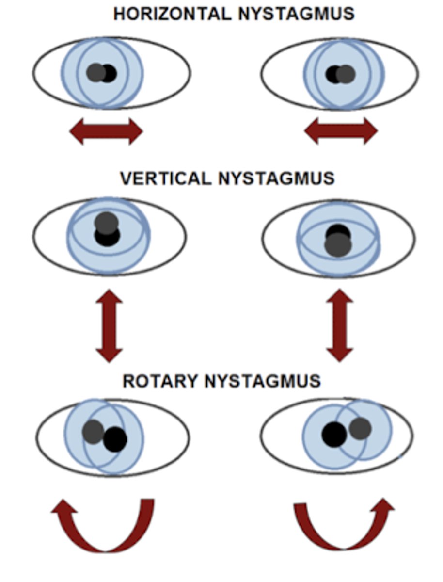
3 types of nystagmus
- Horizontal
- Vertical
- Rotary
Causes of nystagmus (8)
- Stroke
- Inner ear disease
- Multiple sclerosis (MS)
- Alcohol intoxication
- Head trauma
- Brain tumors
- Cerebellar disease
- Medications
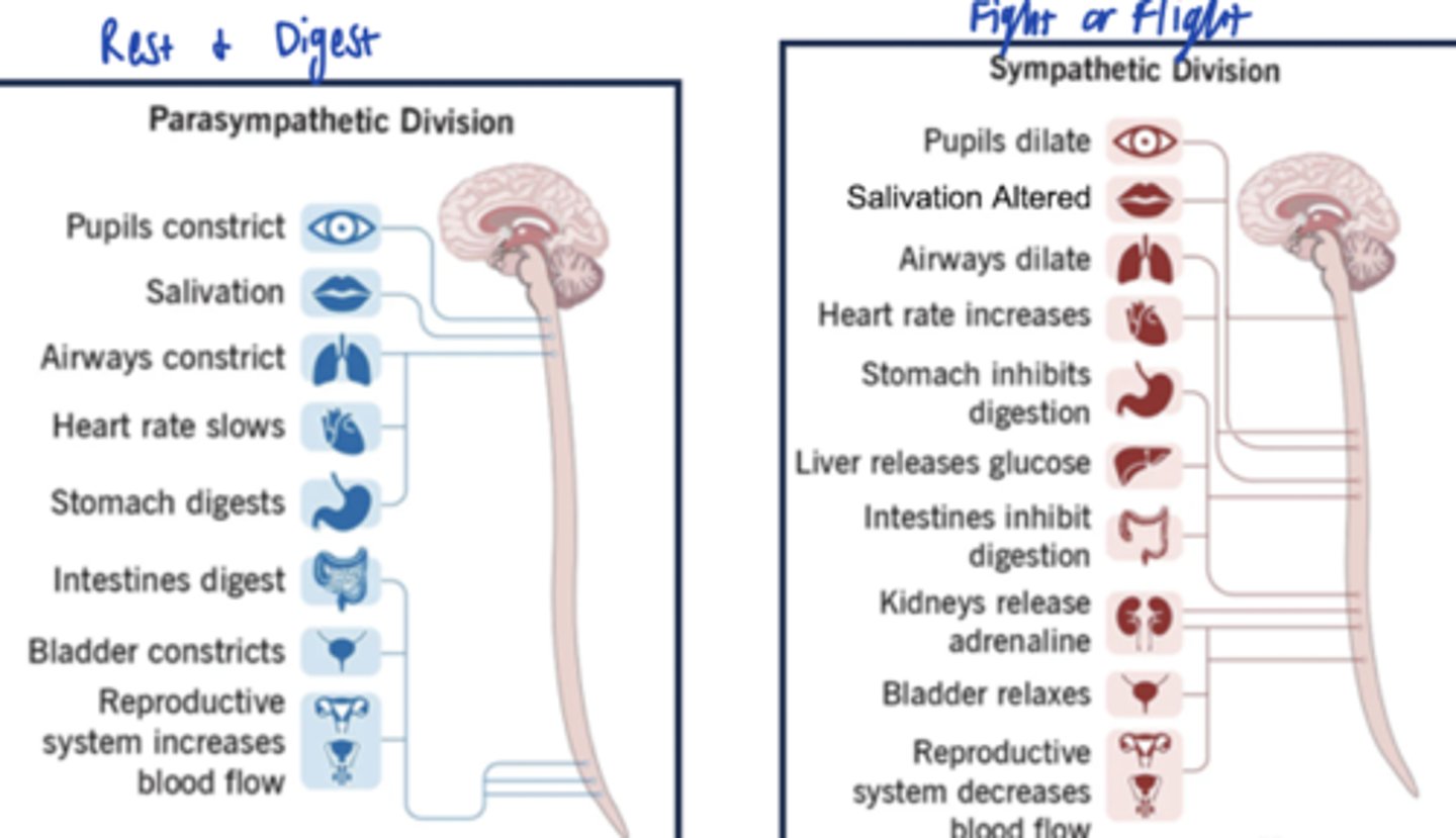
Functions of parasympathetic NS
Predominant tone for resting, digestion, evacuation (excretion), and sexual activity
- rest/digest
- feed/breed
- protective
Which body structures are innervated by the parasympathetic vs sympathetic NS?
Parasympathetic
- Head, thorax, abdominal and pelvic cavities, genitalia
- does NOT innervate most blood vessels or structures in the skin
Sympathetic
- Head, thorax, abdominal and pelvic cavities, genitalia
- DOES innervate blood vessels and skin
T/F: Parasympathetic NS can do mass discharge, but sympathetic NS cannot
False - the opposite
- Parasympathetic: typically, very discrete (separate) control of individual effectors
- Sympathetic NS IS capable of mass discharge --> activation of multiple organ systems concurrently, inhibition of others (decrease blood flow to skin and digestive tract activity)
The parasympathetic NS has a _____________ (short/long) preganglionic axon, and a ____________ (short/long) postganglionic axon
The sympathetic NS has a _____________ (short/long) preganglionic axon, and a ____________ (short/long) postganglionic axon
- Parasympathetic: long preganglionic, short postganglionic
- Sympathetic: short preganglionic, long postganglionic
T/F: The parasympathetic NS inhibits urination and defecation
False - causes urination and defecation
Parasympathetic vs Sympathetic effect on the eyes
- Parasympathetic: Causes pupils to constrict in response to bright light (protective)
- Sympathetic: Causes pupils to dilate in response to dim light (enhance vision)
Parasympathetic vs sympathetic effect on the airways
Parasympathetic:
- Causes constriction of airways (protective)
- Increases bronchial secretions (protective)
Sympathetic:
- Dilates bronchioles
Parasympathetic vs sympathetic effect on the heart
- Parasympathetic: Decreases HR/slows the heart
- Sympathetic: increases HR
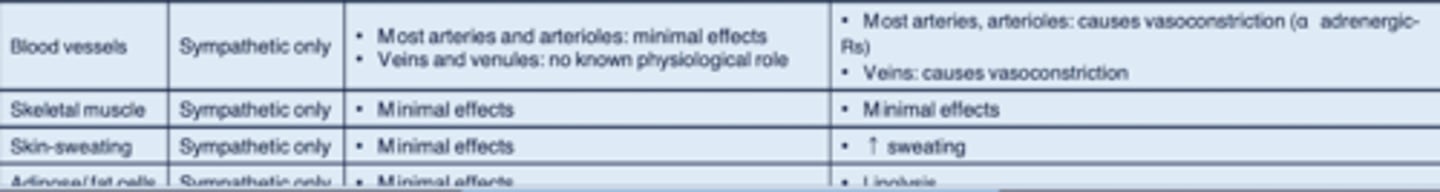
Parasympathetic vs sympathetic effect on the blood vessels
- Parasympathetic: Minimal effects on arteries and veins bc little innervation in these areas
- Sympathetic:
-- most arteries, arterioles: causes vasoconstriction (alpha-adrenergic receptors)
-- veins: vasoconstriction
Parasympathetic effect on skeletal muscle
Minimal effects/none
Parasympathetic effect on sweating
Minimal effects (no innervation in the skin)
Parasympathetic effect on the sexual response
- In males, causes erection
- In females, increases blood flow and lubrication
Parasympathetic vs sympathetic effect on salivation
- Parasympathetic: Increases watery secretions
- Sympathetic: increases thick mucous secretions
Parasympathetic effect on the GI system
Activity is INCREASED
- Increased motility in stomach and intestines
- Sphincters: relaxation
- Increased GI secretions (saliva, HCl)
- Increased pancreas secretions (pancreatic enzyme secretion (exocrine), insulin secretion (endocrine))
- Gallbladder: contraction
- Liver: glycogen synthesis (glucose storage)
Functions of sympathetic NS
Predominant tone under conditions of physical activity or stress
- Fight or Flight = stress response
Parasympathetic vs Sympathetic NS efferent activity compared - Chart
Adrenergic receptos - Functions:
- Alpha 1
- Alpha 2
- Beta 1
- Beta 1 and Beta 2
- Beta 2
- Alpha 1: peripheral blood vessels --> vasoconstriction
- Alpha 2: inhibits GI secretions
- Beta 1: SA node --> increases HR
- Beta 1 and Beta 2: cardiomyocytes --> increased contractility
- Beta 2: lungs --> bronchodilation and vasodilation
Usually, parasympathetic tone is ________________ to sympathetic tone on the same effector, but sometimes, dual innervation is ___________
- ex of each scenario
opposite; synergistic
- Opposite ex: increased sympathetic = increased HR, and increased parasympathetic = decreased HR
- Synergistic ex: Salivation; increased PS = normal saliva, and increased sympathetic = altered saliva
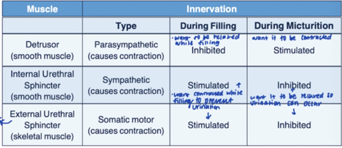
Destrusor, Internal urethral sphincter, and external urethral sphincter
- filling vs micturition
Parasympathetic regulation of micturition
- Detrusor: contraction
- Internal and external urethral sphincter: relaxation
2 types of sweat glands
- Epocrine: located all over body, function for thermoregulation
- Apocrine: located in armpits, genitals, perianal; active from puberty onward
Targets with sympathetic innervation only
- Sweat glands
- Pilomotor muscles of skin (surround hair follicles)
- Arterioles (most)
- Veins
- Adipose fat cells
T/F: An autonomic reflex travels through the cerebral cortex
False
- it is an involuntary response to a stimulus, cerebral cortex is not directly involved
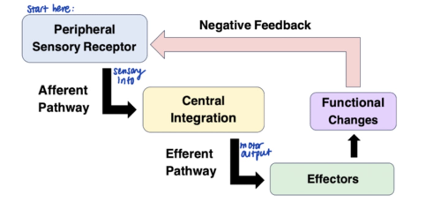
3 components of a reflex arc
1. Afferent limb = sensory input
- receptor --> afferent pathway
2. Central integration center (brainstem)
3. Efferent limb = Autonomic input
- efferent pathway --> effector (SM, glands, GI, neurons)
Exs of autonomic reflexes (7)
- Pupil diameter
- Salivation
- Swallowing
- BP
- Respiration rate
- Hormone release (ex: ADH)
- GI motility
Describe the ANS blood pressure reflex
- Afferent limb: sensory receptors = baroreceptors; detect stretch
- Integration center: Brainstem (or HTM)
- Efferent: sympathetic and parasympathetic (CN X) innervation - autonomic flow to blood vessels, SA node, ventricles, endocrine tissues
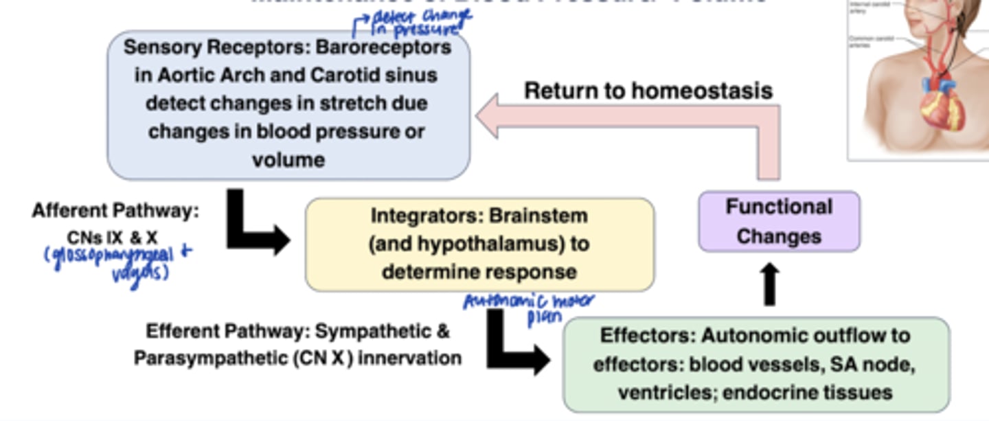
What are baroreceptors and where are they located?
= Mechanoreceptors, respond to stretch
- in wall of aortic arch and carotid sinus