PSYC 121 Midterm 1
1/18
There's no tags or description
Looks like no tags are added yet.
Name | Mastery | Learn | Test | Matching | Spaced |
|---|
No study sessions yet.
19 Terms
Cornea
Does most of the focusing (80%)
Rigid and cannot adjust its focusing power for objects at different distances → lens come into play
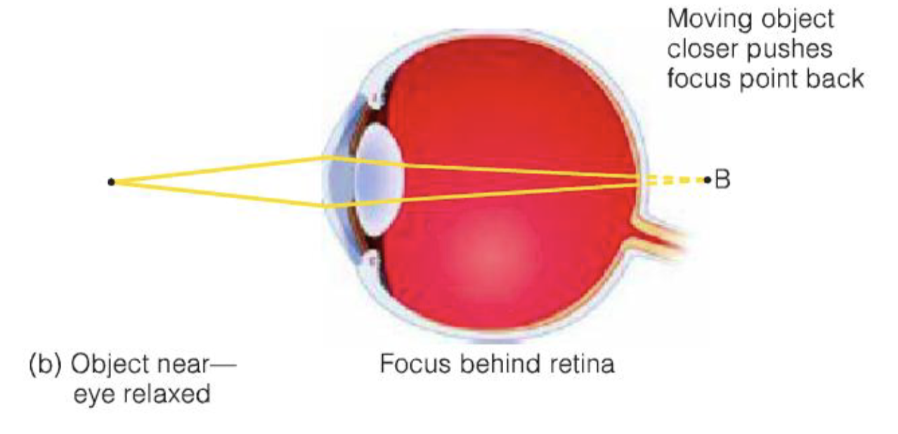
Lens
Can accommodate for CORNEA by contracting (getting thicker) or dilating (getting flatter) to do the remaining focusing work
Looking at a far-away object, the lens is flat
closer → if it doesn’t contract → blur
After the lens contracts/thickens, the object is focused correctly
Cones
3 types (sensitive to different wavelengths)
Most useful for color vision in bright light
High resolution
Most dense in fovea
Rods
Only one type (sensitive to light vs. dark)
No color information, useful in dim light
Low resolution
Most dense in periphery
Fovea which contains only
cones
In most of retina, there are many more ____ than ____
rods; cones
Blind spot
The region on the retina where ganglion cells exit the eye via the optic nerve, sending signals to the brain
No receptors at all in this area
Superior colliculus
The remaining 10% of ganglion cells go to here which is a region involved in controlling eye movements.
Binocular disparity
The difference between the images on the left and right eyes
Functions of the LGN
Organizes the information from retina
Receives feedback (top-down) signals from cortex (e.g. attention modulation)
Regulate the signal from retina, sending fewer impulses to cortex
LGN organizes the info from the retina according to:
Which eye it came from
Which type of receptor it came from (rods or cones)
Receives feedback (top-down) signals from cortex (e.g. attention modulation)
Regulate the signal from retina, sending fewer impulses to cortex
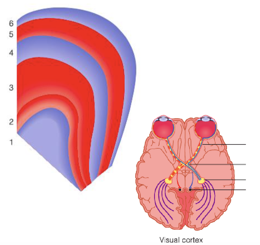
Lateral Geniculate Nucleus (LGN)
Organized into 6 layers, each layer receiving information from one eye
Layers 2, 3, 5 ipsilateral (same side)
Layers 1, 4, 6 contralateral (opposite side)
LGN → V1 Process
Sends most of its axons to the primary visual cortex (V1), and then the signal gets relayed to V2, V3, and so on.
Visual Processing
Light → pupil → cornea + lens → retina → rods and cones receptors → signals emerge from the back of the eye in the optic nerve → LGN → V1 cells break down different orientations of light using simple cells, complex cells, and hyper-complex cells
Simple cells
Excitatory and inhibitory areas arranged side by side, respond best to bars of a particular orientation
Complex cells
Respond best to movement of a correctly oriented bar across receptive field, direction tuned
Hyper-complex cells
respond best corners or angles, or particular length bars moving in particular direction.
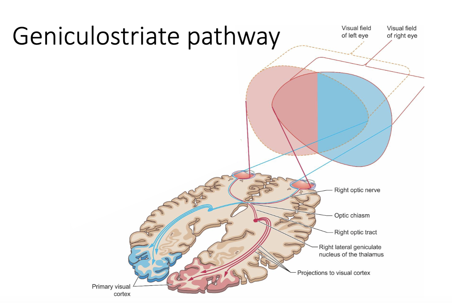
What is the LGN pathway? Draw a diagram of it.
90% of optic nerve fibers go
Left and right visual field → LGN (thalamus) → V1 (primary visual cortex)
Signals from each half of each retina meet at the optic chiasm.
Half stay in the ipsilateral LGN, and half go to the contralateral LGN
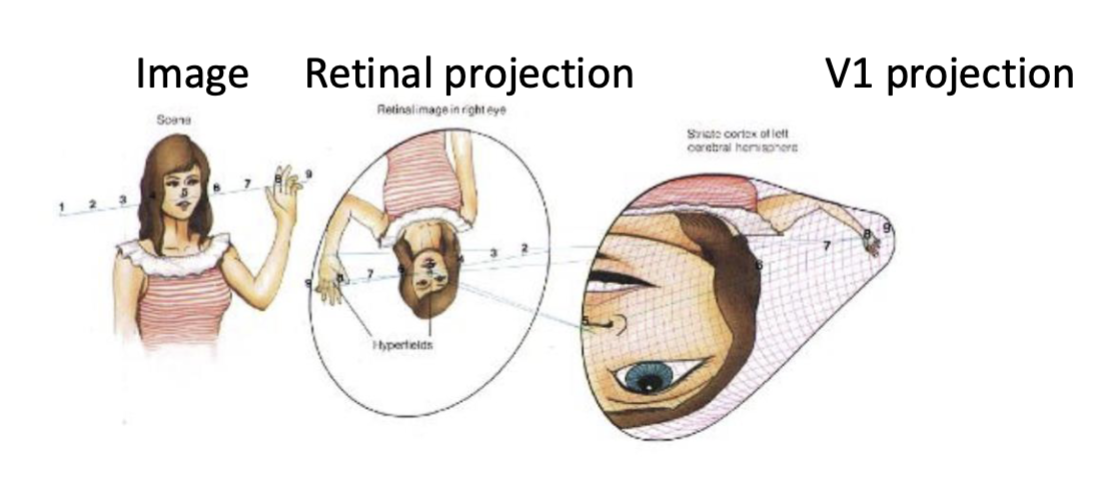
Cortical magnification
A phenomenon whereby the central visual field, particularly the fovea, is represented by a disproportionately large area of the primary visual cortex (V1) relative to the peripheral visual field.
Higher density of photoreceptors and greater processing demands of foveal vision, resulting in finer spatial resolution and more detailed visual processing for stimuli presented near the center of gaze.