Lecture 16: Chemical Senses Central
1/33
There's no tags or description
Looks like no tags are added yet.
Name | Mastery | Learn | Test | Matching | Spaced | Call with Kai |
|---|
No analytics yet
Send a link to your students to track their progress
34 Terms
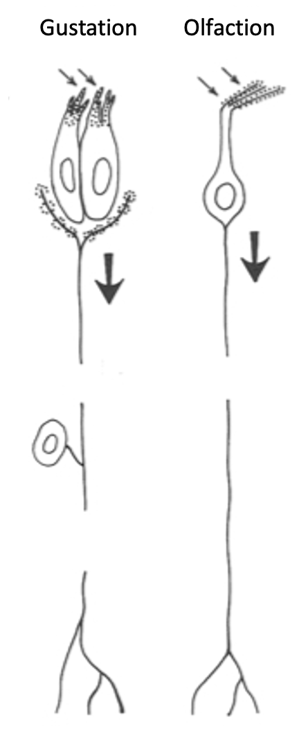
What is unique about olfactory sensory neurons (OSNs) compared to most sensory systems?
OSNs are both receptor cells (transduction) AND neurons that conduct APs to the CNS (no separate ganglion cell needed).
Gustatory system has TRCs for transduction + separate gustatory ganglion cells for conduction to CNS
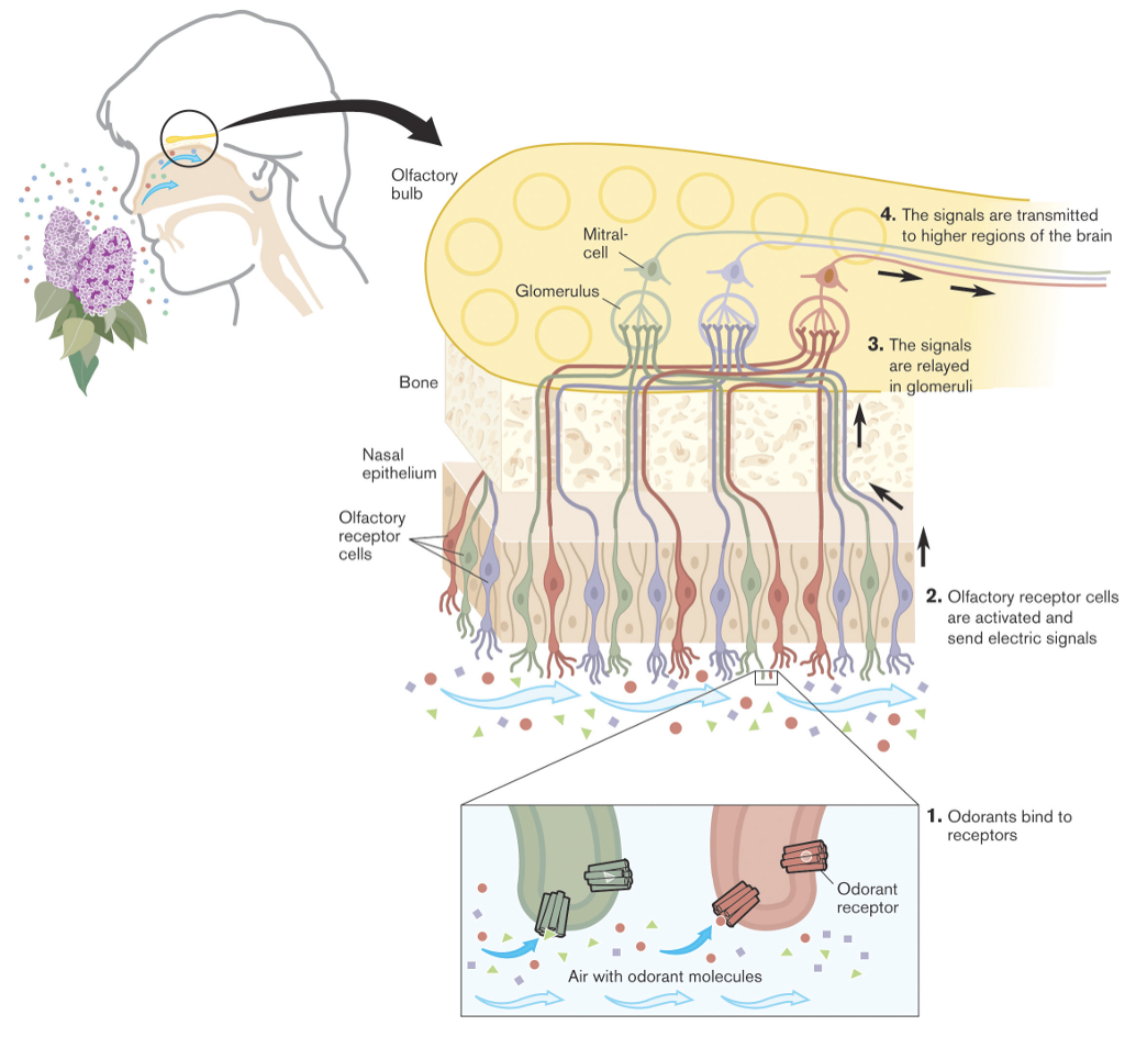
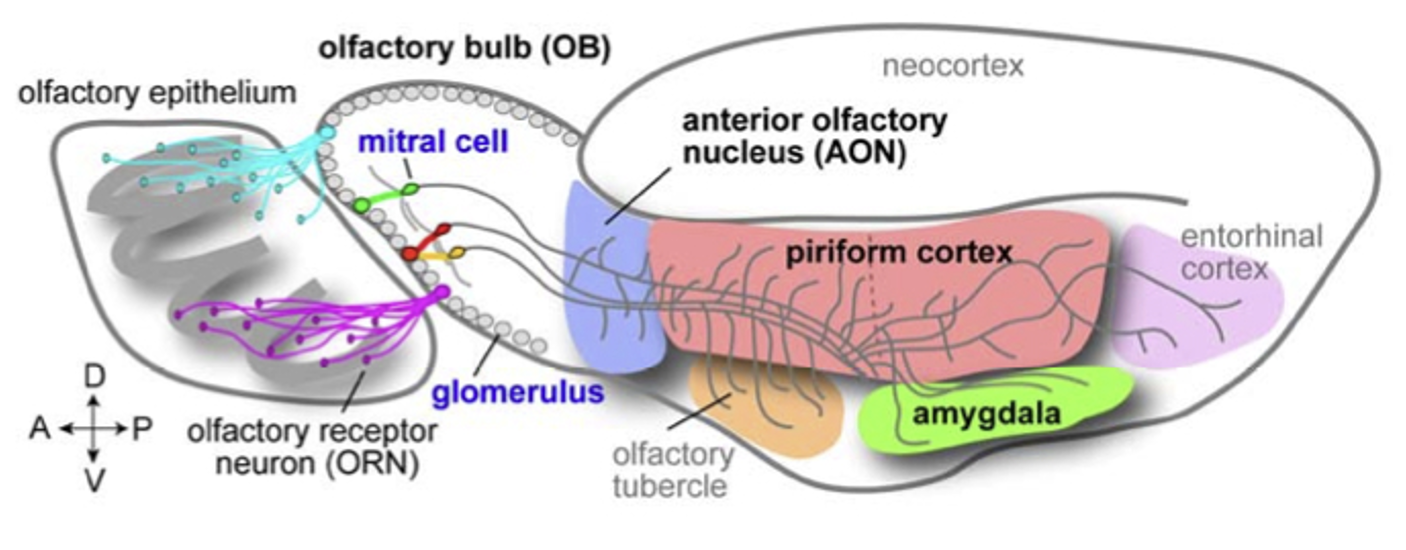
Trace the olfactory pathway from odor to brain
Odorant → OSN receptor → OSN AP → glomerulus (OB) → mitral cell (OB) → higher CNS areas
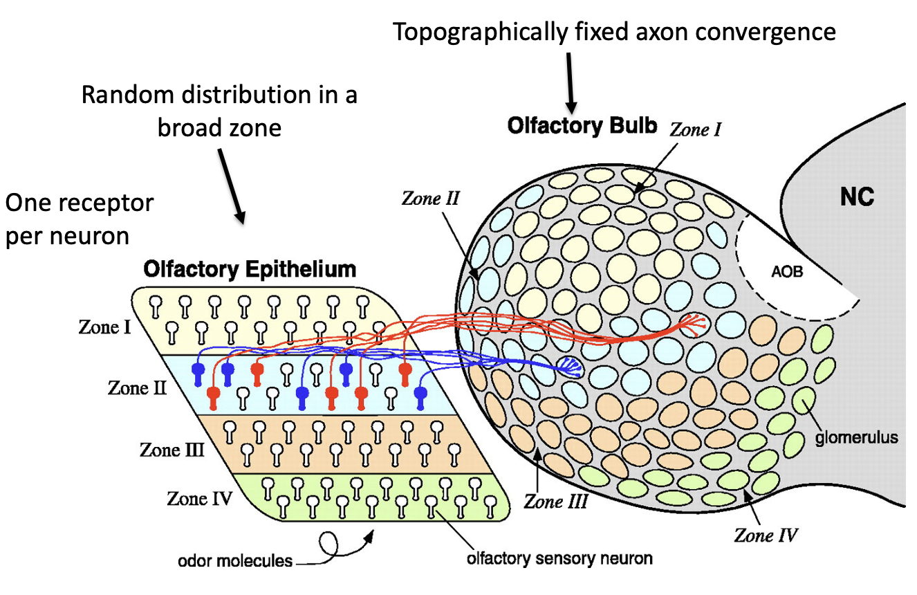
How are OSNs distributed in the olfactory epithelium?
Divided into 4 zones (I–IV)
Each OSN expresses 1 receptor → restricted to ONE zone
Within that zone: random distribution
What is the accessory olfactory bulb (AOB) and do humans have it?
AOB receives vomeronasal (pheromone) input from vomeronasal sensory neuron axons
Humans do NOT have AOB or a functional vomeronasal organ
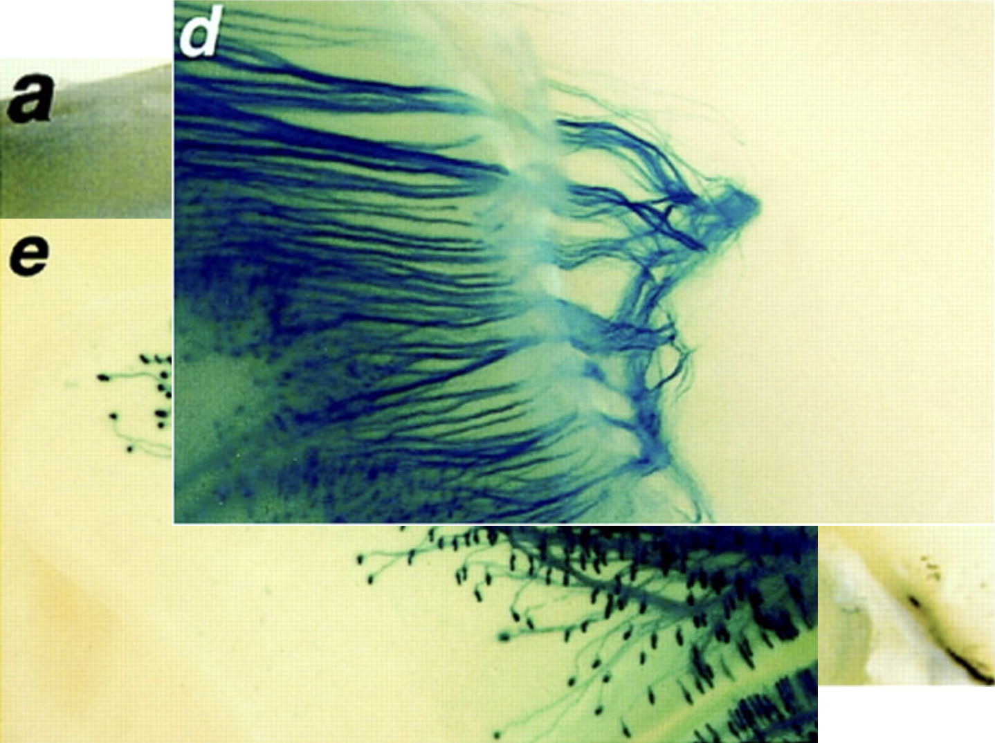
What does this image show?
a: Olfactory epithelium (location of OSN cell bodies)
d: Bundled OSN axons projecting toward the olfactory bulb
e: Convergence of axons into glomeruli in the olfactory bulb

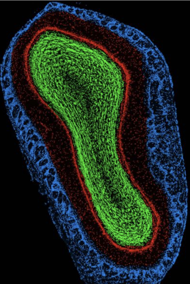
How is the olfactory bulb structured?
Laminar organization
Layered structure
Glomerular layer (blue), mitral cell layer (red), granule cell layer (green)
What do axons from the glomeruli form synapses with?
Mitral cells (M), tufted cells (T), periglomerular cells (PG)

What neurotransmitter do OSNs use?
Glutamate (excitatory)
What kind of synapse occurs between mitral cells and granule cells?
Dendrodendritic synapses
Granule cells are GABAergic inhibitory interneurons → provide lateral inhibition b/w mitral cells
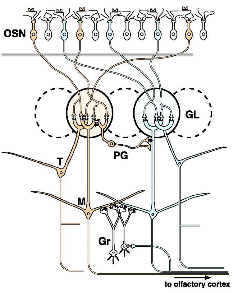
What is the function of periglomerular (PG) cells?
Inhibitory interneurons
Modulate input at the glomerulus level
What is the overall circuit logic in the olfactory bulb?
Excitatory input (OSNs → mitral/tufted) + inhibitory modulation (PG + granule cells) → refined odor signal via lateral inhibition
Why is lateral inhibition needed in the olfactory bulb?
Because OSNs are broadly tuned, lateral inhibition sharpens output → more specific odor coding
What is lateral inhibition in the olfactory bulb?
Inhibitory interactions between neighboring glomeruli/mitral cells that suppress weaker signals
A/B/C (OSNs): broad responses
D (mitral cell): narrow, refined response
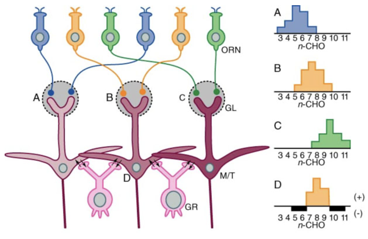
How is odor identity coded in the olfactory bulb?
By unique spatial patterns of activated glomeruli in OB
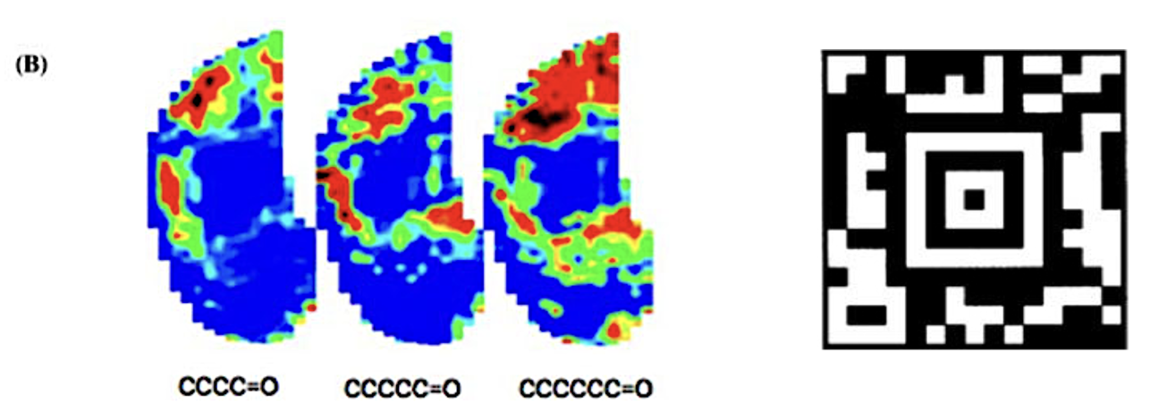
What is the “2D barcode” analogy in olfaction?
Shown in (B) is neural activity imaging of a mouse olfactory bulb to aldehydes of increasing carbon chain length. Each aldehyde elicits a unique spatial activity pattern across the bulb. This activity pattern resembles a 2-D barcode in the amount of information that exists in a complex spatial map.
Where do mitral cells send their axons to?
Primary olfactory cortex: Made up of anterior olfactory nucleus (AON), piriform cortex, olfactory tubercle
Amygdala (emotion)
Lateral entorhinal cortex (memory)
What type of processing happens from OSNs → mitral cells → cortex?
Convergence → divergence
(many OSNs → ~25 mitral cells → divergent output to 5 distinct targets)
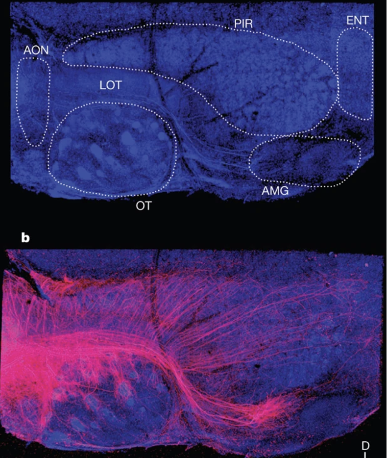
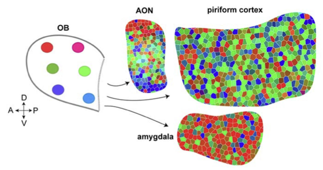
Is mitral cell output to piriform cortex topographic?
❌ No — it is non-topographic (diffuse)
A single piriform cortex neuron receives input from multiple glomeruli/mitral cells
How do mitral cells from a single glomerulus project to different brain regions?
Piriform cortex: diffuse, widespread projections → convergence from many glomeruli onto single neurons
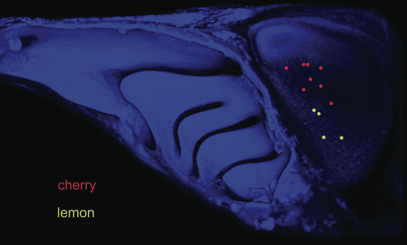
Amygdala: stereotyped, patchy projections → more structured and glomerulus-specific
👉 Output pattern depends on target
Functional roles of piriform cortex vs amygdala in olfaction?
Piriform cortex: integrates inputs from multiple glomeruli → complex odor perception (combinatorial coding)
Amygdala: receives more specific patterns → drives innate behaviors (attraction/aversion)
How do olfactory bulb outputs differ across anterior olfactory nucleus (AON), piriform cortex, and amygdala?
AON: topographic organization → preserves spatial relationships, AON in 1 hemisphere sends axons to contralateral AON + OB.
Piriform cortex: widely overlapping input from all regions of OB → integrates inputs from many glomeruli (no map)
Amygdala: linked to animal’s stereotypical response to predators + other dangers (ex. contaminated food, decaying flesh)
What are the major output pathways from the primary olfactory cortex?
OB → Thalamus → Orbitofrontal cortex → conscious perception
OB → Amygdala + Hypothalamus → emotional & autonomic responses
OB → Entorhinal cortex → Hippocampus → memory
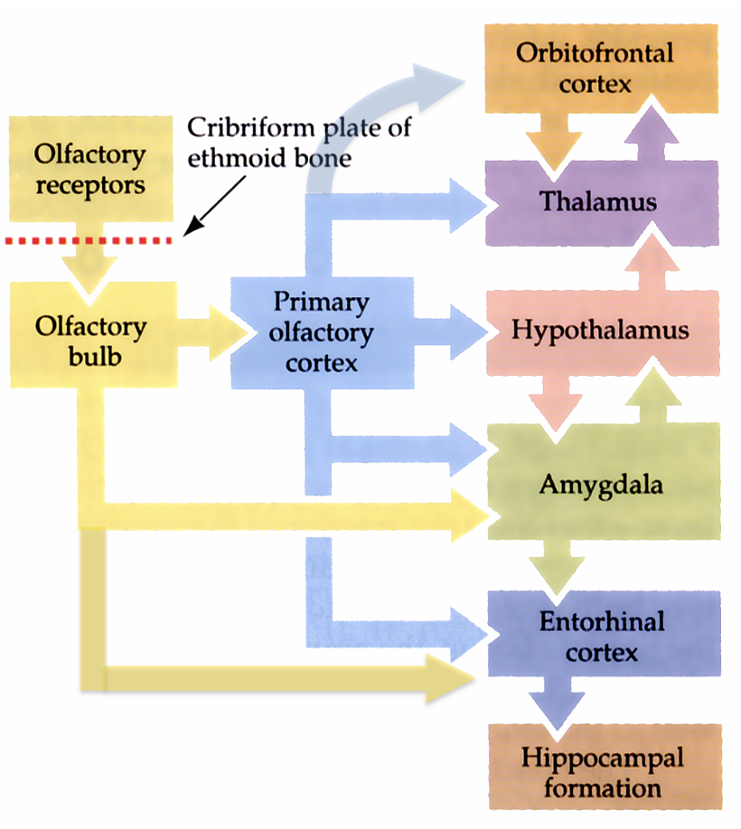
What is thalamic relay of olfactory input to medial orbitofrontal cortex necessary for?
Conscious perception of odorants
What is hypothalamic output to autonomic nuclei in brainstem responsible for?
Reflexive response to pleasant + unpleasant odors
What is the vomeronasal organ (VNO) and its pathway?
Specialized olfactory structure in many mammals (not humans)
Detects pheromones (social/chemical signals)
OSNs in VNO send axons → accessory olfactory bulb (AOB) (NOT main OB pathway)
How are pheromones detected in the VNO?
VNO sensory neurons express V1Rs (~30–50) and V2Rs (~100) (both GPCRs)
OSNs expressing V1Rs innervate anterior AOB
OSNs expressing V2Rs innervate posterior AOB
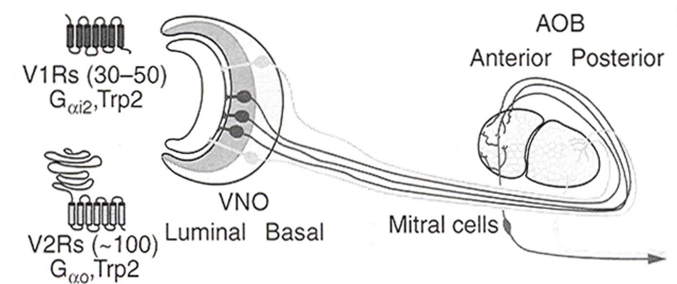
Where does VNO/AOB signaling go and what about humans?
AOB → strong projections to amygdala (innate behavior circuits)
Does NOT overlap with main olfactory bulb pathways
In humans:
V1R/V2R = pseudogenes
VNO = vestigial (nonfunctional)
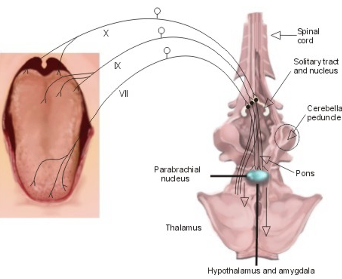
Outline the central gustatory (taste) pathway.
Peripheral taste neurons = ganglion cells in 3 cranial nerves ganglia (VII, IX, X)
Their central axons converge on nucleus of the solitary tract (brainstem)
From there splits:
Thalamus → gustatory cortex → conscious perception & discrimination of tastants
Hypothalamus/brainstem circuits → autonomic/reflex responses (feeding, salivation, etc.)
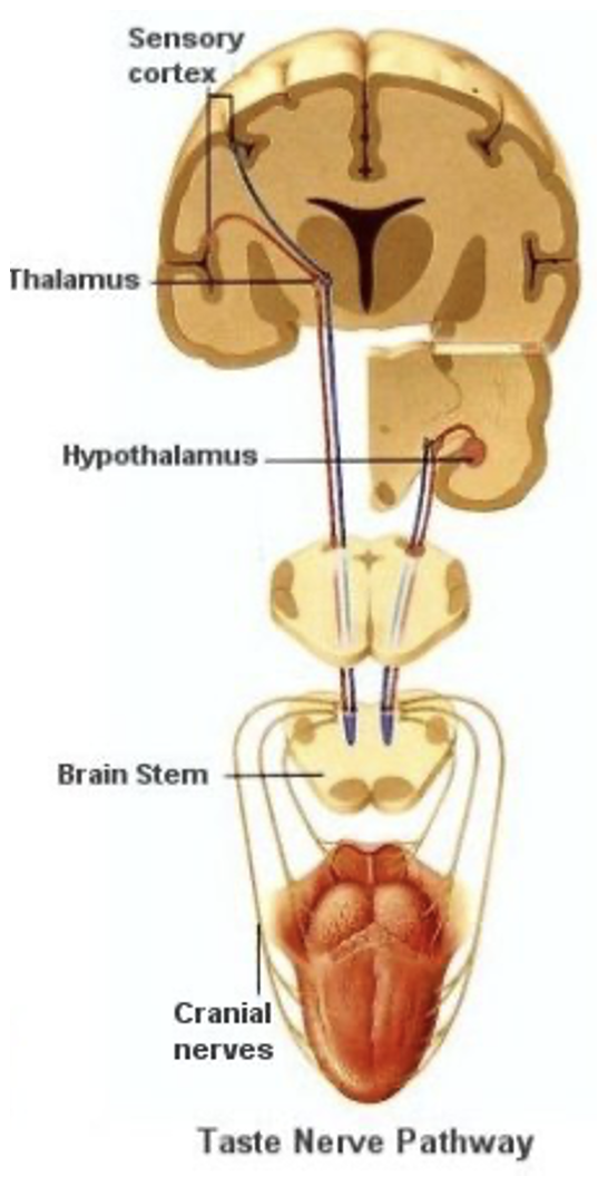
Where do the axons of ganglion cells in 3 ganglia for cranial nerves VII, X, X go?
Join the solitary tract + terminate in anterior 1/3rd of nucleus of the solitary tract in the medulla
A second-order gustatory nucleus in rodents is the parabrachial nucleus in the pons
Describe the gustatory pathway in humans (serial processing).
NTS axons bypass parabrachial nucleus (in humans) → project to VPMpc (thalamus)
VPMpc → primary gustatory cortex (rostral insula + frontal operculum) → responds to physical properties (ex. concentration of sugar)
Primary gustatory cortex → medial orbitofrontal cortex (OFC) (secondary area) → encodes reward/pleasantness
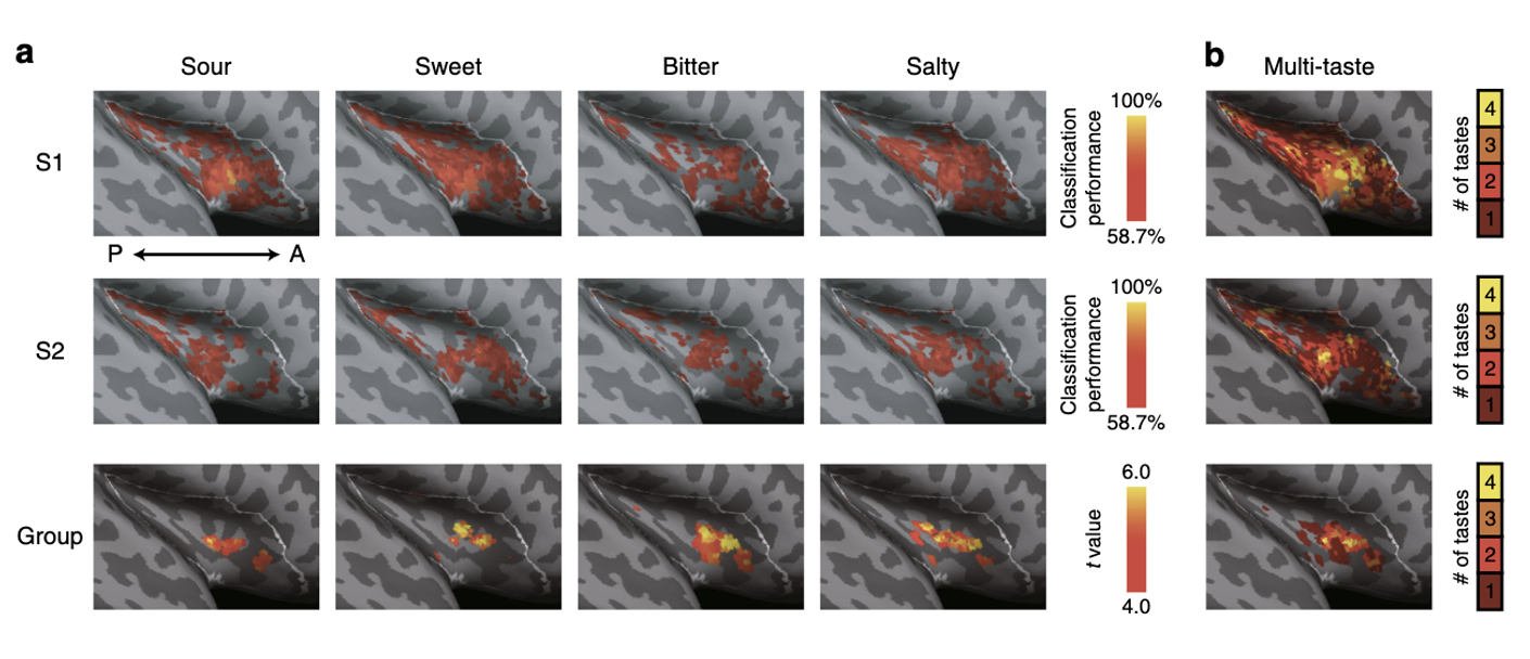
Is there a gustotopic map in primary gustatory cortex?
Evidence suggests partially spatially distinct representations for tastants (sweet, bitter, etc.)
Supports “labeled-line” model (specific pathways for each taste)
BUT maps are overlapping + variable across individuals → not as clean as visual/retinotopic maps
Key idea: biased spatial organization, not strict map
What is the evidence for convergence in gustatory coding?
Recent fMRI shows strong overlap in cortical responses to primary tastants
data indicates that groups of neurons respond to 2, 3, even 4 tastants.
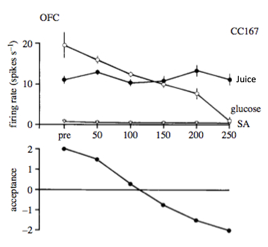
What does the orbitofrontal cortex (OFC) encode in gustation?
OFC (secondary gustatory cortex) encodes food value/pleasantness, not just identity
Neuronal response decreases with satiety (sensory-specific satiety)
Monkey rejects glucose solution following 100-150ml (unappealing)
At that point, even tho primary cortex continues responding, secondary cortex OFC progressively stop responding above spotaneous activity (SA) → suggests food value
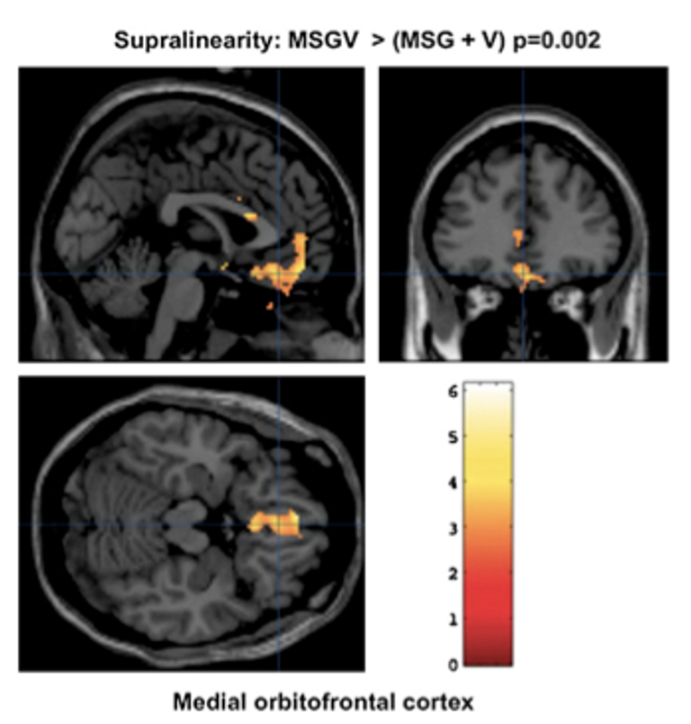
What is a supralinear response?
the medial orbitofrontal cortex (mOFC) shows much greater activity to presentation of MSF + pleasant food odor (V) than when stimuli presented separately.
as pleasantness increased, mOFC activity increased