1415 Principles Exam 2 (Apical, Subcostal)
1/73
There's no tags or description
Looks like no tags are added yet.
Name | Mastery | Learn | Test | Matching | Spaced |
|---|
No study sessions yet.
74 Terms
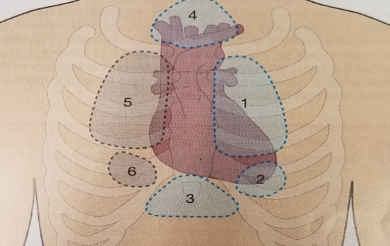
Which of the following window is Apical Window?
2
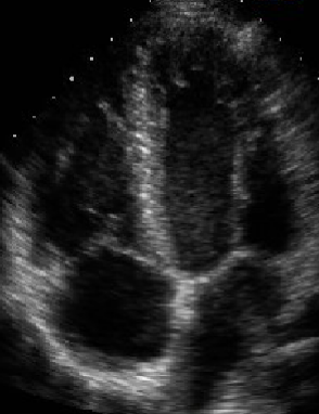
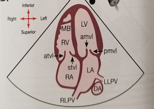
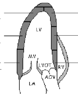
What view is this?
Apical 4 Chamber
What view is this?
Apical 5 Chamber
What view is this?
Apical 2 Chamber
What view is this?
Apical 3 Chamber
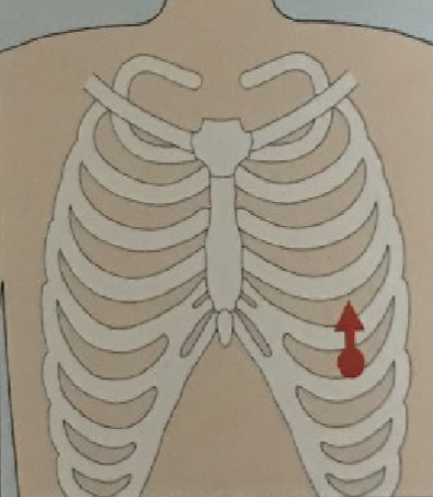
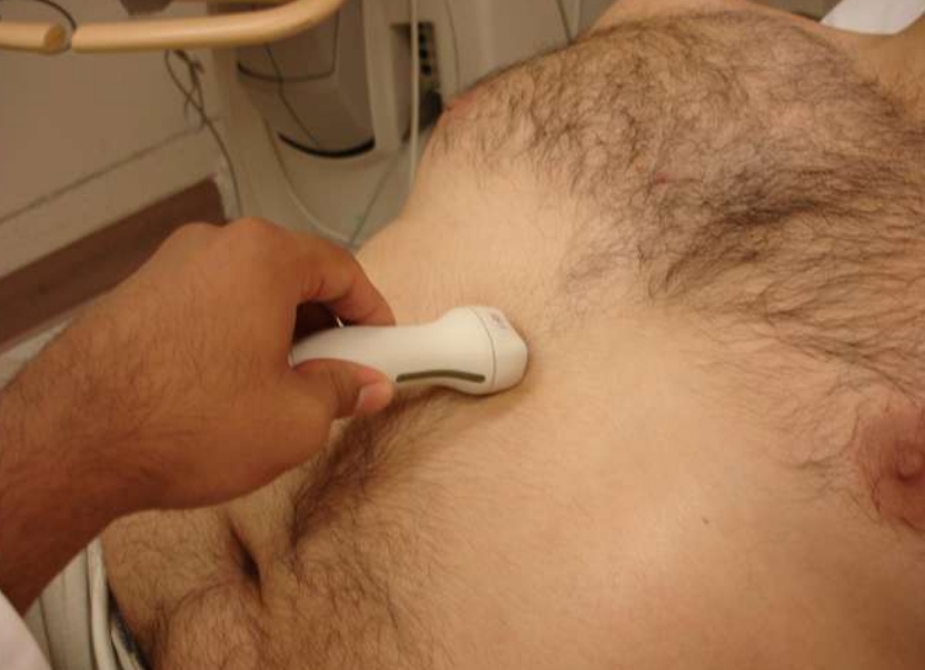
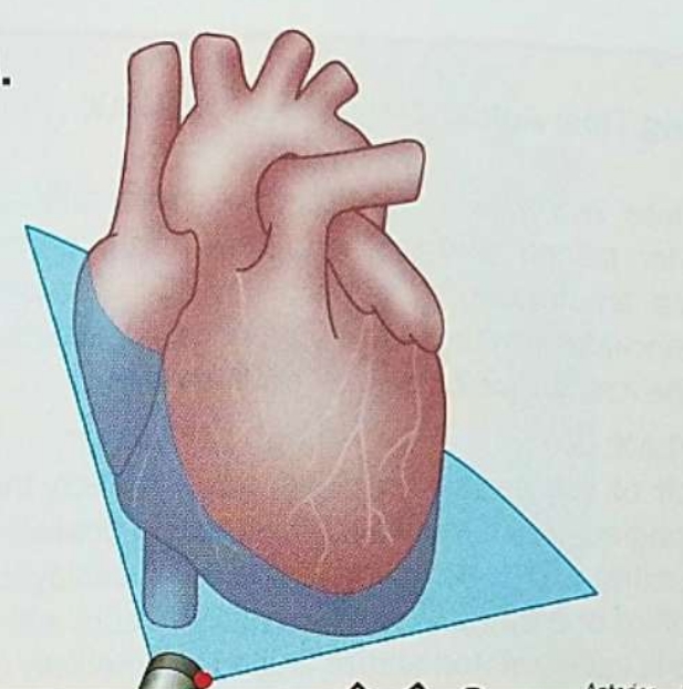
For apical positioning, the patient is on their ___ side.
Left
For apical positioning, probe is aroung the ___ intercostal space in the _________
5th, point of maximum impulse region
For apical 4 chamber position, the POM is point towards ____ and beam directed superiorly towards patient’s head
3’o clock
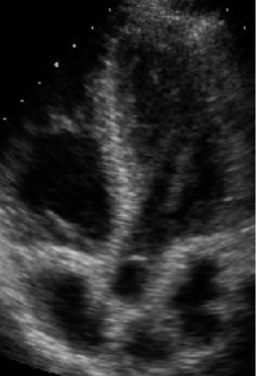
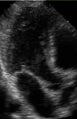
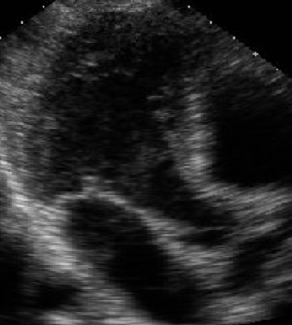
What view is being shown?
Apical 4 chamber
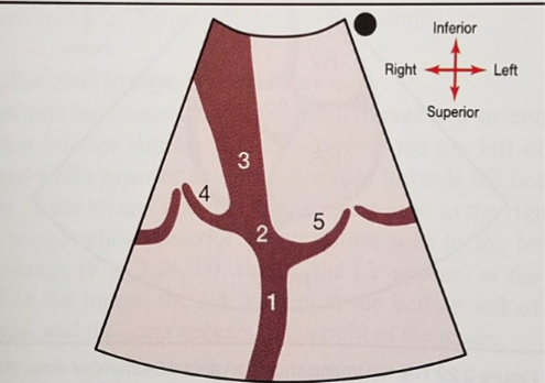
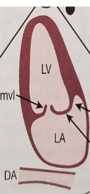
What is number 1?
IAS
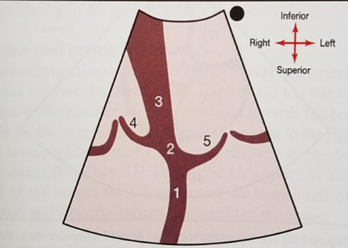
What is number 2?
Crux

What is 5?
AMVL

What is number 4
STVL
What can you see in A4C
Crux, ventricles, trabeculations, moderator band, false tendon, TV position, atrioventricular septm, LAA (rare), Leaflets opening and closing, coronary sinus, pulmonary artery
How can you see Coronary Sinus in A4C
Tilt the transucer posteriorly
How can you see Pulmonary Artery in A4C?
Tilt the transducer anteriorly
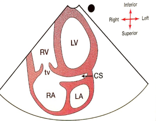
What new structure can be seen if tilted posteriorly?
CS
What view is this?
A4C, Focused Right Ventricle view
What is the white line?
False tendon
What determines a good A4C?
An elongated left ventricle with optimal visualization of all four chambers. Crux is centered
How to move from A4C to A5C?
-Tilt probe anteriorly until LVOT and proximal ascending aorta are seen
-Rotate the probe slightly to the left of the patient to show the mitral valve opening
-Tilting more anteriorly will show RVOT
What is different from A5C compared to A4C?
Can see LVOT, AO
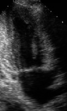
What view is this?
A5C
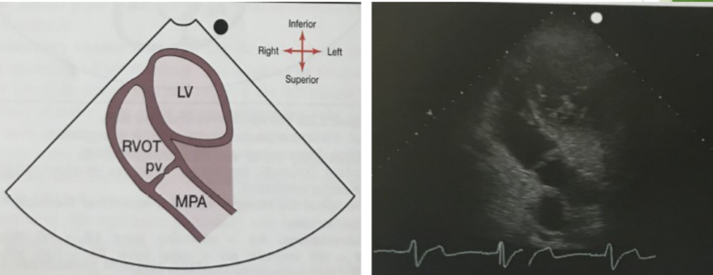
What view is this? Tilting more anteriorly
Pulmonary artery and RVOT view
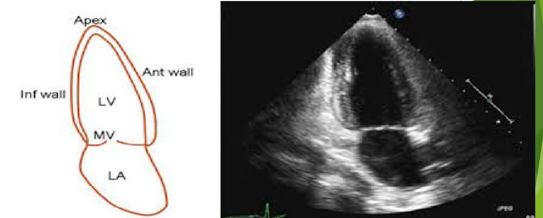
How to move from A4C to A2C?
Rotate index marker to 1’o clock or ~45-90 degrees CCW from A4C
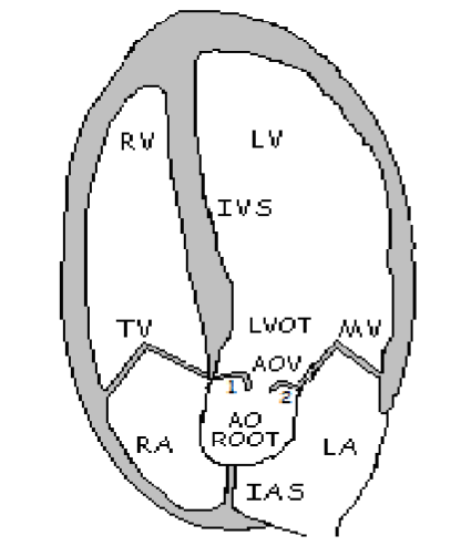
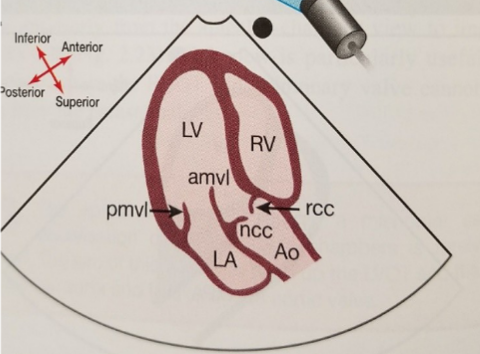
What is 1?
RCC

What is 2
NCC

How can LCC be seen?
In PSAX
Where does A2C image plane lie between?
A4C and Apical Long Axis
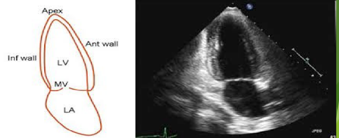
What view is this?
A2C
A2C View Structures
LV –Left Ventricle
LA –Left Atrium
Mitral valve leaflets
True anterior and inferior LV walls
Left atrial appendage
Descending aorta

What leaflet is on the left←?
Posterior MVL
What leaflet is on the right→?
Anterior MVL
What is the right arrow point to?
LAA
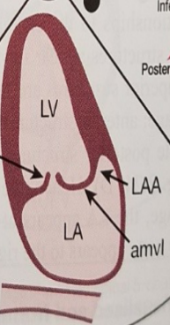
What is the bottom anatomy?
DA
What view will be obtained from this?
A3C
What view is this?
A3C
What view is this?
A3C
What direction is the POM point for A3C?
12o’clock
How to obtain A3C?
Rotating 90 degrees CCW from A4C or 45 degrees from A2C
A3C is similar to the view of?
PLAX

What is bottom left wall of the image?
Basal Infero-Lateral

What is middle left?
Mid Infero-lateral

What is top left?
Apical Lateral
What is the top?
Apical Cap

What is top right?
Apical septum

What is right middle?
Mid Antero-Septal

What is bottom right?
Basal Antero-septal
Heart looks foreshortened (rounded apex) because of
transducer too high or medial
Value of Subcostal views
Used in technically difficult patients
Used if other windows and images are suboptimal
COPD, ICU patients on ventilators
Congenital heart disease (CHD)
IAS, IVS definition
Avoids the lungs
Subcostal view image plane and standard views
Subcostal four chamber
Subcostal short axis
Sometimes included in subcostal
IVC view
Abdominal aorta
For subcostal view patient is in ______ position
Supine
For subcostal view, what can help for a better image?
Relaxing abdominal muscle by bending the knee
For subcostal, the probe is positioned where?
Inferior to the sternum close to the midline
For subcostal, the POM is rotated where?
Approximately 3 o’clock to patient’s left side
For subcostal, how to obtain all four chambers?
Tilt scan plane anteriorly with minor rotational adjustments
For subcostal, what usually helps with image?
Patient’s deep breath, often have to push to go under the ribs
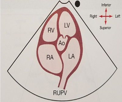
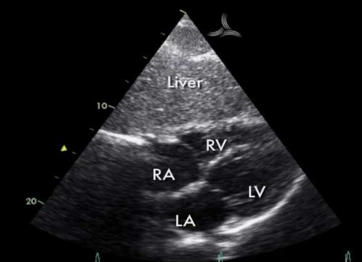
What view is this?
Subcostal
What view would be obtained here?
Subcostal 4ch View
What view is this?
Subcostal 4ch
Why is subcostal 4ch good?
Best view to access RV free wall, IVS, and IAS
Why is subcostal 4ch good for taking measurements of IVS, IAS
The ultrasound beam is perpendicular to IVS and IAS
What can be seen in subcostal 4ch
Liver appearance
4 chambers
IAS (perpendicular), IVS
MV, TV
Tilting anteriorly in subcostal 4ch will see what structures?
ROT and PV
What view is this?
Subcostal 5ch (use only if A5C is not good)
What view is this?
Subcostal view RVOT and PV
What view is this?
Subcostal Short Axis Aorta and IVC trans
How to get Aorta and IVC transverse (SAX)?
Probe perpendicular to abdomen
Which valve is subcostal view parallel to?
Tricuspid
What is subcostal view frequently used to evaluate
RVSP, VSD, ASD and tamponade
What image can be obtained from this?
Subcostal Short Axis
Transducer technique subcostal short axis
Subxiphoid position
POM 12 o’clock
Rotate probe 90 degrees ccw from s4c
Sweep patient’s left to right to get SAX
LV apex
Pap
LV MV
RVOT