A&P 2 Lab Practical
1/102
There's no tags or description
Looks like no tags are added yet.
Name | Mastery | Learn | Test | Matching | Spaced |
|---|
No study sessions yet.
103 Terms
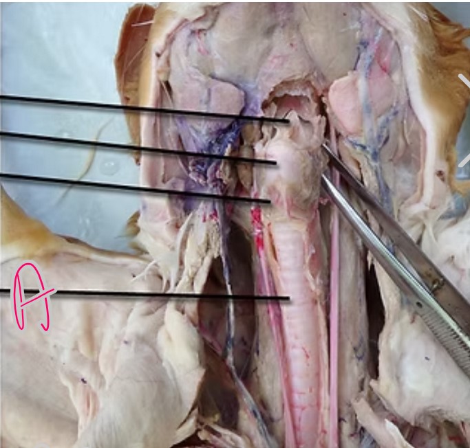
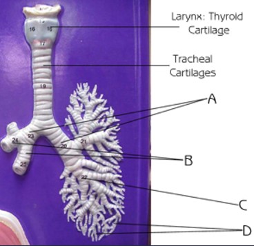
What is A?
Trachea
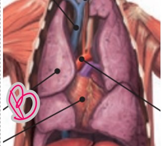
What is B?
Lungs
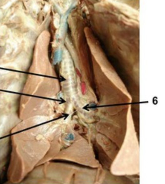
What is 6
Primary Bronchus
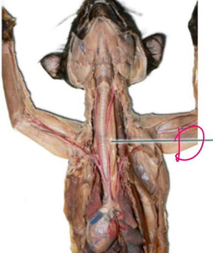
What is D
Esophagus
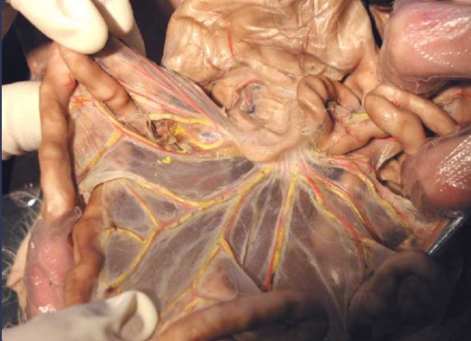
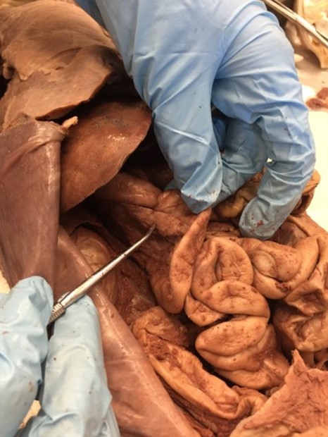
What is shown in the image?
Mesenteries
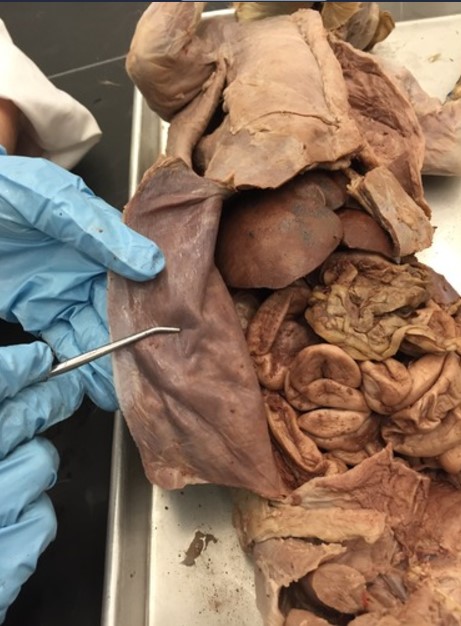
What is being held in the picture?
Peritoneum
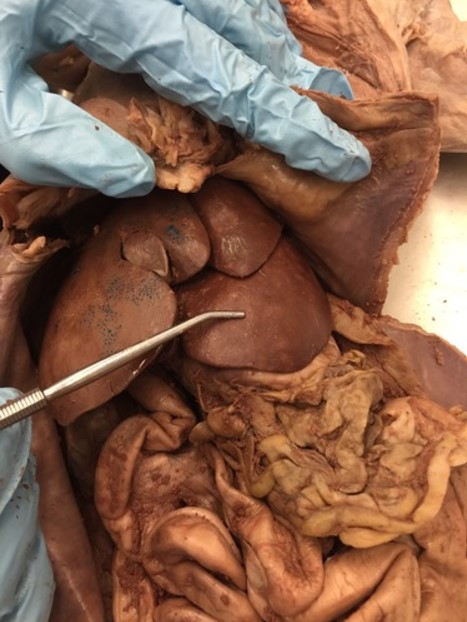
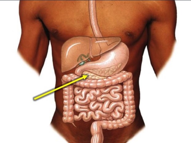
What organ is being pointed to in the image?
Liver
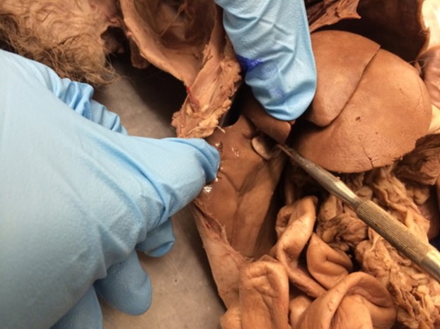
What organ is being indicated in the image
Gallbladder
What organ is indicated in the image
Pancreas
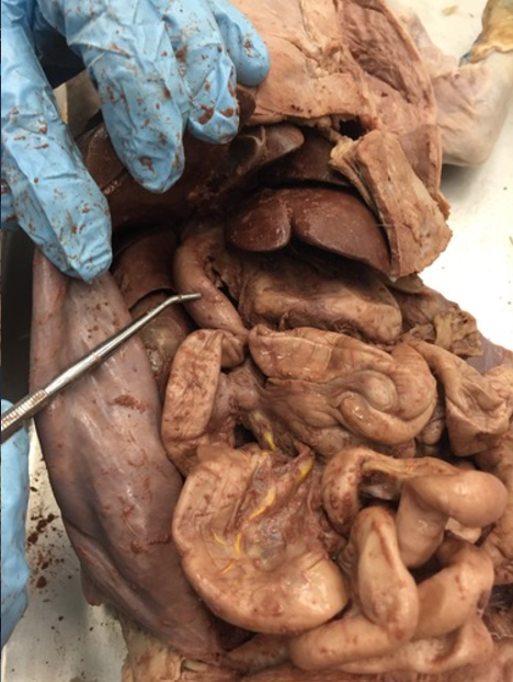
What part of the small intestine is indicated in the image
Duodenum
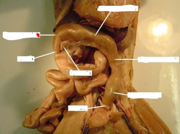
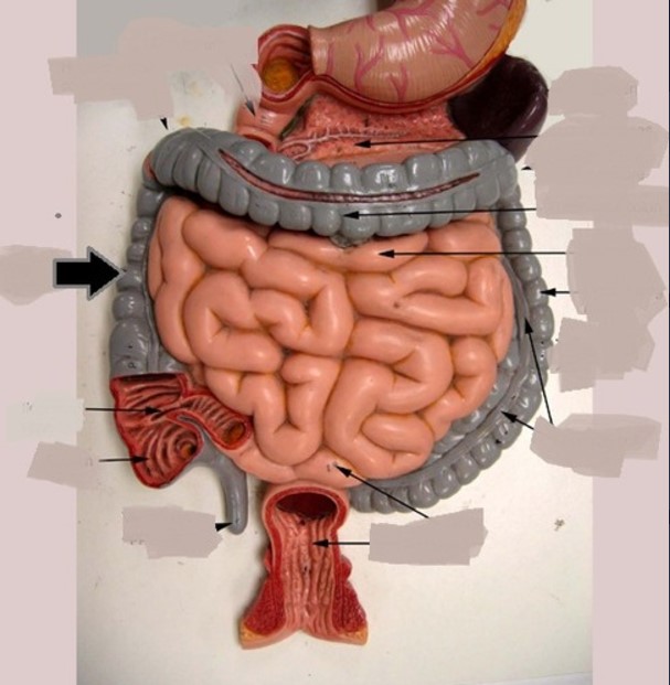
What portion of the large intestine is indicated by the red dot?
Ascending Colon
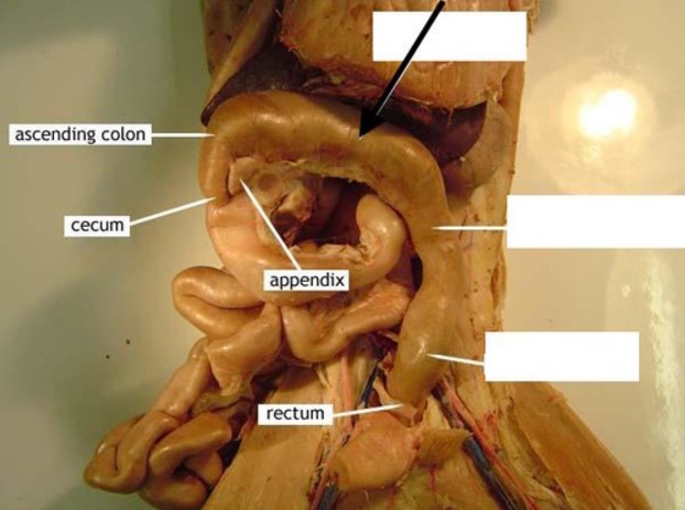
What portion of the large intestine is indicated by the black arrow?
Transverse Colon
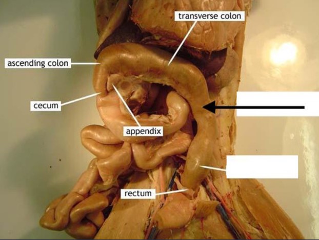
What portion of the large intestine is indicated by the black arrow?
Descending Colon
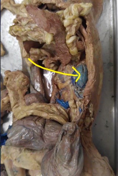
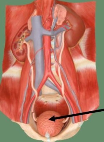
What organ is indicated by the yellow arrow?
Kidney
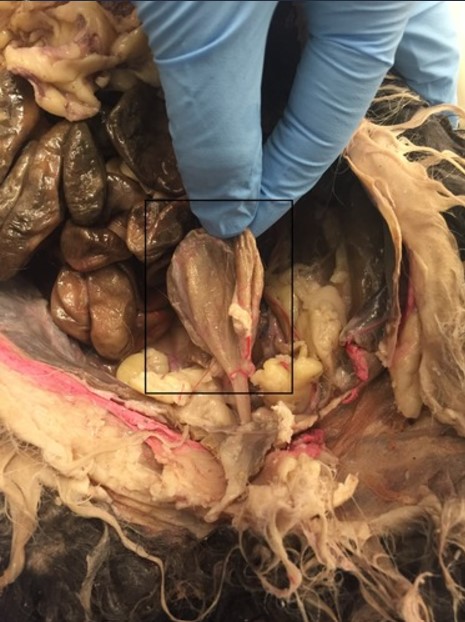
What is indicated by the box?
Urinary Bladder
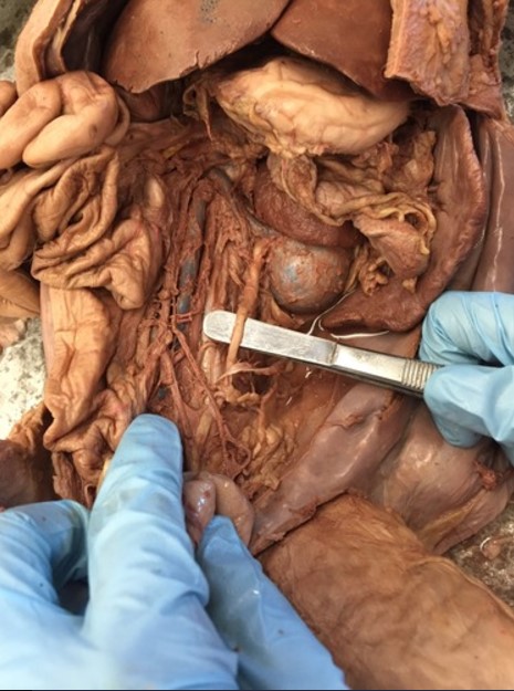
What is indicated by the dissection tool in the image
Ureter
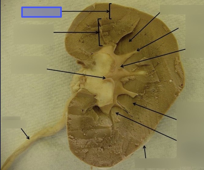
What portion of the kidney is indicated by the blue box
Renal Cortex
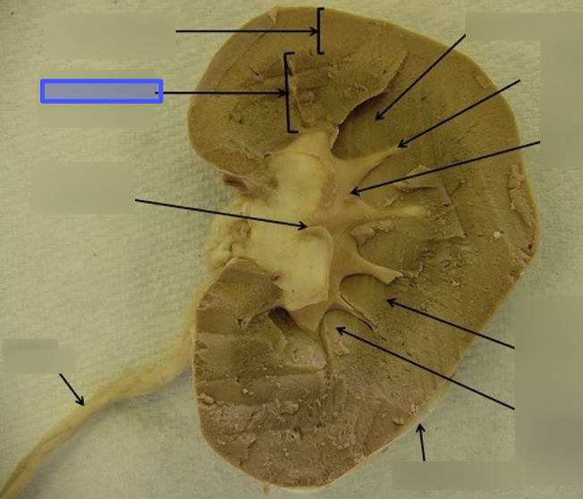
What structure of the kidney is indicted by the blue box?
Renal Medulla
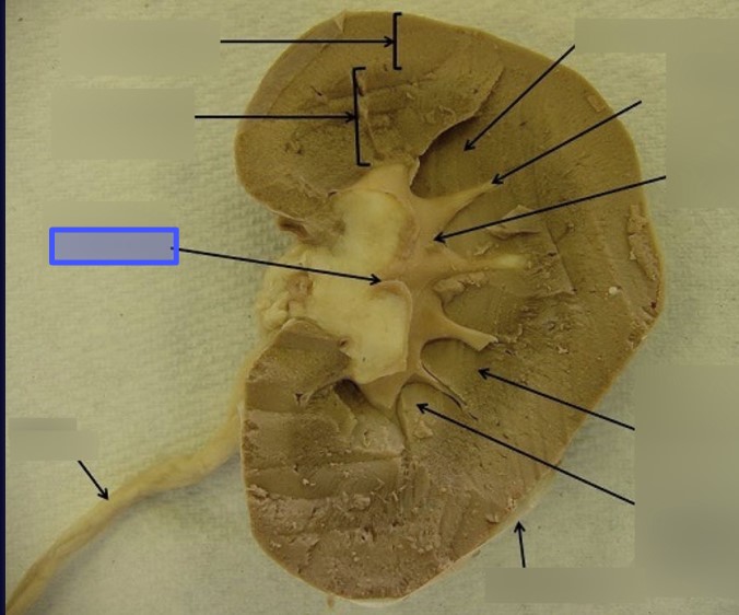
What structure is indicated by the blue box
Renal Pelvis
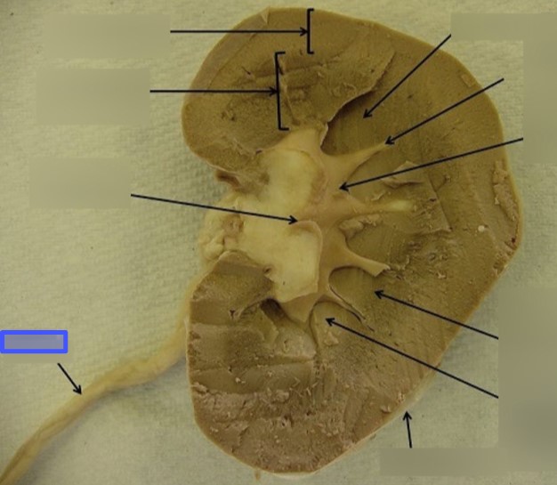
What structure is indicated by the blue box
Ureter
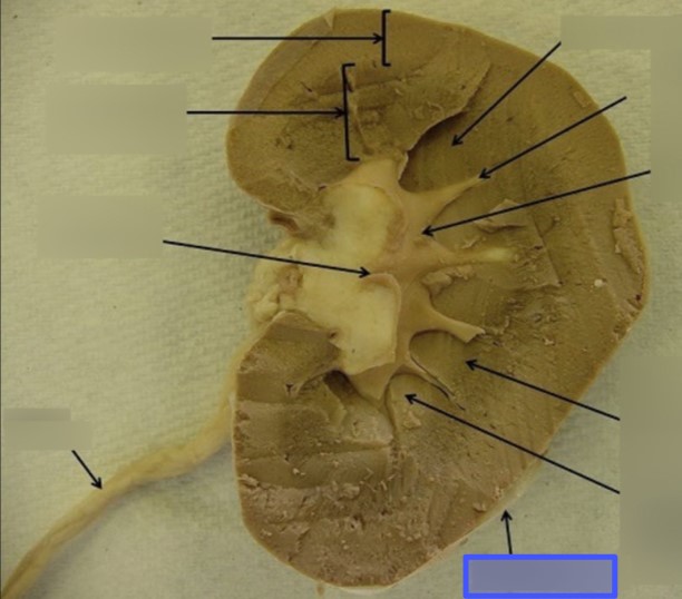
What structure is indicated by the blue box
Fibrous Capsule
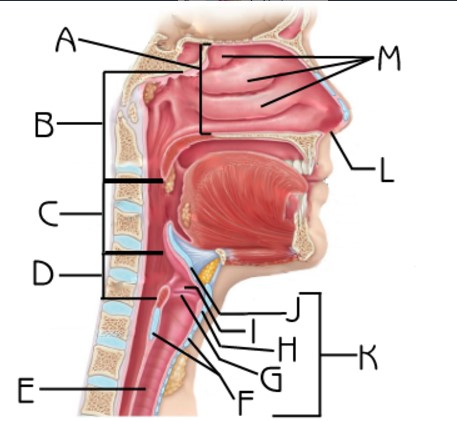
What structure is indicated by K
Larynx

What structure is indicated by J
Epiglottis

What structure is indicated by G
Vocal Chords

What structure is indicated by E
Trachea
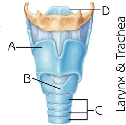
What structure is indicated by D
Epiglottis

What structure is indicated by B
Bronchiole
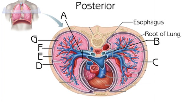
What structure is indicated by D E and F
Pleura

What structure is indicated by A
Primary bronchi
What structure is indicated by B
Secondary bronchi

What structure is indicated by C
Tertiary Bronchi
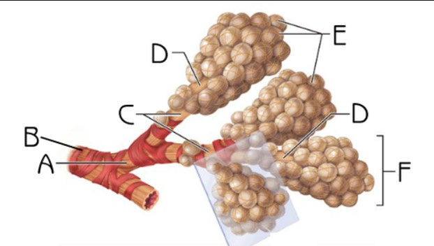
What structure is indicated by E
Alveoli

What muscle is indicated by the black arrow?
Diaphragm
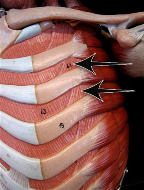
What muscles are indicated by the black arrows
Intercostals
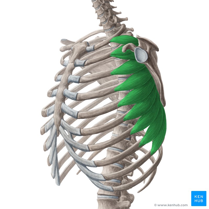
What muscle is indicated in green
Serratus Anterior
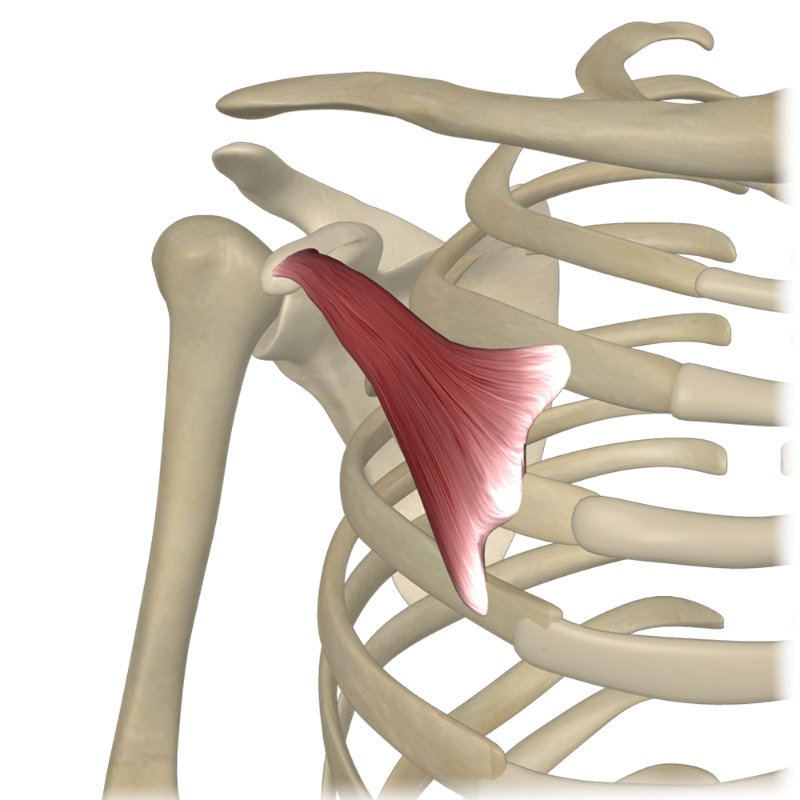
What muscle is pictured
Pectoralis Minor
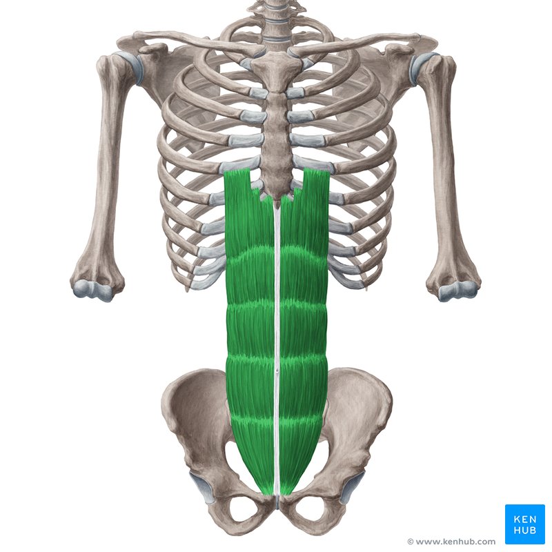
what muscle is pictured
Rectus abdominus
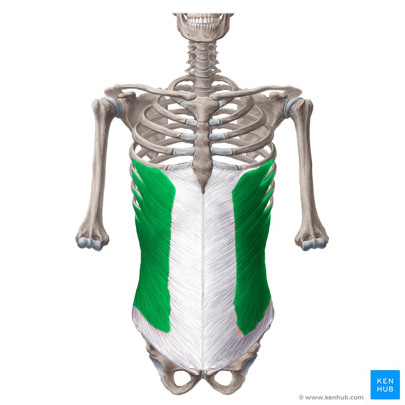
what muscle is pictured?
external oblique
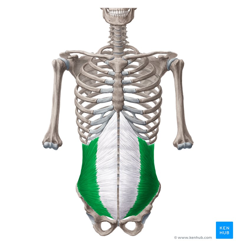
what muscle is pictured
internal oblique
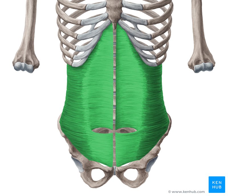
What muscle is pictured
transverse abdominus
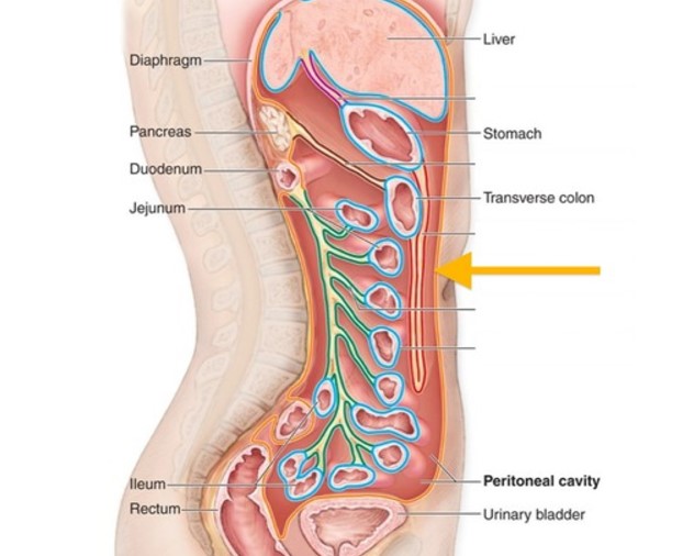
what structure is indicated by the arrow
peritoneum

What structure is indicated by A
Pharynx
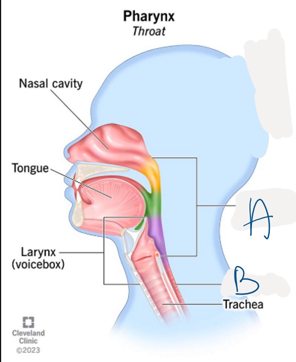
What structure is indicated by B
Esophagus
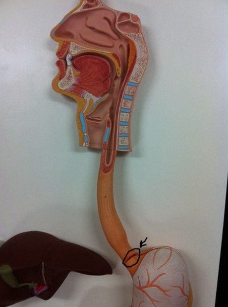
What structure is circled?
Gastroesophageal sphincter
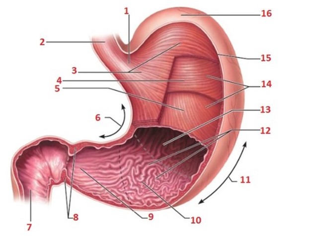
What structure is indicated by 8
Pyloric sphincter
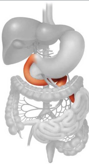
What structure is pictured
Duodenum
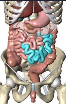
What structure is highlighted in blue?
Jejunum
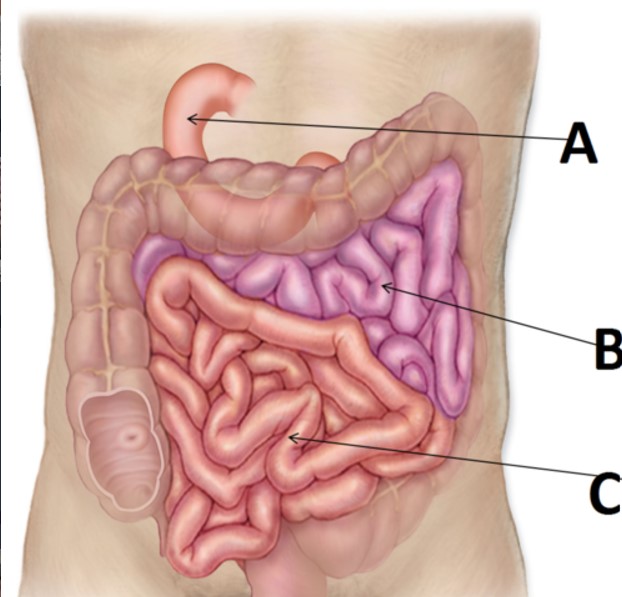
What structure is indicated by C
Ileum
What structure is blue
Large Intestine
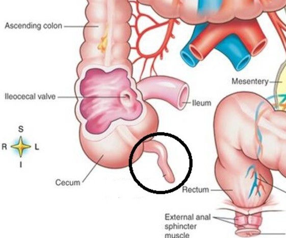
What structure is circled in black
Appendix
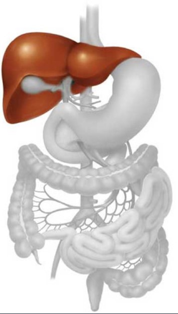
What structure is highlighted
Liver
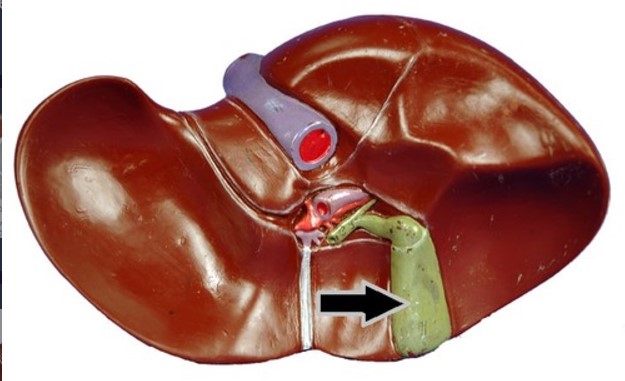
What structure is indicated by the arrow
Gallbladder
What structure is indicated by the arrow
Pancreas
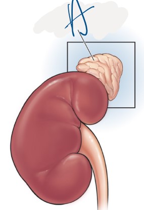
What structure is indicated by A
Adrenal Gland

What structure is indicated by A
Renal Cortex

What structure is indicated by B
Renal Medulla

What structure is indicated by C
Renal Pelvis
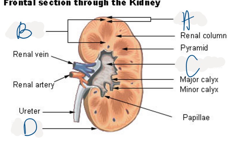
Which structure is indicated by D
Fibrous Capsule
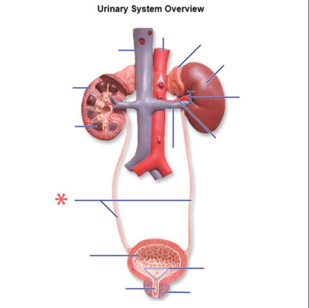
What structure is indicated by the red star?
Ureters
What structure is indicated by the black arrow
Urinary Bladder
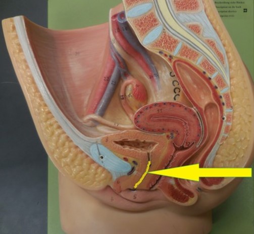
What structure is indicated by yellow
Urethra
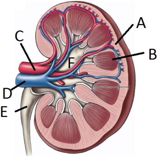
What structure is indicated by C
Renal artery

What structure is indicated by D
Renal vein

What structure is indicated by A
Scrotum
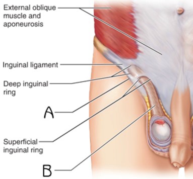
What structure is indicated by B
Spermatic cord

What structure is indicated by B
Testes

What structure is indicated by A
Epididymis

What structure is indicated by B
Seminiferous tubules
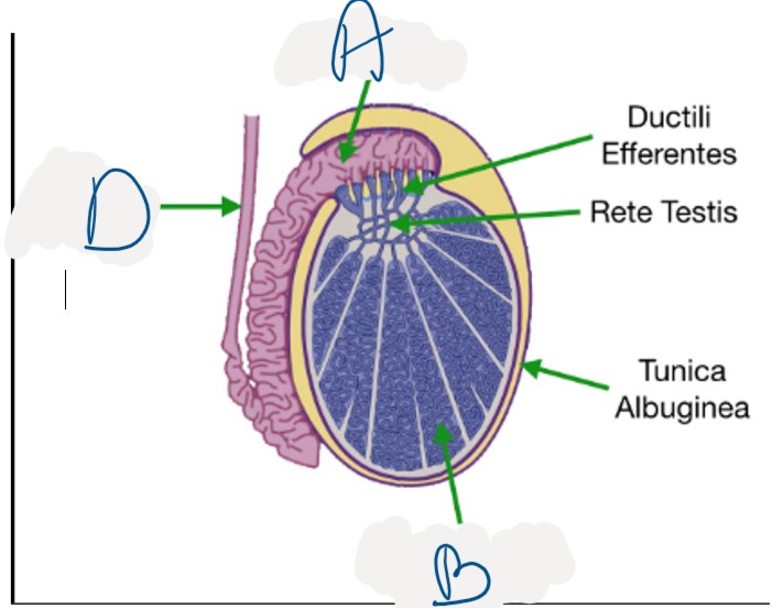
What structure is indicated by D
Vas deferens

What structure is indicated by G
Ejaculatory duct

What structure is indicated by H
Seminal vesicles

What structure is indicated by F
Prostate gland
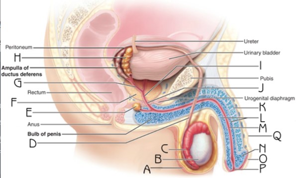
What structure is indicated by Q
Penis
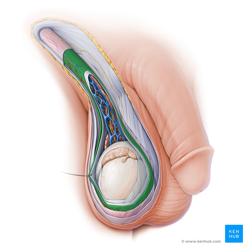
What structure is highlighted in green
cremaster muscle
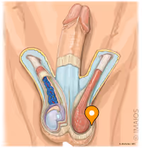
What structure is indicated by the pin
dartos muscle

What structure is indicated by D
Ovaries

What structure is indicated by C
Fallopian tubes
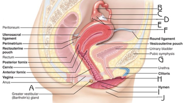
What structure is indicated by F
Uterus
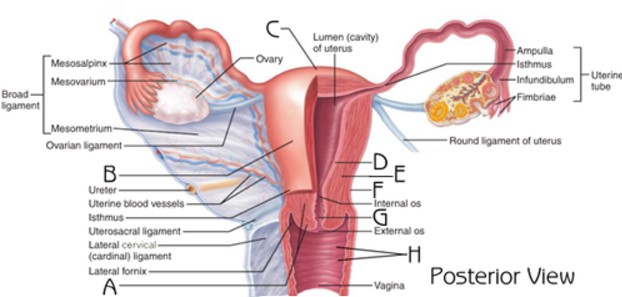
What structure is indicated by A
Cervix

What structure is indicated by J
Labia majora

What structure is indicated by I
Labia minora

What structure is highlighted in green?
Clitoris

What structure is labeled A
Mammary gland lobules
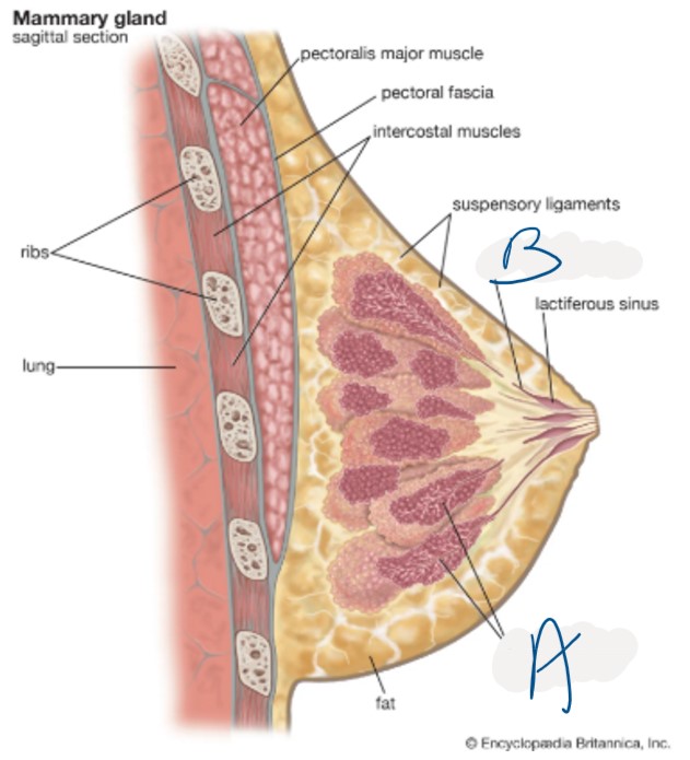
What structure is labeled B
Mammary gland ducts

What structure is labeled by A
Areolas
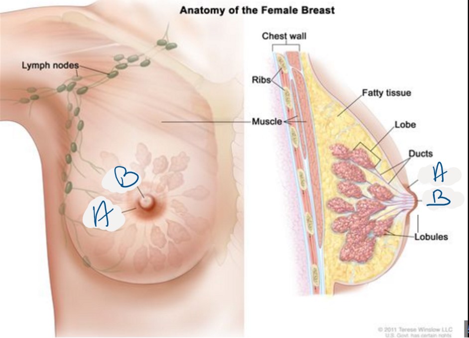
What structure is indicated by B
Nipple

What structure of the trachea is indicated by A
Cilia
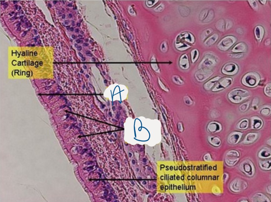
What structure is indicated by B
Goblet cells
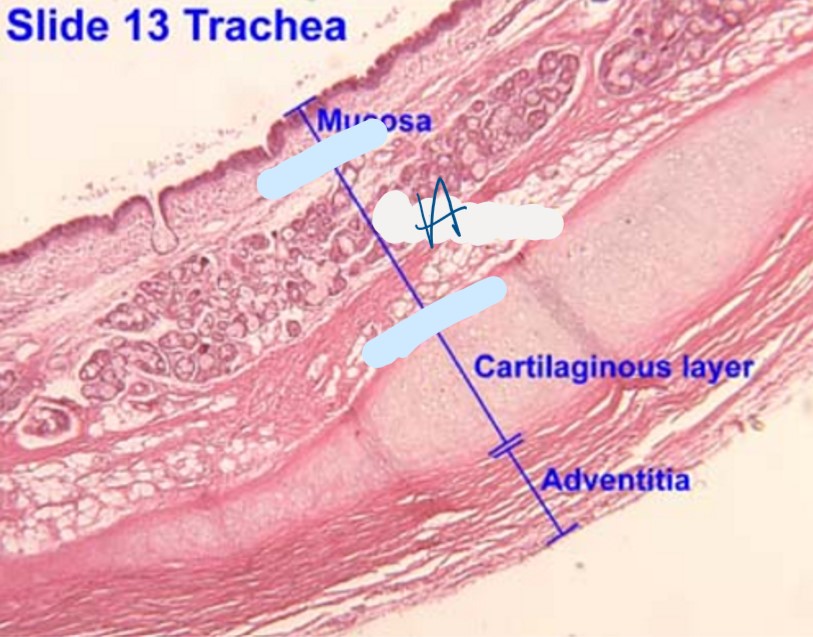
Which layer of the trachea is indicated by A
Submucosa
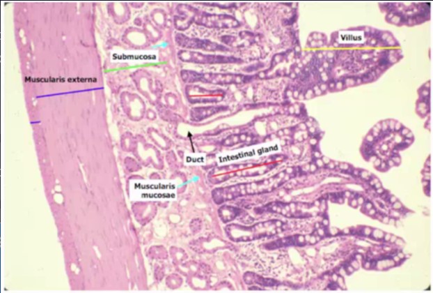
What structure is indicated by the red lines
Simple columnar epithelium
What is the structure outlined in green
Villi
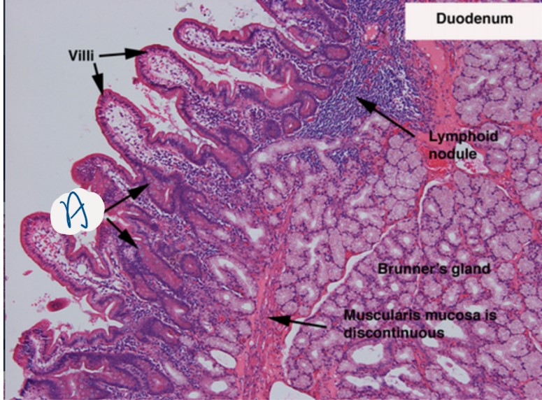
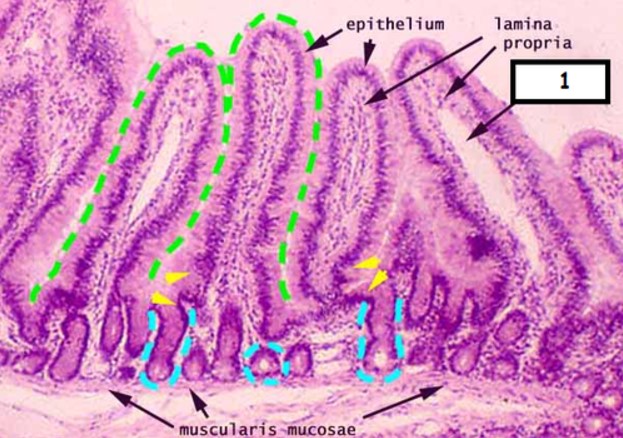
What structure is indicated by A
Goblet cells
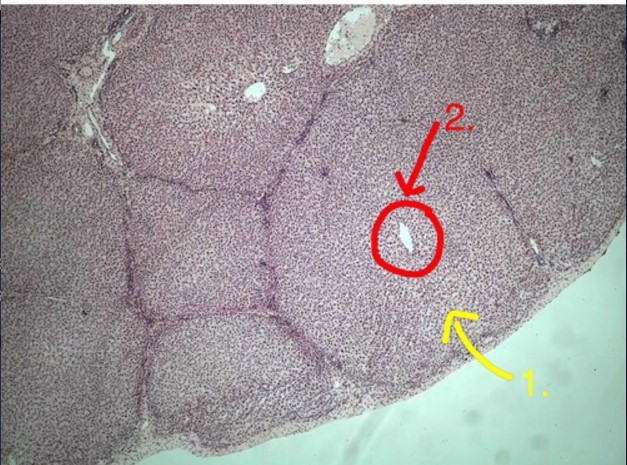
What structure is indicated by 2?
Central vein
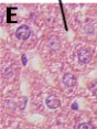
What structure is indicated by E
hepatocytes

What structure is indicated by A
Glomerulus

What structure is indicated by B
Bowman’s Capsule
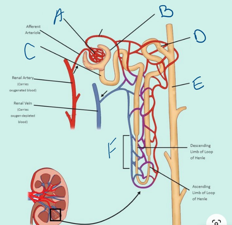
What structure is indicated by C
Proximal Convoluted Tubule (PCT)

What structure is indicated by D
Distal Convoluted Tubule (DCT)

What structure is indicated by E
Collecting Duct

What structure is indicated by F
Loop of Henle