Module 2: Chapter 3: Biological bases of behaviour
1/64
There's no tags or description
Looks like no tags are added yet.
Name | Mastery | Learn | Test | Matching | Spaced |
|---|
No study sessions yet.
65 Terms
Define the nervous system's two classes of cells and their respective functions
Neurons (Newscaster): Information transmission, communication with other neurons, muscles, glands, etc. voluntary movement happen through sending electrical signals and releasing chemicals (neurotransmitters)
Glial Cells (Newsroom: Lighting, Camera, Script, Platform): Aid in structural support, help insulate neurons and have roles in communication, provide nutritional support to neurons, remove debris from the nervous system and change the signalling of neurons
Define Dendrites
branch-like structures that receive messages from other neurons
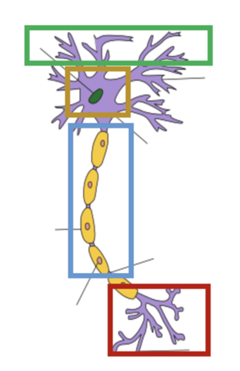
Define Cell body (soma)
contains genetic information: nucleus that determines the cells function, where neurotransmitters are created (synthesized)

Define Axon
Conducts electrical impulses (one-way directional flow originating from cell body.) Can be myelinated (fatty glial sheath) which insulates the axon and speeds up transmission + less signal degradation

Define Axon terminal
Where neurotransmitters are released that then can reach the dendrites of other nearby neurons and influencing transmittion

State the four processes of neuronal activity
1) Resting potential
2) Action potential
3) Synaptic transmutation
4) Graded potentials
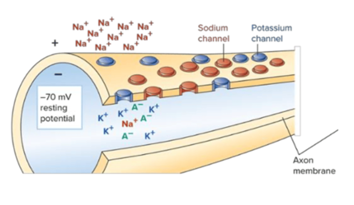
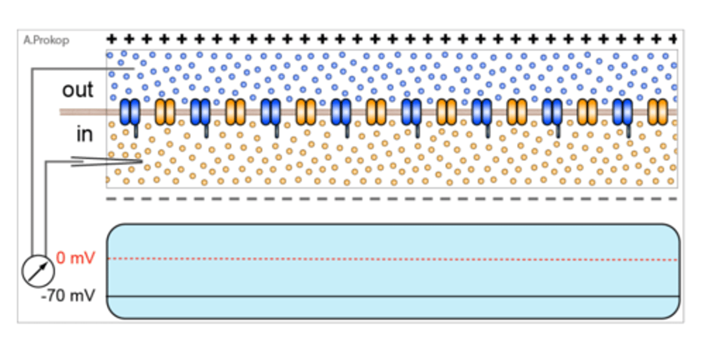
Define Resting Potential
State of the neutron when no other nearby neutron is trying to communicate with the given neuron
- Working hard to maintain an RP: electrical difference between the inside and outside of the cell
- Relative electrical charge inside the neuron vs outside the neuron difference is -70 mV
- To achieve resting potential neuron operates ion channels along the cell membrane
- Sodium and potassium channels are closed at rest to keep the negative charge
- Tiny battery of potential energy: electrochemical difference between the inside and outside of the cell, setting up the situation where ions (electricity) wants to flow
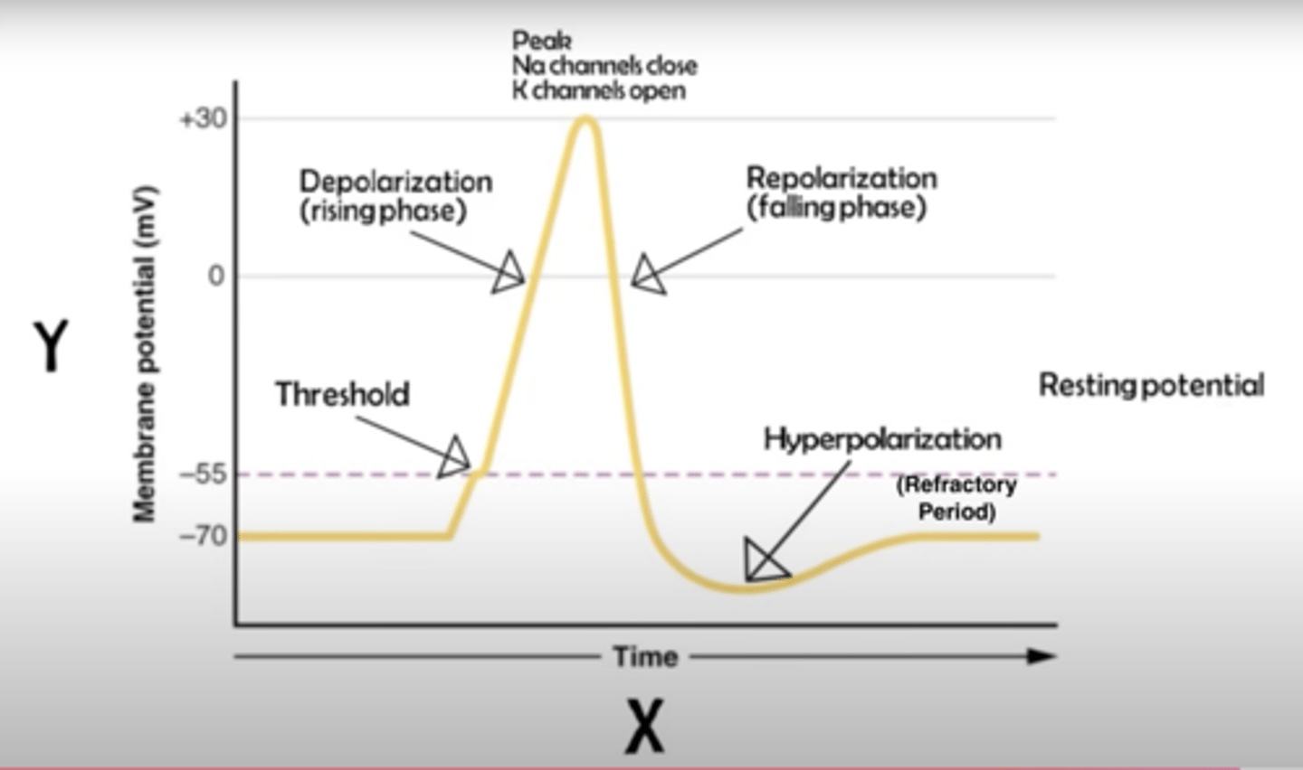
Explain Action Potential
Electrical pulse that is sent down the length of the axon terminating in axon terminals (impulse generation). When a neuron is stimulated by other cells and
1. Crosses the action potential threshold (-55 mV):
2. Depolarization: the neuron "fires"
- Triggers a non-reversible chain reaction that is all or nothing once threshold is reached a neuron will fire exact same way or it won't at all: If the threshold isn't reached, no action potential occurs.
- Ion channels open briefly, allowing positively-charged sodium ions to rush into the cell
- This influx causes the membrane potential to become more positive
3. Repolarization
- Complete, rapid, and brief reversal of the electrical potential across neuron's membrane: potassium (K⁺) channels open, allowing K⁺ ions to leave the cell which restores the negative charge inside the cell
- This rapid decrease in potassium ions restores the membrane potential to a negative value, bringing it back toward the resting potential (-70mV)
- After firing the neuron undergoes an absolute and relative refractory period
Explain what happens after a neuron fires: Absolute refractory Period + Relative refractory period + Repolarization
Absolute refractory period: Milliseconds hyper polarization: no amount of stimulation can make the neuron fire again (needs to recharge)
Relative refractory period: Milliseconds could potentially fire another action potential but harder to make it fire
- Repolarization: potassium ion channels open, allowing positively charged potassium ions (K⁺) to flow out of the cell restores the negative charge inside the cell
Explain Synaptic Transmission: Presynaptic and Postsynaptic
Primary way neurons communicate with each other:
Presynaptic neuron firing an action potential that culminates in neurotransmitters being released into the synaptic cleft and depending on what that neurotransmitter is can change electrical potential of postsynaptic neuron is a graded fashion
- Communicate via synaptic transmission: Neurons dont touch at synapses they are separated by a microscopic gap: synaptic cleft
- Electrical signals can't jump the gap
- Presynaptic (sending): Neuron sending message releasing neurotransmitters into the synaptic cleft
- Postsynaptic (receiving): Neurotransmitters bind to specialized areas of these adjacent neurons and stimulate them

Define Graded Potentials
Neurotransmitters that successfully bind to a postsynaptic neuron, influence its electrical charge via graded potentials. Can make post synaptic neurons more positively or negatively charged, depending on what the neurotransmitters being released are
Excitatory postsynaptic potential (EPSP) Ex: Glutamate: When a neurotransmitter binds to a receptor, sodium channels open (flows into the cell), increasing the positive charge of the neuron
Inhibitory postsynaptic potential (IPSP) ex: GABA: When a neurotransmitter binds to a receptor, potassium channels open (flows out of the cell), increasing the negative charge of the neuron
If EPSPs outweigh IPSPs, the neuron may reach the action potential threshold -55mV

Describe the 3 step process of neurotransmitter deactivation
1. Autoreceptor activation: Neurotransmitters bind to special sites on the presynaptic neuron, which acts as a kind of negative feedback loop (inhibiting synthesis of neurotransmitters and slowing the release of new transmitters)
2. Breakdown: Other chemicals in the synapse (eg. acetylcholinesterase) break down neurotransmitters into their chemical components
3. Reuptake: Neurotransmitters are taken back into presynaptic axon terminal
Define Neurotransmitter Glutamate
Found throughout the central nervous system (excitatory) If glutamate successfully binds to a post synaptic cell its going to make it more likely to fire: depolarization getting closer to the -55mV threshold more likely to create action potential
Define Neurotransmitter GABA
Found throughout the central nervous system (inhibitory) If glutamate successfully binds to a post synaptic cell its going to make it less likely to fire: hyperpolarization getting further from the -55mV threshold less likely to create action potential
Define Neurotransmitter Acetylcholine (ACh)
Memory and muscle activity (generally excitatory) motor neurons synapsing with muscles releasing acetylcholine into the synapse b/w neuron and the muscle causing it to relax or contract. Botox blocks functioning of acetylcholine stops voluntary muscle control
Define Dopamine
Operates as both an excitatory and an inhibitory neurotransmitter depending on where in the nervous system it is operating. Critical for voluntary movement, learning and motivation, pleasure, arousal
Define Agonist
A compound that mimics the activity of neurotransmitters
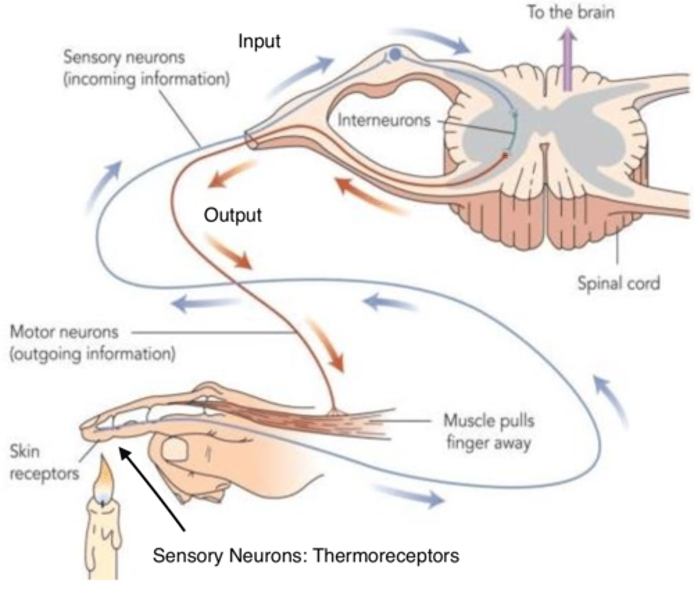
Define the 3 classes of neurons within the nervous system
1) Sensory neurons: specialized neurons carry input messages from the sense organs (eyes, ears, nose, tongue, and skin) to the spinal cord and brain ex: Photoreceptors: modulate pattern of neural activity based off of the amount of light and quality of light
2) Motor neurons: Synapse with muscles. Transmit output impulses from the brain and spinal cord to the body's muscles and organs
3) Interneurons: Synapse with other neurons. Outnumber sensory and motor neurons, perform connective or associative functions within the nervous system, interneuronal activity makes it possible for the complexity of our higher mental functions, emotions and behavioural capabilities
*DIAGRAM: All three classes interacting
State the two major divisions of the nervous system
1) Central Nervous System
2) Peripheral Nervous System
Define central nervous system
the body's primary information processing system; consists of all the neurons in the brain and spinal cord
Define peripheral nervous system
consists of the remaining nerves that fan out throughout the body connecting to the central nervous system via our spinal cord, connected to muscles, glands and sensory receptors. Divided into the somatic and autonomic nervous system
Define Efferent Signal
Ex: Brain signal send to hand
Central to Peripheral: Top down
Define Afferent Signal
Ex: Registering heat in hand
Peripheral to Central: Bottom up
Describe the Central nervous system
Brain: Organ containing 1.4kgs of protein, fat and fluid, most active energy consumer of all bodily organs, consumes 20% of oxygen you use in a resting state. Never rests; rate of energy metabolism is relatively constant day and night
Spinal Cord: Nerve superhighway, nexus between the central and peripheral nervous system, nerves enter/leave through the spinal cord (axon travels through on its way to being interpreted by the brain). Spinal reflexes do not involve the brain
Describe the Two categories of the Peripheral Nervous System
Somatic Nervous System:
- Allows you to sense and respond to your environment: sensory neurons, motor neurons: take in sensory information and respond accordingly
- Voluntary control of body movements
- Sensory and motor neurons bind to create nerves
- Transmit messages to sensory receptors
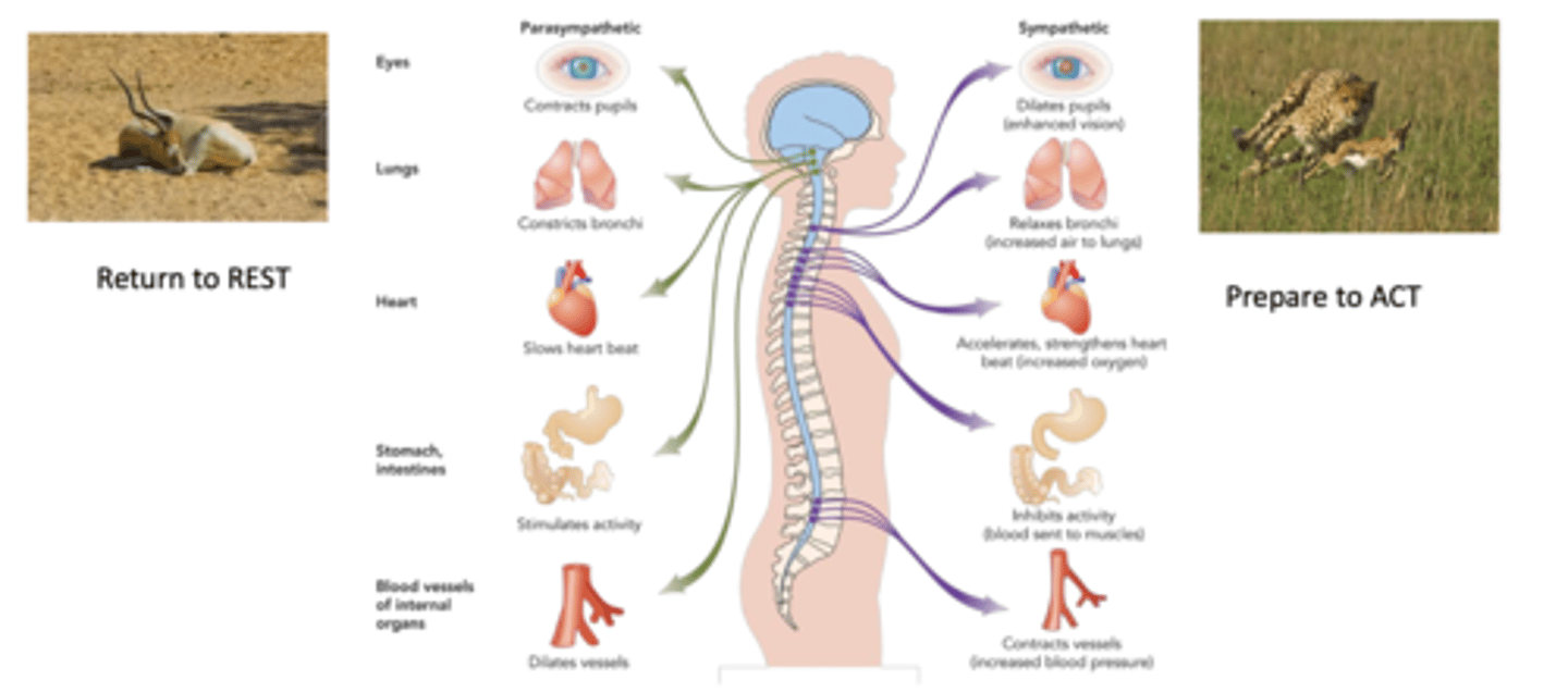
Autonomic Nervous System:
Controls involuntary movement of glands and smooth muscles in body organs: digestion, pancreas, respiration, heart rate
1. Sympathetic Nervous System: Arouse body "fight or flight" and acts as a centralized unit: activation results in cascade of physiological changes to prepare for quick movement
2. Parasympathetic Nervous System: Slows down body processes (rest or digest) and can operate more specifically: homeostasis: keeping the body steady
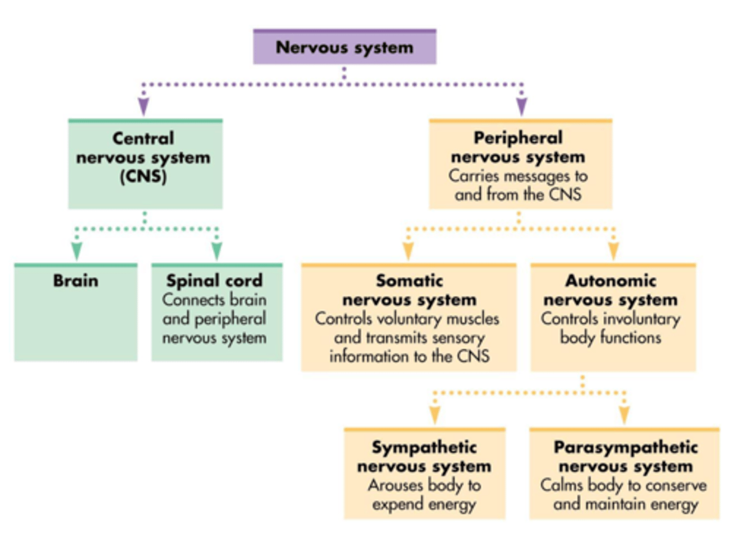
Describe the organization of the nervous system
1. Nervous system = divided into CNS (brain and spinal cord) and PNS (nerves and ganglia outside of the brain and spinal cord)
2. PNS = divided into autonomic NS (involuntary, smooth glands, involuntary bodily functions) and somatic NS (voluntary, allows you to sense and respond to your environment: )
3. Autonomic NS of the PNS: divided into sympathetic (away from homeostasis; fight or flight response) and parasympathetic towards homeostasis (rest or digest)
4. PNS is also divided into affarent (sensory) NS which sends sensory info to the PNS and the efferent (motor) NS which is then broken down into autonomic and somatic, and from autonomic to sympathetic and parasympathetic
What is a misguided method to study brain-behaviour?
Phrenology: the measurement of bumps to predict mental traits
What is the first of four tools to study the brain and explain it....
1. Destruction and Stimulation Techniques: Stimulating areas with electricity/chemicals: probe small areas of the brain with micro electrodes, stimulating action potentials in a particular area and examine corresponding changes in behaviour
Drawback: Invasive
Benefit: Provides precise, systematic mapping of brain functions: motor and somatosensory regions correlating specific areas of cortical stimulation to corresponding behavioral or sensory changes, demonstrating how manipulating neuronal activity can influence movement, sensation, and thought, advancing understanding of brain organization and function.
Possible in Exceptional Circumstances: Ethical for Humans: Ex. Dr Wilder Penfield Performing experiments on people awaiting surgery for alleviating epilepsy: portions of their brain exposed: surgeons probe specific parts of brain to figure out where to stimulate to induce a seizure then lesion that area
Dr Penfield systematically stimulated parts of the exposed cortex with small electrical currents action potential within a localized area: documenting corresponding changes: stimulation changes in movement, thought patterns, bodily sensation
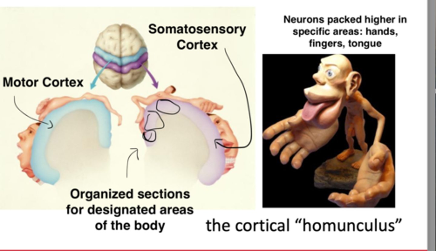
Understood the mapping of motor cortex and somatosensory cortex: as you probe the somatosensory strip, people would feel corresponding changes on different parts of the body, done in an intentional way: stimulate particular parts: ex feeling sensation separately on thumb, then index finger: systematicity: organized mapping to how the somatosensory cortex represented spaces on the body "Sensory of the cortical "homunculus" overrepresentation in specific parts of the body
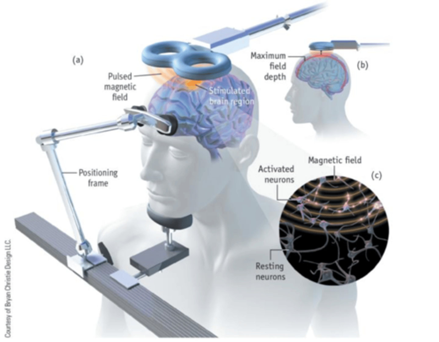
What is the second of four tools to study the brain and explain it....
2. Transcranial Magnetic Stimulation (TMS):
Uses focused magnetic field: pulses to stimulate neural activity in targeted regions of the brain
Depending on strength and timing of magnetic pulses you can alter action potentials: firing/communication between neurons in that specific region being stimulated
Manipulating neural activity, can TMS in specific area then look for corresponding changes in behaviour, then look at a secondary area and if corresponding changes aren't replicated then maybe its something about the first area of the brain that is implicated in some behaviour
- Can cause temporary enhancements or depression of neural activity depending on timing and duration of magnetic pulse has capability of temporary virtual lesion has different applications
Drawback: TMS cannot penetrate deeper brain structures: outermost layer only (cerebral cortex)
Benefit
Benefit: (TMS) offers a non-invasive method to establish causal relationships between brain regions and behavior by temporarily enhancing or suppressing neural activity, enabling targeted investigation of cortical functions and promising research applications, such as alleviating symptoms of depression.
What is the third of four tools to study the brain and explain it....
Electroencephalography (EEG): Technique that uses many passive electrodes on the scalp (receiving signals amplify them), measures excitatory and inhibitory postsynaptic potentials across large region off the brain: electrical activity from populations of many neurons in the brain.
Benefit: Non invasive, EEG offers high temporal resolution, allowing precise tracking of rapid changes in brain activity over time, which is particularly useful for studying dynamic processes like sleep stages and memory consolidation
Drawback: EEG has poor spatial resolution, making it difficult to pinpoint the exact brain regions responsible for the observed activity
ex: stranger things: elle's brain waves
- Can uncover different stages of sleep: Sleep: static behaviour + reduce responsively to external stimuli, slow down breathing, EEG can highlight the different sleep processes: summed excitatory and inhibitory post synaptic potentials, varying across a large portion of the brain, sleep stages: demonstrates dramatic differences, staging is due to the ability of using eeg: characterize different components: deeper stages 3 and 4: is implicated in memory consolidation
What is the fourth of four tools to study the brain and explain it....
Functional Magnetic Resonance Imaging (fMRI): Variant of MRI. Exposed to strong magnet (fixed magnetic strength) Measures localized patterns of brain activity. Activated neurons provoke increased blood flow, which can be quantified by measuring changes of oxygenated and deoxygenated blood to strong magnetic fields: give clues into the brain to see which parts are metabolically greedy: ex: listening to music auditory cortex would be greedy consuming resources
Blood oxygen level dependent (BOLD) signal: The ratio of oxygenated to deoxygenated hemoglobin that permits the localization of brain neurons that are most involved in a task
Benefit: fMRI provides good spatial resolution, allowing precise localization of brain regions involved in specific tasks or mental states, with critical applications like communicating with individuals in vegetative states.
Drawback: fMRI has sluggish temporal resolution, as the BOLD signal reflects changes over several seconds, making it less effective for capturing rapid neural dynamics.
Tennis imagery: activated supplementary motor area
Spacial Navigation Imagery: parahypocampul place area, posterior parietal cortex
Dr Adrian Owen
"Think about playing tennis" "yes"
"Think about walking around your house" "no"
Why is this important?? Clinical implication: communicating with individuals in vegetative / locked in states: 1 in 5 nations in vegetative states are conscious and able to respond to questions in this manner
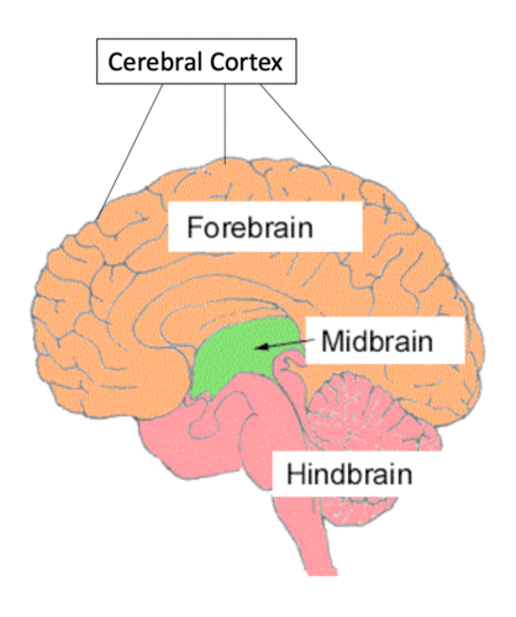
What are the four major areas of the brain?
1. Hindbrain Pons, Medula, Cerebellum
2. Midbrain
3. Forebrain: Thalamus, Hypothalamus, Amygdala, Hippocampus, Basal Ganglia
4. Cerebral Cortex: Four lobes: Frontal, Parietal, Occipital, Temporal
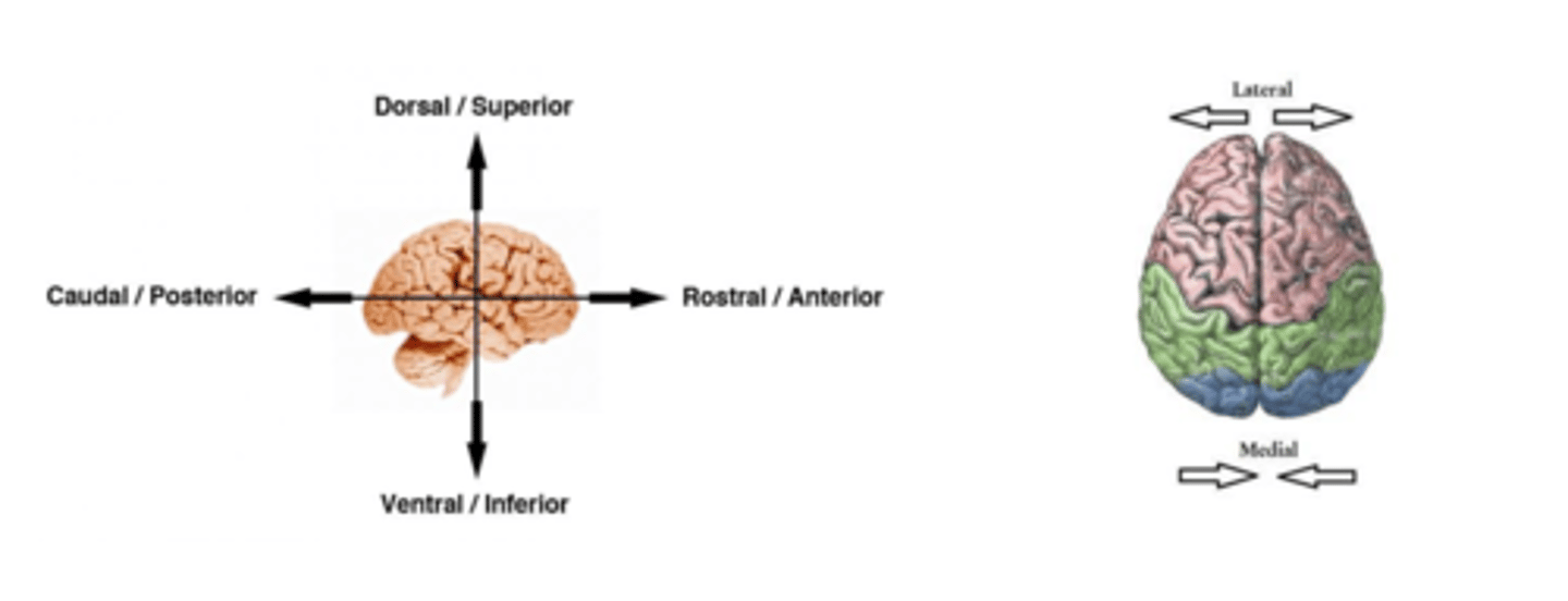
State and define the six orientations of structures in the brain
Dorsal/Superior: Top of the head
Ventral/Inferior: Below the head
Caudal/Posterior: Back of the head
Rostral/Anterior: Front of the head
Medial: Occurring close to the midline of the brain
Lateral: Occurring towards the edges of the brain
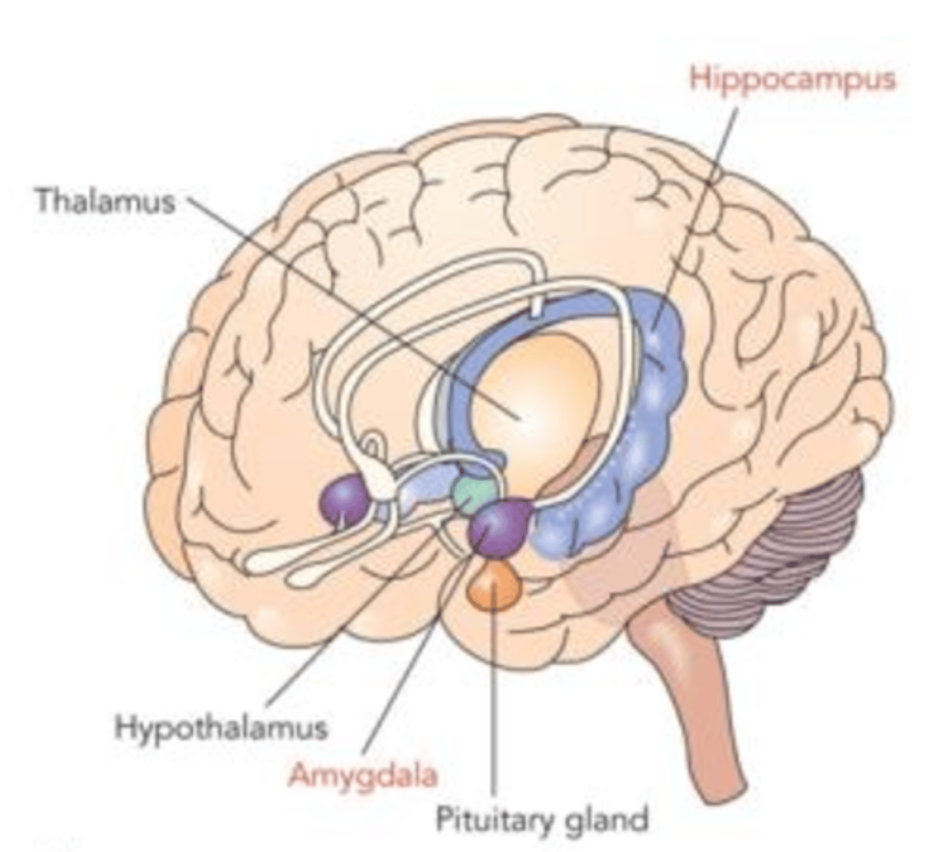
What is the limbic system responsible for and where is it located? Describe the Hippocampus + Amygdala as well as their associated experiments and case studies..
Within the forebrain: Facilitates memory storage and retrieval, represent emotional states, conduit between higher brain areas and autonomic nervous system: fight or flight
Hippocampus: Forming and retrieving memories, Damage to hippocampus can result in profound amnesia: Patient H.M.
Patient H.M. Case Study: Singular study examining behaviour consequences: Underestimated own age, apologized for forgetting names of who he'd been introduced to, describes current state as "waking from a dream, everyday alone in itself"
Severe Anterograde Amnesia: Inability to form new long term memories
Amygdala: Organizes emotional responses especially aggression and fear
fMRI scan experiment: 2 types of visual stimuli: red circle paired with a painful shock, blue circle paired with nothing. Fear conditioned response: seeing the red circle = strong activation of the amygdala: fear of getting shocked
Stimulation experiment: Monkey: Snarling and aggressive behaviour as a result of probing/stimulating this area of the brain, suggests activation in the amygdala is linked to these emotional responses: fear + aggression
Patient S.M. Case Study: Urbach-Wiethe disease resulted in near complete bilateral destruction of the amygdala from late childhood onwards. Called "the woman with no fear": no behavioural or physiological fear response to handling snakes or spiders, haunted houses, scary movies
- Did have fear response to C02 inhalation: slowly reducing oxygen and slowly suffocating, not an associated fear response.
- Suggests that amygdala might be selectively involved in conditioned fear
- Innate fear responses to physiological danger, like suffocation, are managed by other brain regions outside the amygdala
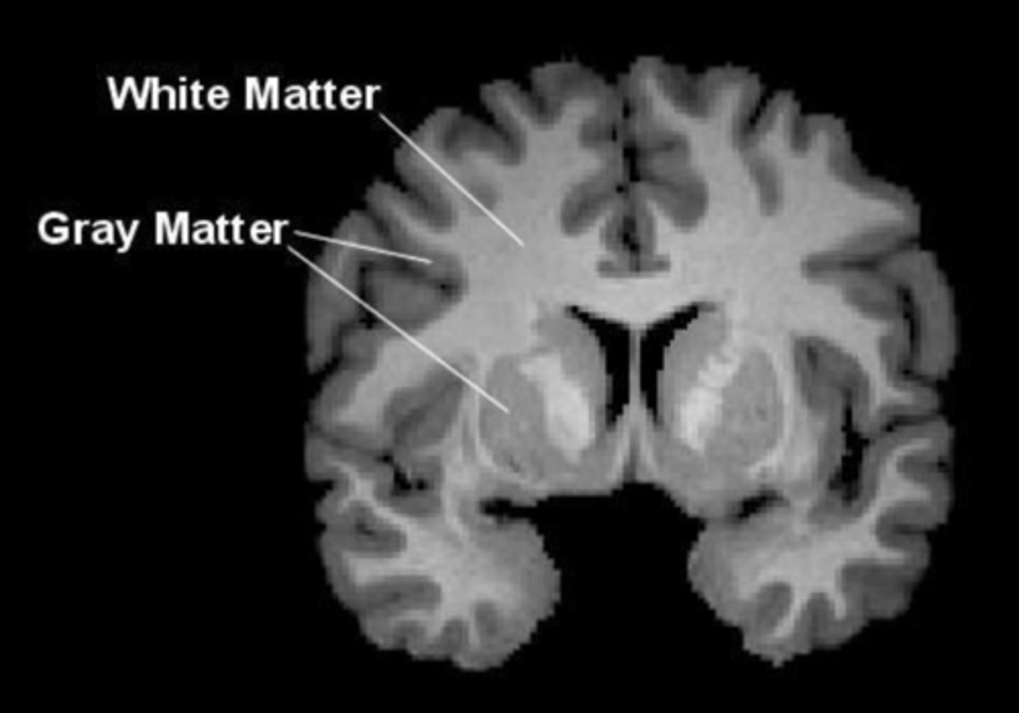
What is the Cerebral Cortex? How does it connect to 2 tools that study the brain?
- 2/3cm sheet of gray matter (unmyelinated cells) outermost layer of the brain, consists of neurons and cell body, 75% of area lies within folds maximizing brain space
- white matter militated axons linking neurons to different areas of the brain
- Provide landmarks for dividing cortex into major areas
Connection to methodology:
- fMRI can image cerebral cortex as well as deeper subcortical areas: amygdala (conditioned fear)
- EEG captures electrical signalling from neurons in the cerebral cortex, outermost layer of the brain
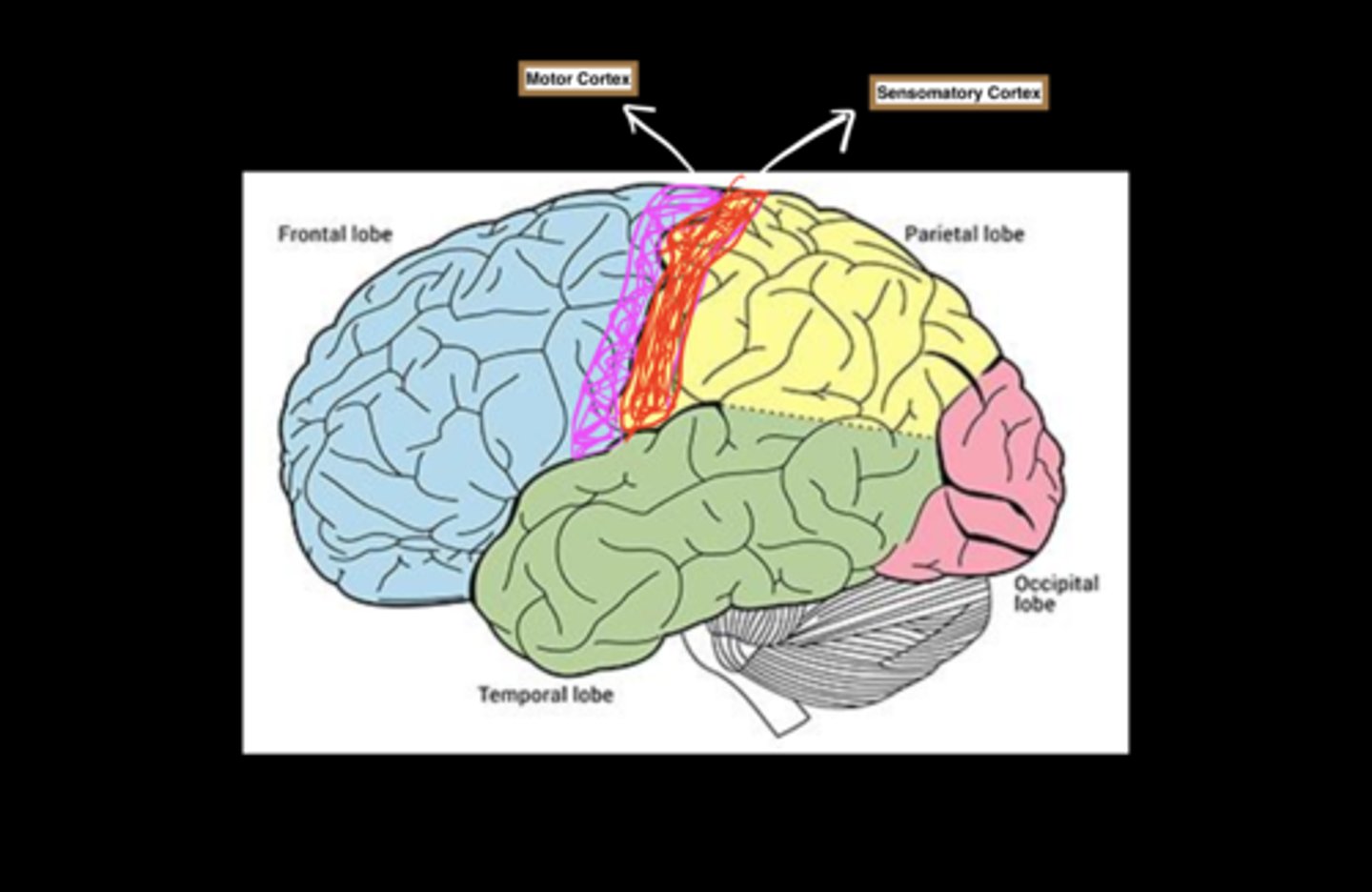
Cerebral Cortex: Four Lobes and their functions
1. Occipital Lobe: Primary cortical visual processing
2. Temporal Lobe: Memory function: formation and retrieval + primary auditory processing
3. Parietal Lobe: Attention, spacial awareness
Somatosensory cortex: most anterior part, corresponds to registration of touch
Contralateral projections: touch sensations from the left side of the body (e.g., left hand) are processed in the right hemisphere, touch sensations from the right side of the body are processed in the left hemisphere
4. Frontal Lobe: Higher order functions: plan, flexibly adapting behaviour, working memory and mental storage
Motor cortex: located at rear of frontal lobe planning, controlling, and executing voluntary movement
Contralateral projections: movements of the left side of the body are controlled by the right hemisphere, and movements of the right side are controlled by the left hemisphere
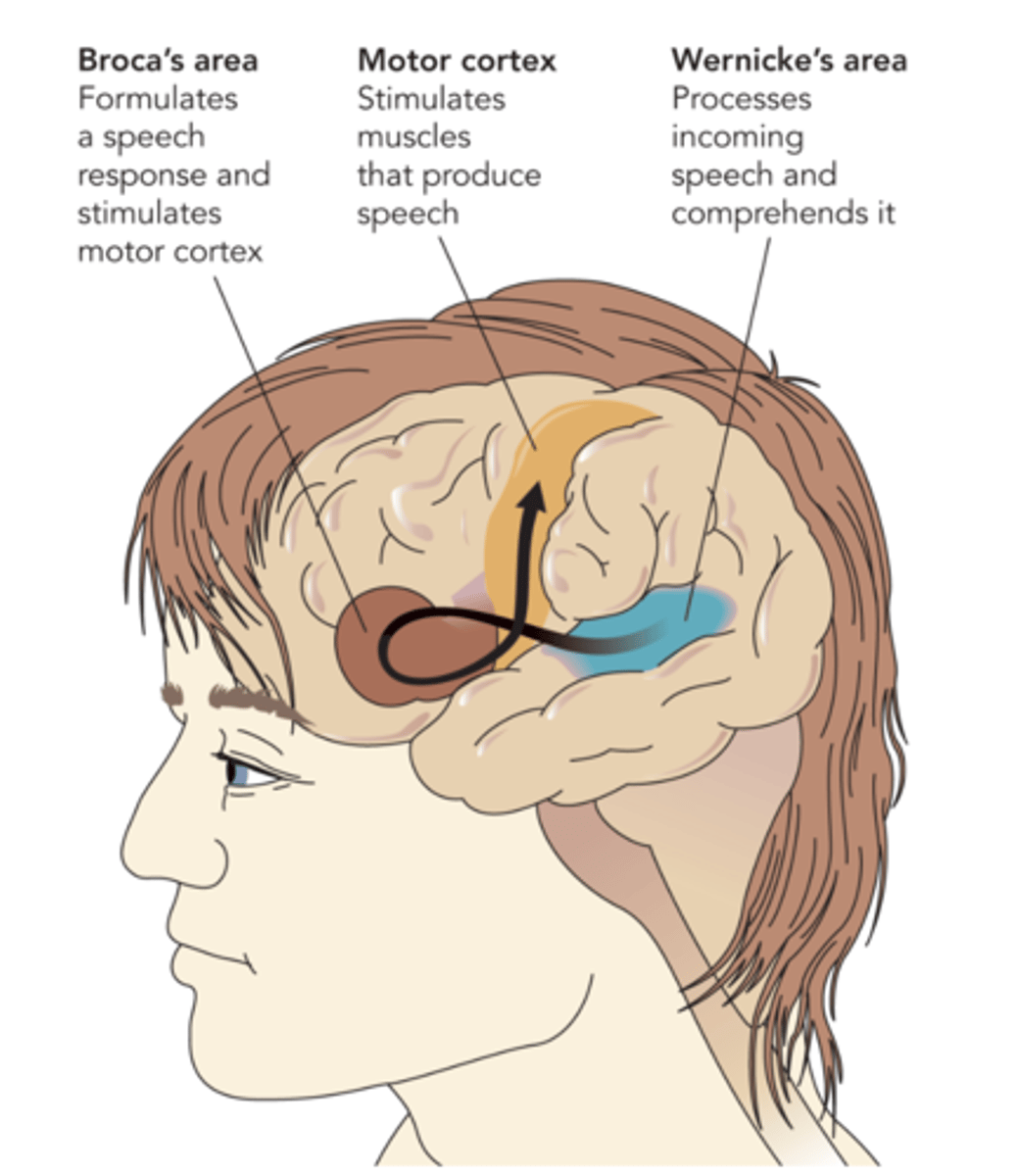
Describe the two critical areas for speech comprehension and production (multiple areas and lobes)
Temporal lobe
Wernickes Area: Processes incoming speech and comprehends it
Damage results in Werniche's Aphasia: Ability reproduce sounds but would be gibberish
Frontal lobe
Broca's area: Formulates a speech response and stimulates motor cortex
Damage results in Broca's Aphasia: Language deficit: speech comprehension but can produce speech
Motor cortex: stimulates muscles that produce speech
Describe the Association Cortex
- Found within all lobes of the brain: Parietal, Frontal, Occipital and Parietal
- Involved in highest level of mental functioning (perception, language, thought, reasoning)
- Referred to as the "silent areas"
- Electrically stimulating association cortex does not give rise to either sensory experience or motor response: but it functions to connect incoming information across the brain
- Accounts for 75% of the human brain
Describe the Frontal Lobe and Associated Case Study
- Most overdeveloped area relative to non-human species
- Responsible for executive functioning: manages planning, adaptability, selective attention, and working memory to organize, focus, and adjust behavior effectively
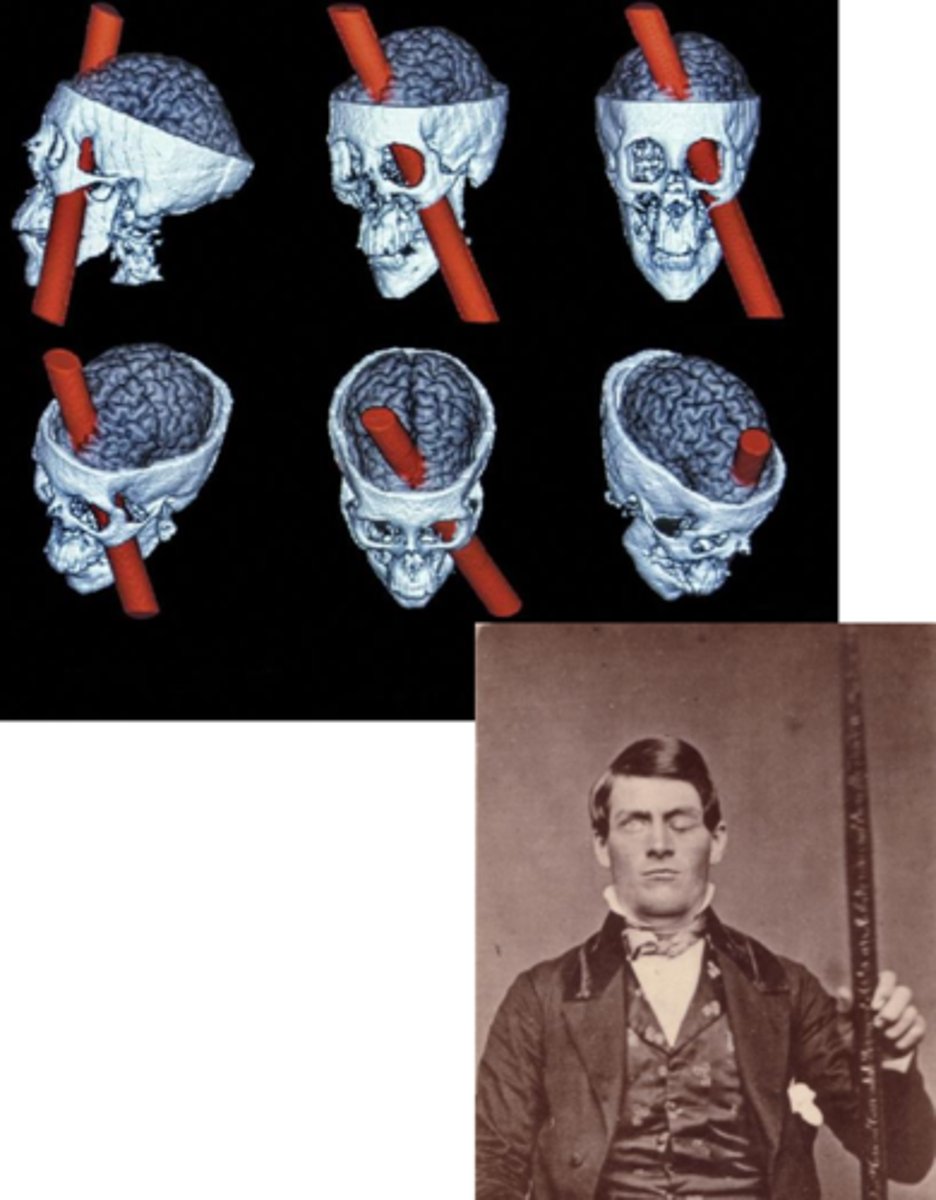
Case Study Example: Phineas Gage
- 1848 railroad accident, spike through prefrontal cortex
- Resulted in corresponding behavioural changes: self awareness, planning, initiative, responsibility, emotional reactivity changed: impulsive, irritable, and prone to inappropriate emotional outbursts, critically implicated in the frontal lobe
Prefrontal Cortex:
- Seat of executive functions
- Goal-setting, judgement, planning, adaptive behaviours: ability to control impulses: profound changes in personality
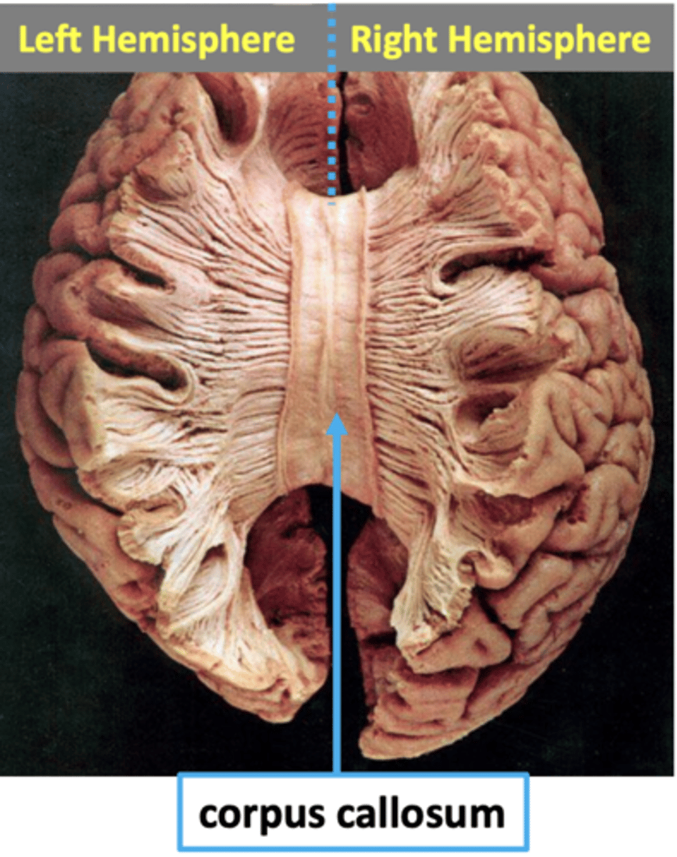
Explain how the human brain has two hemisphere and how lateralization works...
- Two hemispheres connected by the corpus callosum
- "Mirrored" structures in both hemispheres (left and right side)
- Ex: Information processed in the right hemisphere communicates to the left by crossing over the corpus callosum: white matter tract: bulk of information crossover
- Lateralization: Behaviours tend to be strongly processed/represented in one hemisphere versus the other
- Left: Lateralization of Verbal and logical abilities: although language processing occurs in both hem. stronger representation in left for most individuals
- Right: Lateralization of Spatial relations, music processing
Explain why the notion "left brained" and "right brained" individuals is a myth...
- Lateralization in specific processes differ amongst individuals
- Evidence for being "left brained" doesn't exist:
fMRI Study: Mathematically gifted: Greater density in left hemisphere? compare to control... no difference.
Complex personality traits: increased creativity vs. logic show no evidence of lateralization
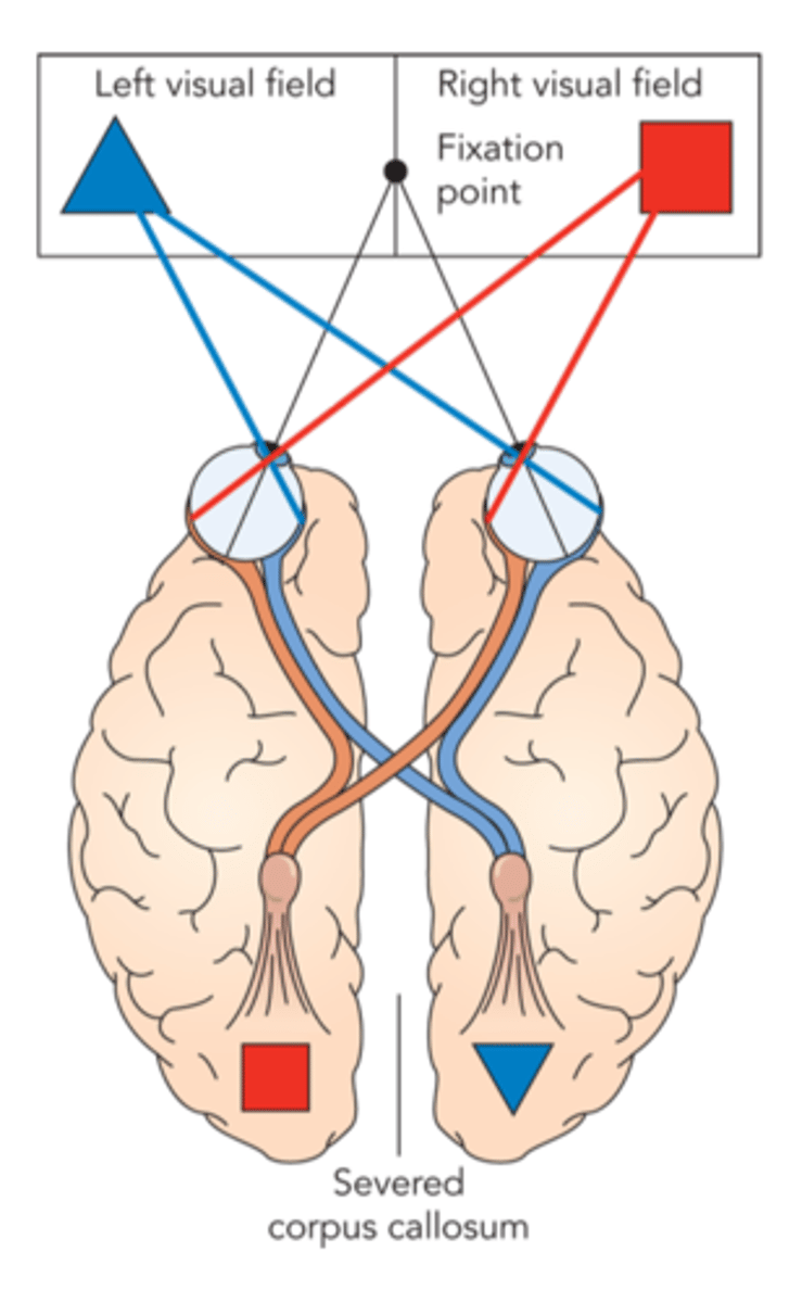
What is a split brain experiment?
- Parts of brain removed to alleviate symptoms of epileptic seizures: Callosotomy: severing corpus collosum, if seizures are lateralized it prevents the seizure from crossing over to the opposite hemisphere
- Severed corpus collosum these two hem. no longer have main pathway to convey information from one side of the brain to the other, two separate brains in one
- optic nerve remains unsevered: contralateral processing: left visual field processed in right hemisphere, right visual field processed in left hemisphere
information processed in one hemisphere then transmitted to the other hemisphere but for...
Callosotomy patients: Information locked in one hemisphere and transmittion cannot occur because of severed corpus collosum
Procedure: Image is flashed to the left of fixation point, asked what they saw. If the information is being processed in the right hemisphere + can no longer communicate, they can't verbalize what they've seen... Left hand being controlled my right hemisphere asked to reach down and grab the object they are able to correctly reach the object.
What is neural plasticity?
Adapted changes in structure and function to the nervous system
Studied through injury and recovery
Brain damage or sensory loss: individuals who are born blind, occipital cortex adapts and responds to stimuli that aren't perceptually processed, hearing spoken language occipital lobe begins to respond to sound due to lack of visual input, if there are sensory losses, doesn't mean the area of the brain goes dark, it will respond to other kinds of stimulation
Explain the Role of experience and environment in relation to neural plasticity...
Musicians:
- No single music area of the brain: involves auditory perception, visual perception, motor coordination and planning, distributed across the lobes in a complex network
- When you examine professional pianists: view enhancement and changes in somatosensory areas: greater cortical representation for fingers than control group
- Better representations of auditory pitch compared to non-musicians: musical training may not cause these changes, could encompass other factors
Describe the Endocrine System
Dictates the regulation, synthesis and release of hormones
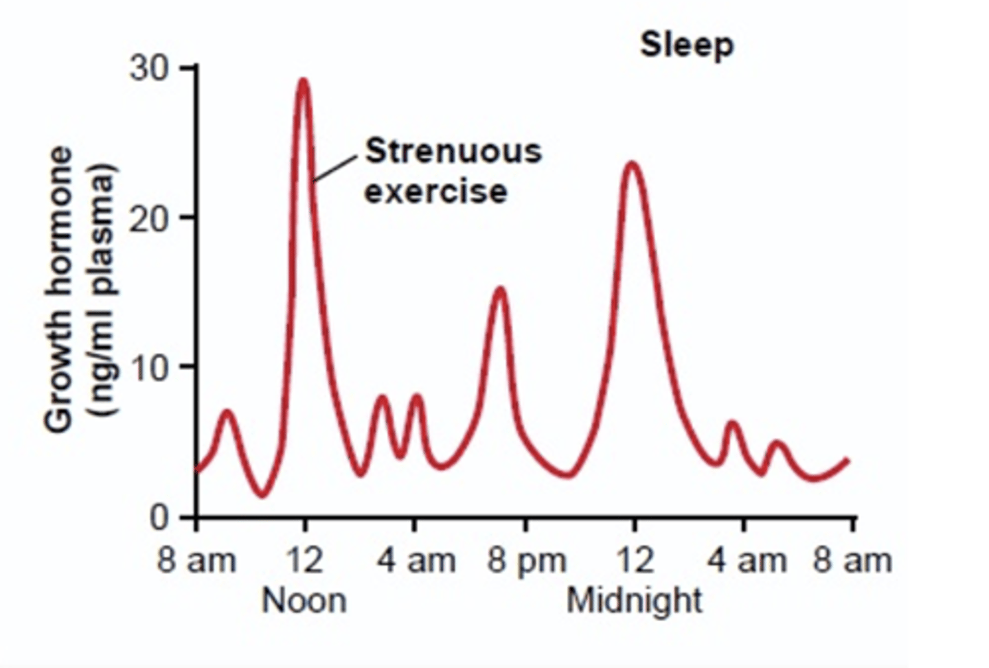
Define Hormones
Chemical messengers in the bloodstream
- Pulsatile release: not a steady pattern: environmental factors: working out + early on in the sleep cycle (release of growth hormones)
- Negative Feedback System: Hormones are released until a set amount is detected in the bloodstream then the release is stopped
Hormones versus neurotransmitters
Similarities and Differences
Similarities:
- Both chemical messengers, binding to receptor sites on cells
- Sometime a chemical can serve "double duty": acting as a neurotransmitter in one context and a hormone in another context
Example: Epinephrine (Adrenaline): Neurotransmitter: can change graded potentials, when opporating in the nervous system, can have very different effects acting as a hormone in the bloodstream
Differences:
- Different timescales and places of operation within the body
- When hormones are released they travel through the bloodstream and have more diffused effects across the body: site of action is less specific, decreased temporal precision in the release (seconds to hours)
- Neurotransmitter released is precisely timed action potentials (millisecond-level effects) and operate much more locally
What is the Hypothalamus + 7 Major Endocrine Glands
Forebrain structure involved in regulating basic biological needs: hunger + thirst, intricately connected to the
1. Pituitary gland: The master hormonal gland that releases hormones to stimulate activity in other endocrine (hormone) glands located throughout the body
2. Pineal gland: helps regulate circadian rhythm
3. Thyroid: metabolic rate
4. Thymus: regulation/creation of white blood cells, immune function
5. Adrenal gland: salt and carbohydrate metabolism
6. Pancreas: sugar metabolism
7. Gonads (ovary/testis): sex hormones
Describe the Endocrine response to the Dog Spider Halloween Costume Example
1. Fight or flight response: Input from retina
2. Amygdala: fear response initiated
3. Hypothalamus: Signals to pituitary gland
4. Pituitary gland signals to Adrenal Gland
5. Adrenal Gland releases epinephrine (adrenaline)
6. Kick starts autonomic/sympathetic nervous system
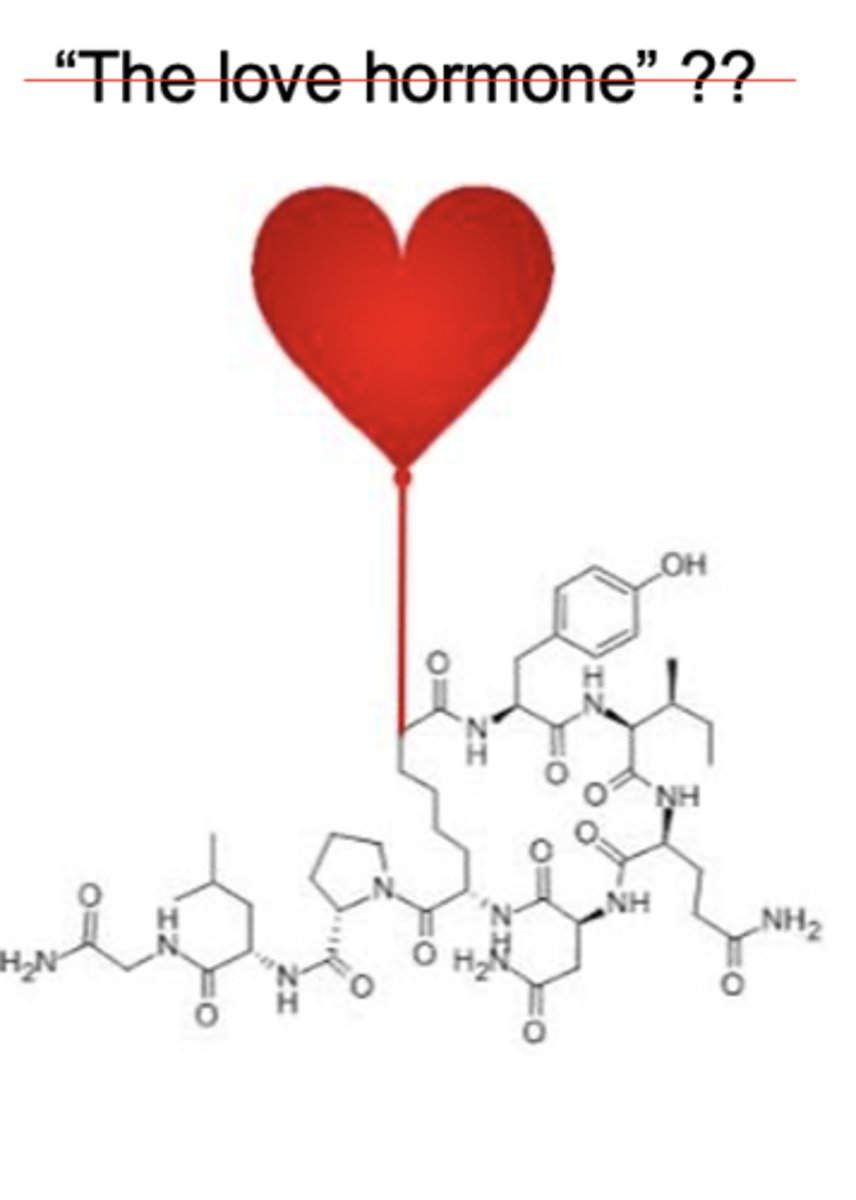
Hormones and social psychology: Define Oxytocin and its Social Consequences
Oxytocin: Hormone released by the pituitary gland
Functions with social consequences:
1. Triggers contractions during childbirth and lactation for breastfeeding
2. Pair-bonding and fidelity: Comparative studies: monogamy and oxytocin release and receptors
3. Heightened senses of empathy for others
4. Increased trust for others
Love hormone: half truth: mechanism of action: contribution to effects; increasing awareness to social cues: attending to someones experience and an empathic connection
To what extent are characteristics genetically determined? Heredity (Genetic) vs. Environment: Jim Twins
Case study providing intriguing evidence of the influences of hereditary factors (Genes) on behaviour: Twins grown up separated: Reunited after realizing they lived almost identical lives: Same name, same wives name Linda, divorced, Both remarried wives named Betty, both had sons named James Allen, both worked jobs in security, heavy smokers, drove the same car, same dogs, same vacation spot, both had brother named Larry
Medical history and brain wave tests almost identical
Is behaviour entirely environmentally or genetically determined?
NO, behaviour is determined by multiple causes, behaviour is shaped by cultural heritage and behaviour is jointly influenced by heredity and environment. Suggests complex interplay between genetics and environment
What is Behavioural genetics?
The study of the influence of genetic factors on behavioural traits
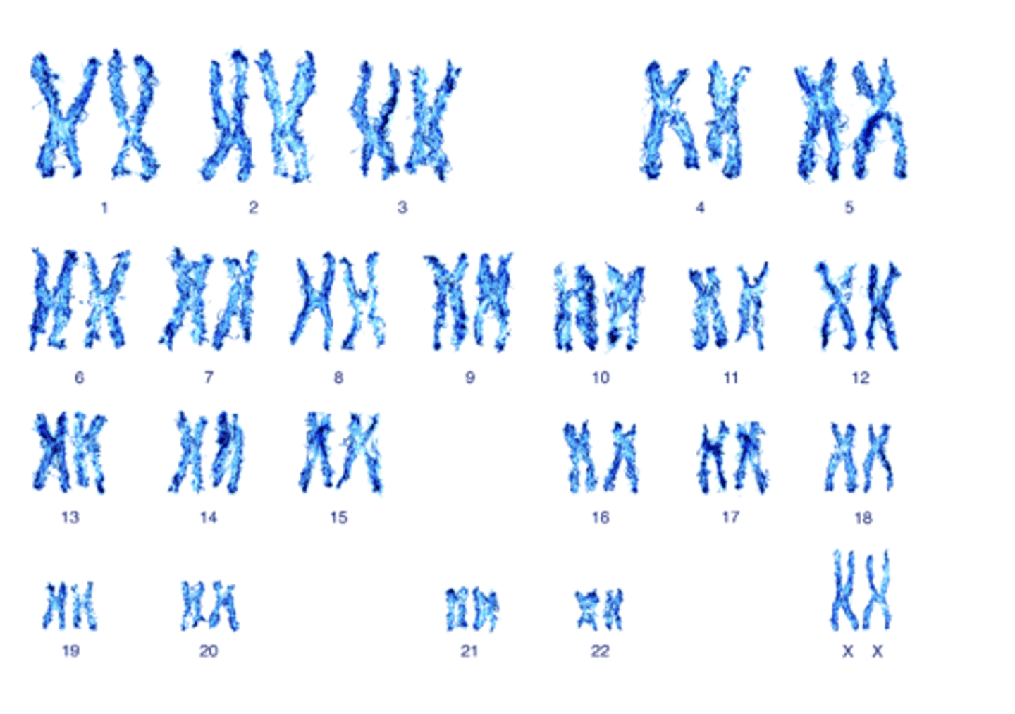
What are chromosomes?
Strands of DNA carrying genetic information
- Human cells: 46 chromosomes in 23 pairs except sex cels (not paired)
- Each chromosome contains thousands of genes, also in pairs
Define Dominant and recessive genes
When one gene pair overrules its pair, it is said to be dominant. Gene pair that is overruled is said to be recessive
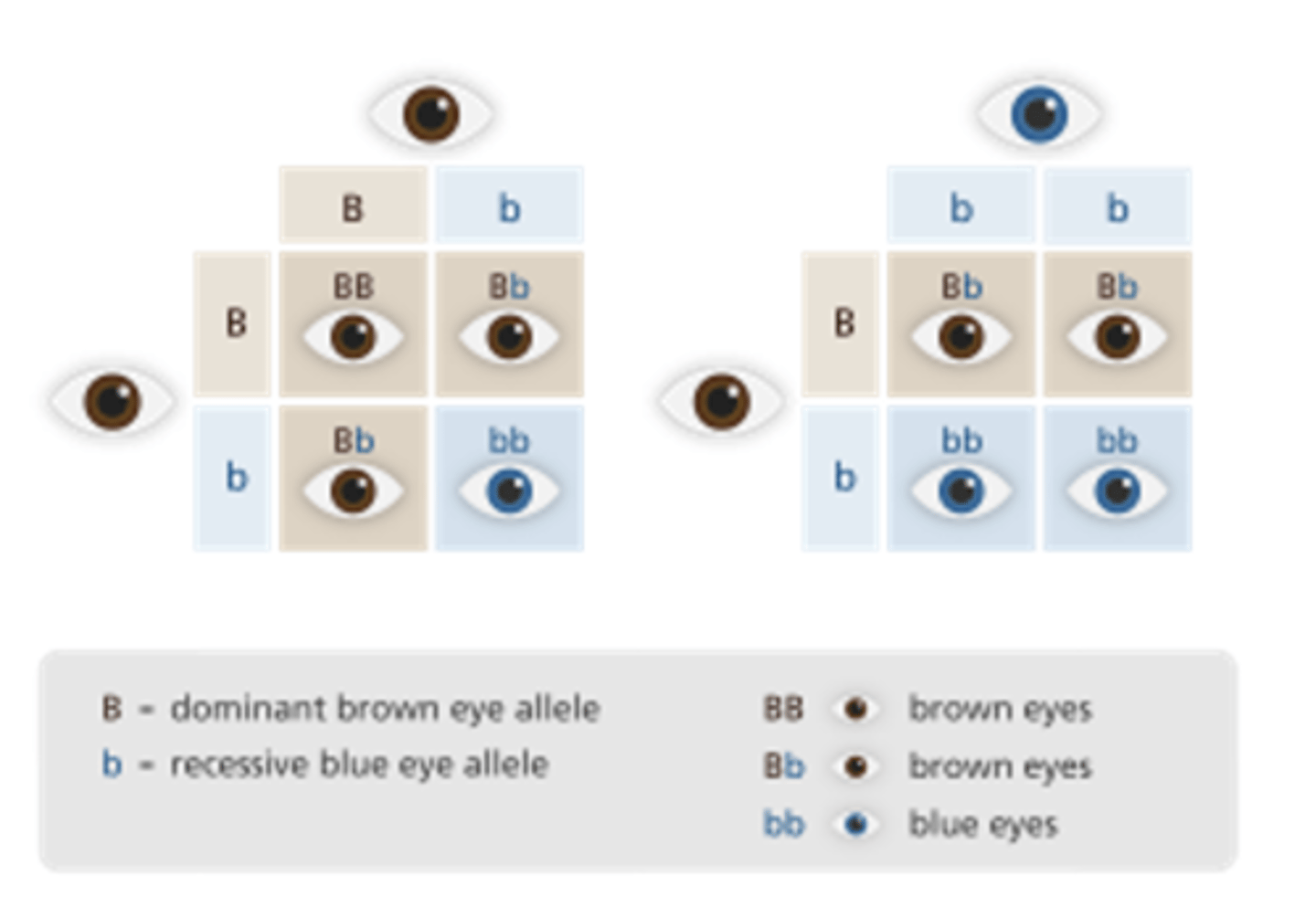
What does it mean to be Homozygous for a trait?
If someone has two genes in a specific pair that are the same (Dom/dom) or (Rec/rec) then that person is considered homozygous for that trait
What does it mean to be Heterozygous for a trait?
If genes are different (Dom/rec) that person is considered heterozygous for that trait.
Define genotype
Refers to someone's genetic makeup: composition of (Dom/rec) genes
Define phenotype
how someones genotype is manifested in observable characteristics
ex: Brown eyes manifested
Define polygenic
when several genes influence a trait ex: at least eight gene pair have been implicated in determining eye colour
How do measure heredity and Behaviour... Highlight the 4 Processes
1. Family Studies: assess hereditary influence by examining blood relatives to see how much they resemble one another on a specific trait to determine whether it runs in the family. Many things run in families, that are not genetic: ex dessert recipes or speaking certain languages
Ex: Familial impairments on speech/language ability have been associated with mutations on the FOXP2 gene. Individuals with such mutations may struggle with articulating words (speech) or forming and understanding language (grammar and syntax).
2. Twin Studies: Yield better evidence about the possible influence of heredity because identical twins share 100% of the same genes and fraternal twins share 50% of genetic relatedness. If twins of both types are raised in similar environments and identical twins are more similar on a given trait the assumption would be it is probably a genetic explanation
Correlation between intelligence and personality. Findings suggest that identical twins tend to be more similar than fraternal twins with regard to intelligence and specific personality traits (extraversion) suggests that intelligence and personality are influenced by heredity
3. Adoption studies: Access genetic influence by comparing adopted children with their biological and adoptive parents: if they are more like their biological parents on a trait, It is probably genetic
4. Genetic Mapping: Process of determining the location and chemical sequence of specific genes on specific chromosomes. Theorists suspect that thousands of genes may influence complex behavioural abilities and trraits with each of these specific genes exerting a tiny effect
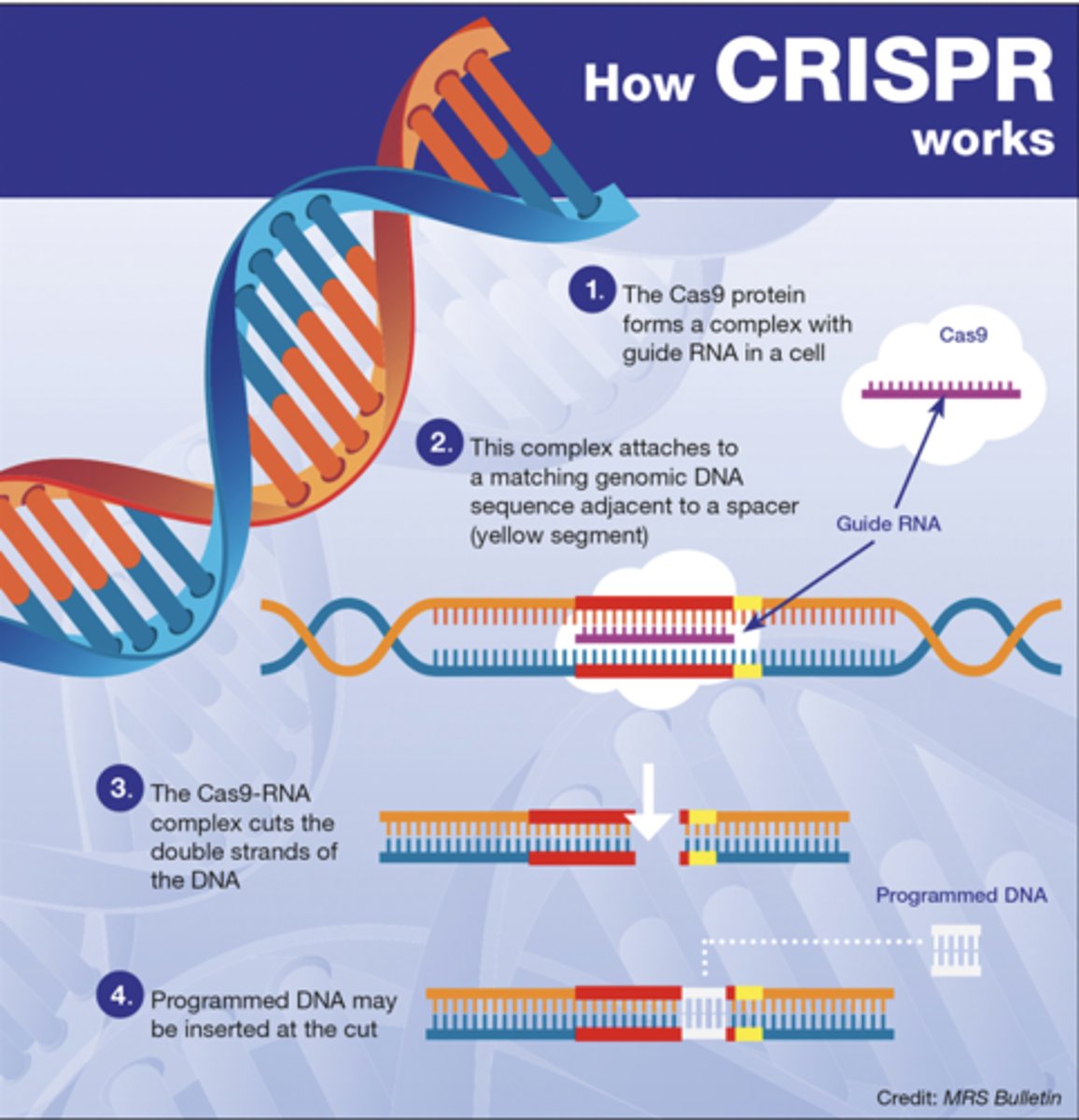
Explain what CRISPR is, how it functions and an associated case study
Clustered regularly interspaced short palindromic repeats (CRISPR): Experimentally and causally manipulate the genome. Gene editing tool, stores a small section of harmful viruses in order to recognize them when attacked. Scientists use these to modify the genomes of animals including humans in order to prevent genetic diseases: cystic fibrosis + Alzheimers disease
Oxytocin and Love hormone study: Highlights complexity: If we remove oxytocin receptors, should see corresponding changes in behaviour, experiment showed they still demonstrated pair bonding and did not interfere with monogamy
What is Epigenetics? How Does It Work?
Why Is It Important?
- Study of changes in gene expression without altering the DNA sequence.
Environmental factors like nutrition, stress, and drug use can turn genes "on" or "off."
These changes control how active or inactive certain genes are.
Some epigenetic changes can be passed to future generations.
This process, called gene tagging, allows environmental influences to impact offspring
What does this mean: "We inherit dispositions, not destineis"
While Genetics and hormones set the stage for our potential behaviors, environmental factors, life experiences. Personal choices play a crucial role in shaping who we become. Our genes give us a starting point, but we have the ability to influence the outcome.
What is evolutionary psychology? Who is it based in? What do these theorists study? Give an example
New field in psychology focusing on analyzing human behaviour in terms of adaptive significance
Based on Charles Darwin + his ideas of natural selection/reproductive fitness: variation in reproductive success are what fuels evolutionary change
Evolutionary theorists study adaptations or inherited characteristics that increase in a population because they help solve a problem of survival or reproduction upon emergence
Adaptations may extend when no longer needed: Ex: fight or flight response may have been helpful in primitive times but now relate to several stress related diseases