L37 (Lovelace) - Derm Manifestations of Systemic Disease
1/54
There's no tags or description
Looks like no tags are added yet.
Name | Mastery | Learn | Test | Matching | Spaced | Call with Kai |
|---|
No analytics yet
Send a link to your students to track their progress
55 Terms
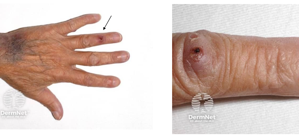
Osler nodes
seen in bacterial endocarditis - due to septic emboli
painful
erythematous nodule with pale center
on fingers and toes
osler node
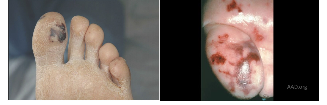
janeway lesion
seen in bacterial endocarditis - due to septic emboli
not painful
angular hemorrhagic lesion of palms and soles
janeway lesion
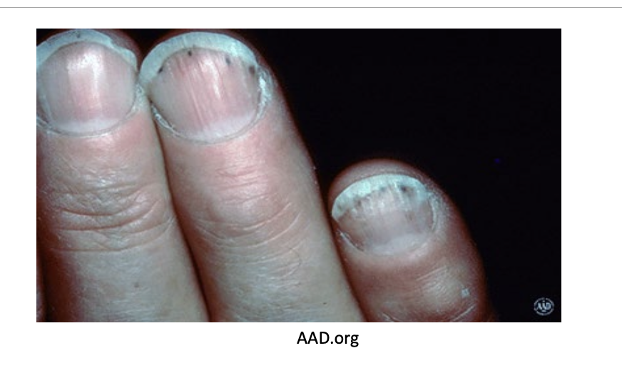
splinter hemorrhages
spilnter hemorrhages
seen in bacterial endocarditis - due to septic emboli
black linear lesion under nail plate
Jones criteria
met in rheumatic fever
i
Carditis
Polyarthritis
Sydenham Chorea
Erythema Marginatum
Subcutaneous nodules
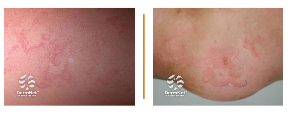
erythema marginatum
seen in acute rheumatic fever
trunk and upper arms and legs - almost NEVER on face, palms, or soles
pink or red macule or papules that spread outwards in a circular shape
as lesions advance, edges become raised and red and central clearing forms
erythema marginatum
subcutaneous nodule
seen in rheumatic fever
painless
over joints, back of scalp, over vertebrae
firm, round, mobile nodules between .5-2cm
usually only when severe carditis is present
appear in later phase of rheumatic fever
subcutaneous nodules
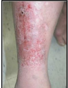
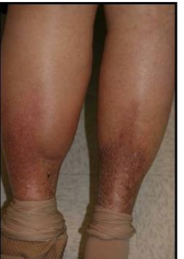
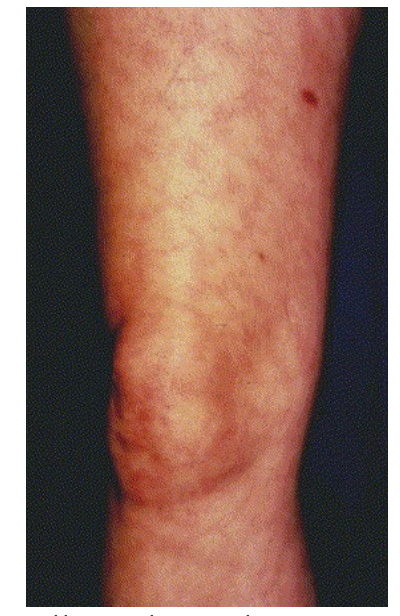
stasis dermatitis
chronic LE edema —> hyperpigmentation and LE swelling
typical presentation: erythema, scale, pruritus, erosions, exudate, crust
lower 1/3 of legs, superior to medical malleolus
bilateral or unilateral
may develop lichenification
often has varicose veins and hemosiderin deposits (pinpoint yellow-brown macules)
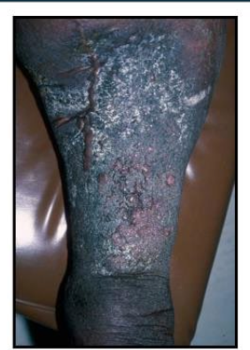
lipodermatosclerosis
Long-term uncontrolled edema (stasis dermatitis) pay progress to lipodermatosclerosis
fat necrosis —> permanent sclerosis
“inverted champagne bottle” legs
may have acute inflammatory episodes with pain and erythema (can be mistaken for cellulitis)
Elephantiasis verrucosa nostra
Long-term uncontrolled edema (stasis dermatitis) may progress to elephantiasis verrucosa nostra
inflammation of draining lymphatics —> damaged vessels —> lymphatic insufficiency
pebbly, hyperkeratotic, rough overlying skin
ulceration in this scenario would be super hard to treat
stasis dermatitis
lipodermatosclerosis
elephantiasis verrucosa nostra
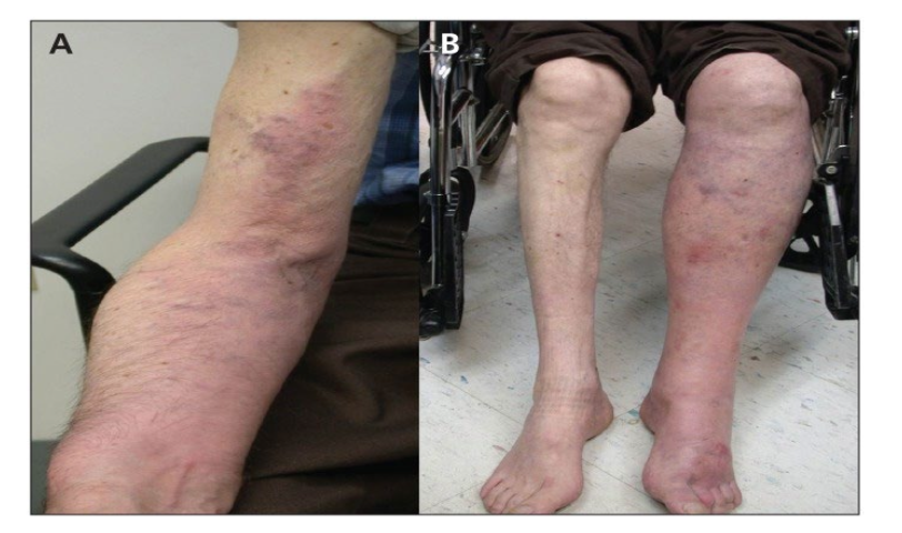
cholesterol embolus
result of cholesterol mobilization from atherosclerotic plaques lining walls
can be sporadic, but usually with invasive vascular procedures or therapies (anticoagulation, thrombolytics)
scattered, violaceous, reform (net-like) vascular patches
cholesterol embolus
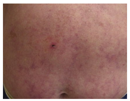
livedo reticularis
livedo reticularis
well-know, common
macular, violaceous, connecting rings in net-like pattern
caused by anatomy and physiology of cutaneous microvascular system
associated with
antiphospholipid syndrome
sneddon’s syndrome (rare, livedo reticularis + cerebrovascular lesions)
cryoglobulinemia, cryofibrinogenemia
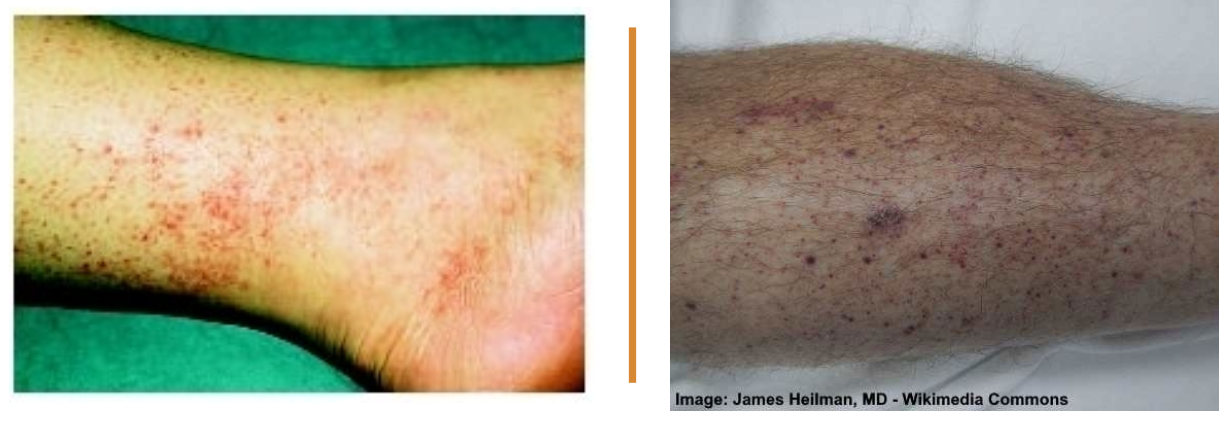
petechiae
petechiae
tiny hemorrhage spots under skin or in mucous membranes
purple, red, or brown dots
not palpable!!!
caused by:
infection
RMSF
hemolytic uremic syndrome
viral hemorrhagic fevers (ebola)
thrombocytopenia
TTP
ITP
leukemia
nutritional deficiency
Vit C (scurvy!)
Stewart-Treves Syndrome
development of an aggressive lymphangiosarcoma at site of chronic lymphedema
(lymphedema is consequence of radical mastectomy)
90% upper limb
chronic lymphedema + multiple red-blue macule or nodules
Stewart-Treves Syndrome
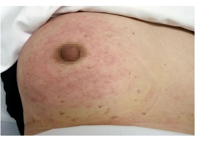
Peau d’orange
inflammatory brest carcinoma
type of cancer in which breast cells block lymph vessels in skin —> red and swollen breast
may also see dimpling or pitting
may also see inverted nipple
peau d’orange
rough, bumpy, pitted skin appearing on breast
looks like an orange peel
caused by
inflammatory breast cancer
mastitis
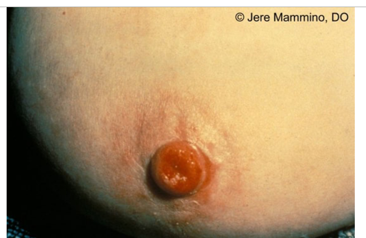
Paget’s Disease
Paget’s Disease of the Breast
MALIGNANT
one breast (usually)
similar to eczema on outside: dry, scaling nipple and areola
eczema usually affects both breasts!!
MC in F >50
***associated with underlying invasive breast cancer or ductal carcinoma in situ
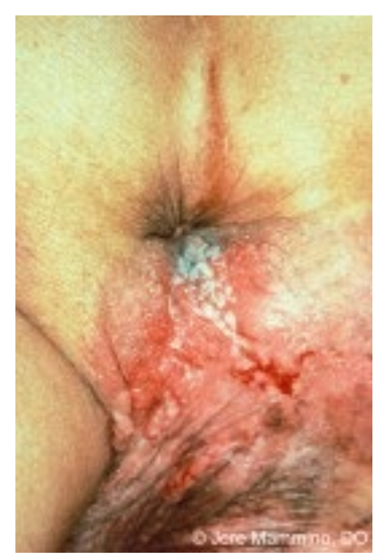
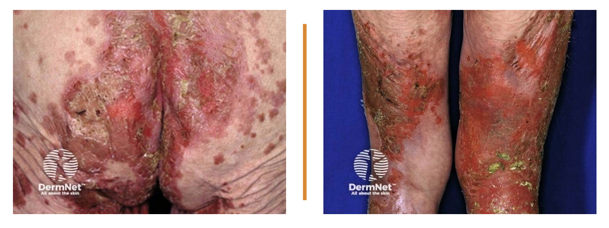
Extramammary Paget’s Disease
Extramammary Paget’s Disease
MALIGNANT (adenocarcinoma)
slow growing, non-invasive
confined to epidermis usually - can extend into deeper dermis
mostly found in GU: vulva, penis, scrotum, perineum
can also be found in armpit/axillary region
looks like a rash - often mistaken for eczema
Necrolytic migratory erythema
associated with glucagonoma syndrome
genital and anal region, butt, groin, lower legs
rash fluctuates in severity (we don’t know why the rash appears)
initially: ring-shaped red area that blisters, erodes, and then crusts
pruritic and painful
sore mouth tongue, sore mouth, cracked dry lips, ridging of nails
Glucagonoma syndrome
adults >50
slow-growing tumor in alpha cells of pancreas —> excessive glucagon excretion —>
diabetes
weight loss
diarrhea
neuro and psych sx
venous thrombosis
anemia
necrolytic migratory erythema
necrolytic migratory erythema
Carcinoid syndrome
neuroendocrine tumor of GI tract
episodic flushing***
diarrhea
wheezing
right valvular heart disease
niacin deficiency (pellagra)
increased urinary 5-HIAA (serotonin metabolite)
Trousseau syndrome
migratory superficial thrombophlebitis in pts with occult or recently diagnosed visceral malignant disease
Trousseau Syndrome
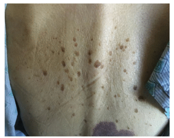
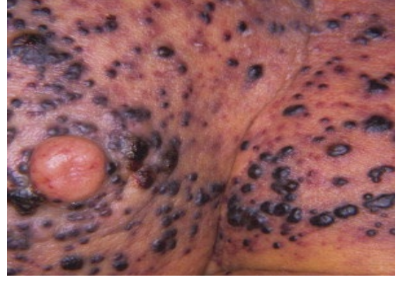
Sign of Leser-Trelat
Sign of LEser Trelat
sudden development of multiple pruritic seborrheic keratoses
often associated with malignancy
roughly half are adenocarcinomas (MC = stomach, breast, colon, rectum)
can also be seen in lymphoma, leukemia, squamous cell carcinoma
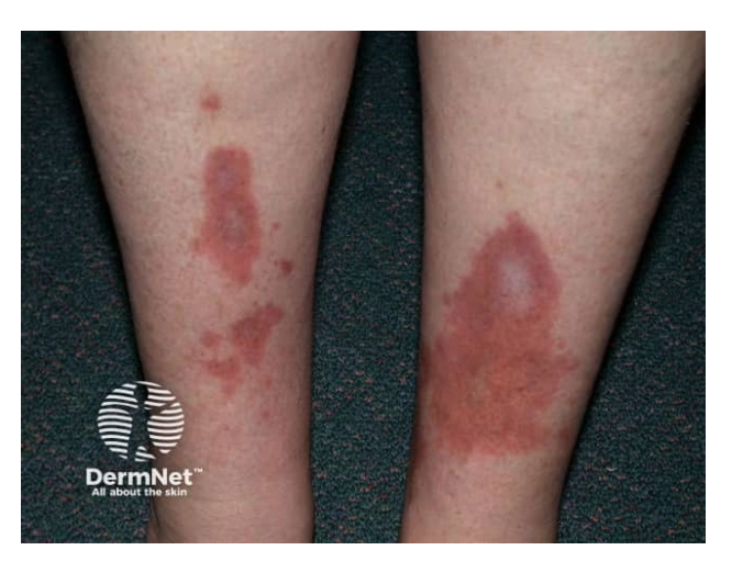
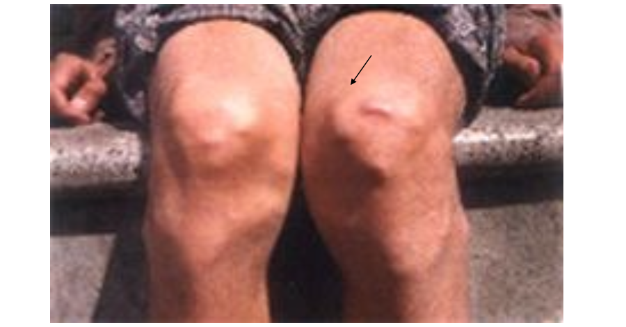
Necrobiosis Lipoidica Diabeticorum
Necrobiosis Lipoidica Diabeticorum
granulomatous skin disorder
shins of diabetics
begins as dull red papule or plaque —> slowly enlarges into one or more yellow-brown patches with a red rim
central atrophy - shiny, pales, thinned, prominent blood vessels
may have reduced sweating and sensation
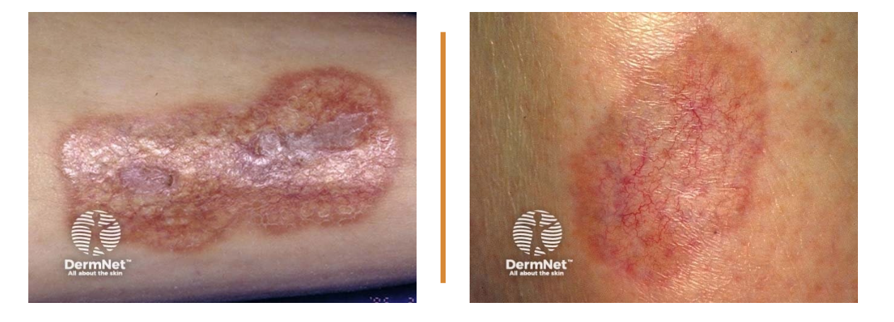
Necrobiosis Lipoidica Diabeticorum
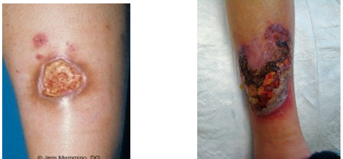
Pyoderma gangrenosum
Pyoderma gangrenosum
uncommon; recurrent and chronic ulcer
NOT ACTUALLY INFECTIOUS OR GANGRENOUS —> associated with systemic disease (has have underlying conditions like IBD)
pathergy: exaggerated skin lesions in response to minor trauma
undetermined border
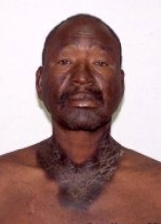
Pellagra
Pellagra
Niacin (Vit B3) deficiency
begins as rash without defined borders that resembles sunburn —> become severe with darker pigmentation, blisters, skin sloughing on face, neck, arms, legs
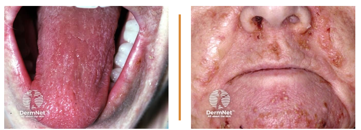
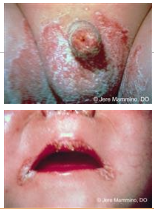
Vit B2 deficiency
deficiency in riboflavin
causes:
stomatitis of mouth and tongue (magenta tongue!!!!!!!!!)
cheilosis: inflammation of lips, scaling and tissues at corners of mouth
Scurvy
deficiency of ascorbic acid (Vit C)
swollen guns
easy brusing
petechiae
perifollicular hemorrhages***
“corkscrew” hears***
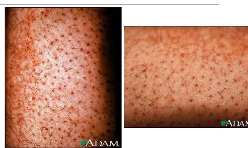
scurvy
Zinc deficiency
acquired:
angular chelitis
sclay plaques on areas of friction
nail dystrophy
hair loss
acrodermatitis enteropathica (inherited): defect in zinc absorption
angular chelietis
gluteal, perineal, and aural burn-like psoriasiform lesions
zinc deficiency
Kwashiorkor
protein malnutrition
edema
anemia
fatty liver
hyperkeratosis***
dyspigmentation****
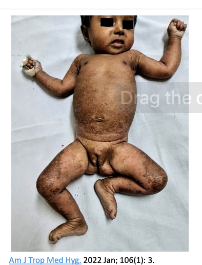
Kwashiorkor