Chapter 2 - Basic components of living systems
1/68
There's no tags or description
Looks like no tags are added yet.
Name | Mastery | Learn | Test | Matching | Spaced |
|---|
No study sessions yet.
69 Terms
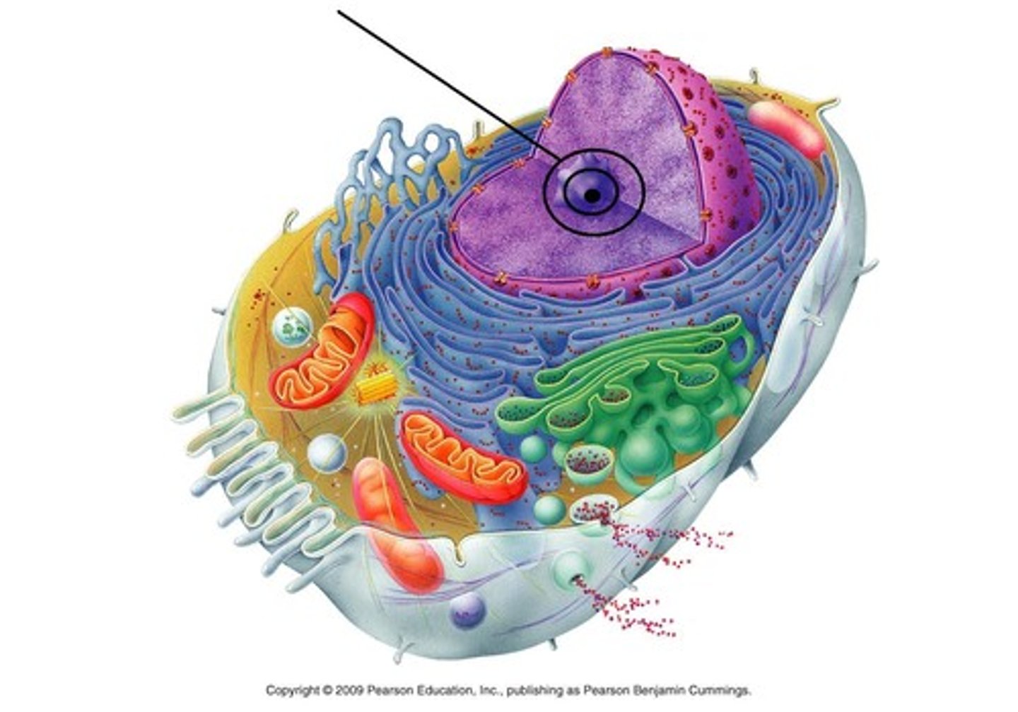
What is the main function of the nucleus?
It controls the cell's activities by containing genetic material such as DNA and proteins.
What is the structure that surrounds the nucleus?
The nucleus is surrounded by a nuclear membrane with pores.
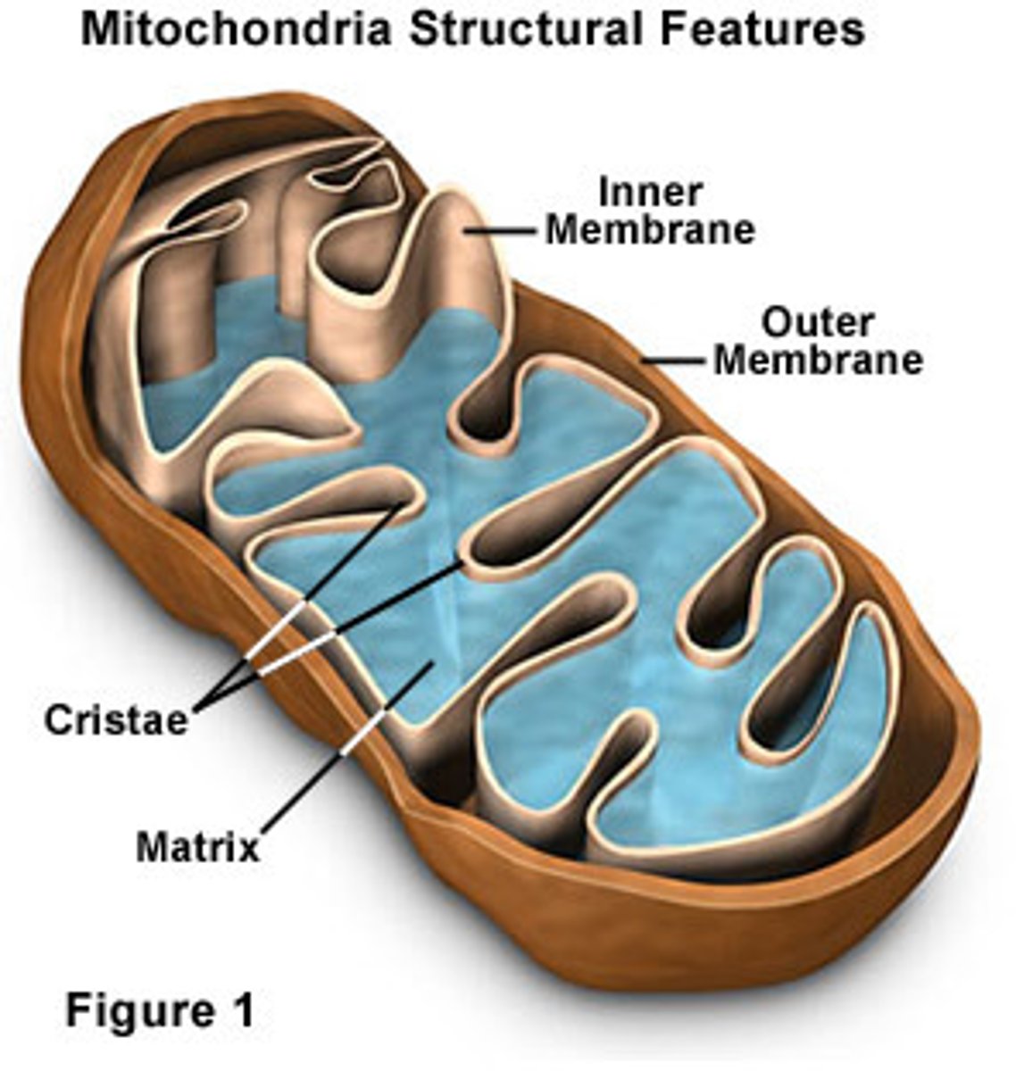
Define mitochondria
The site of cellular respiration, where the energy is stored in the bonds of complex, organic molecules is made available for the cell to use by the production of the molecule ATP
What is the role of the Nucleolus
Responsible for producing ribosomes
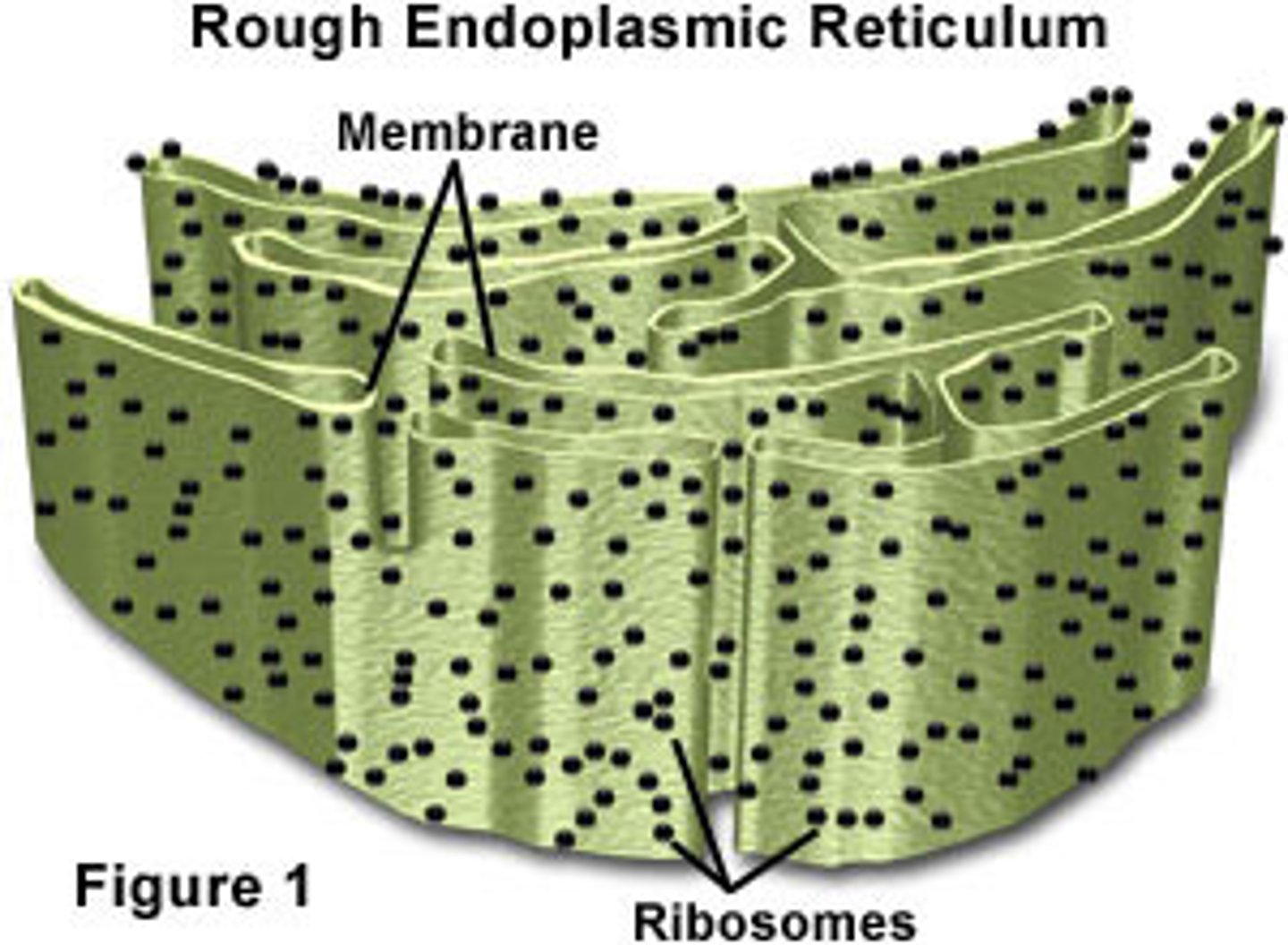
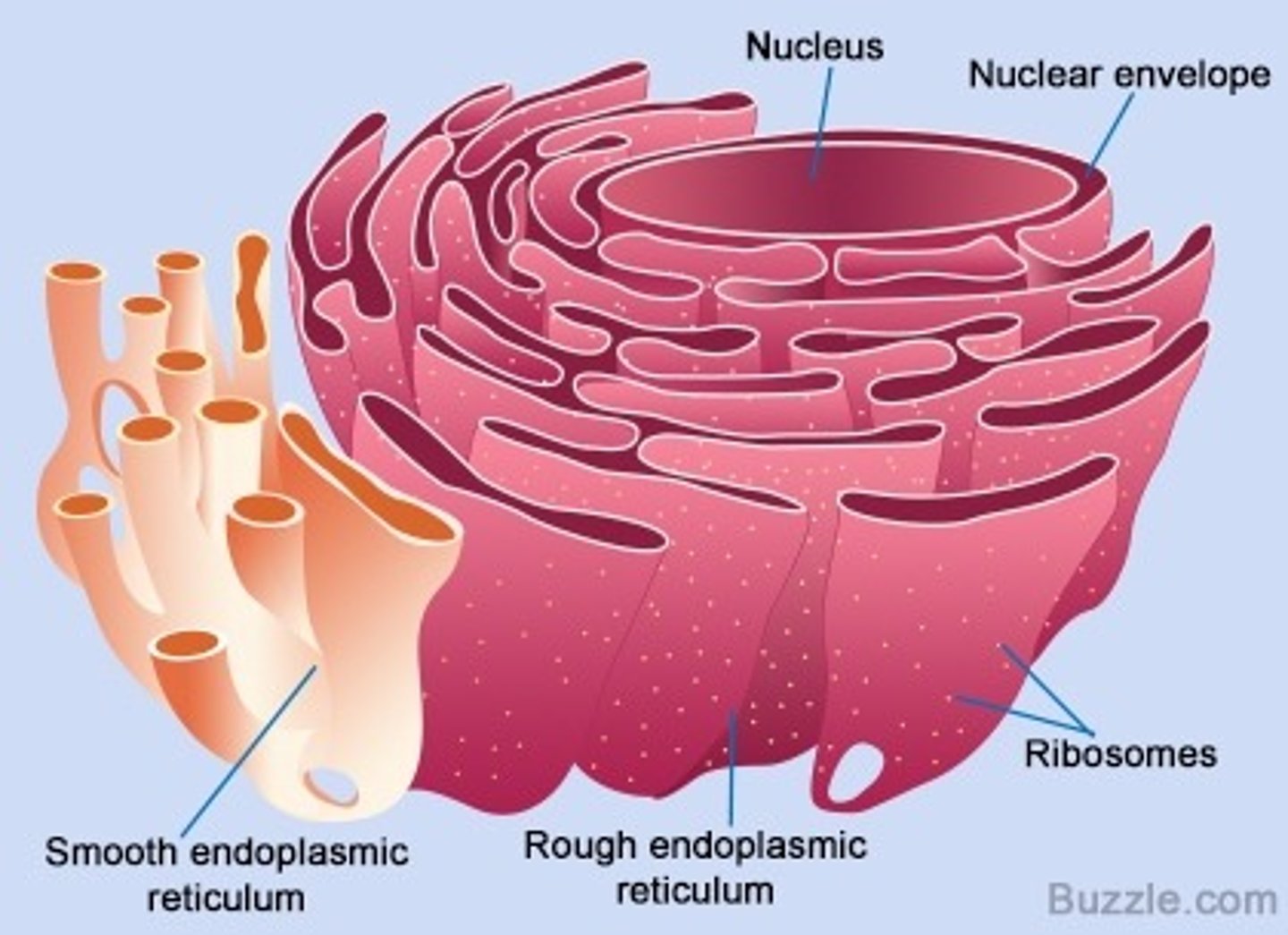
What is the structure of the rough endoplasmic reticulum?
Network of membranes enclosing flattened sacs called Cisternae
What is the function of ribosomes on the rough endoplasmic reticulum?
Responsible for the synthesis of proteins
What is the main function of the rough endoplasmic reticulum?
Transport of proteins
What is the smooth Endoplasmic Reticulum made of?
A network of membranes enclosed flattened sacs called cisternae
What is the function of the smooth Endoplasmic reticulum?
lipid and carbohydrate synthesis and storage
What is the structure of the Golgi Apparatus?
Stack of membrane-bound, flattened sacs called cisternae
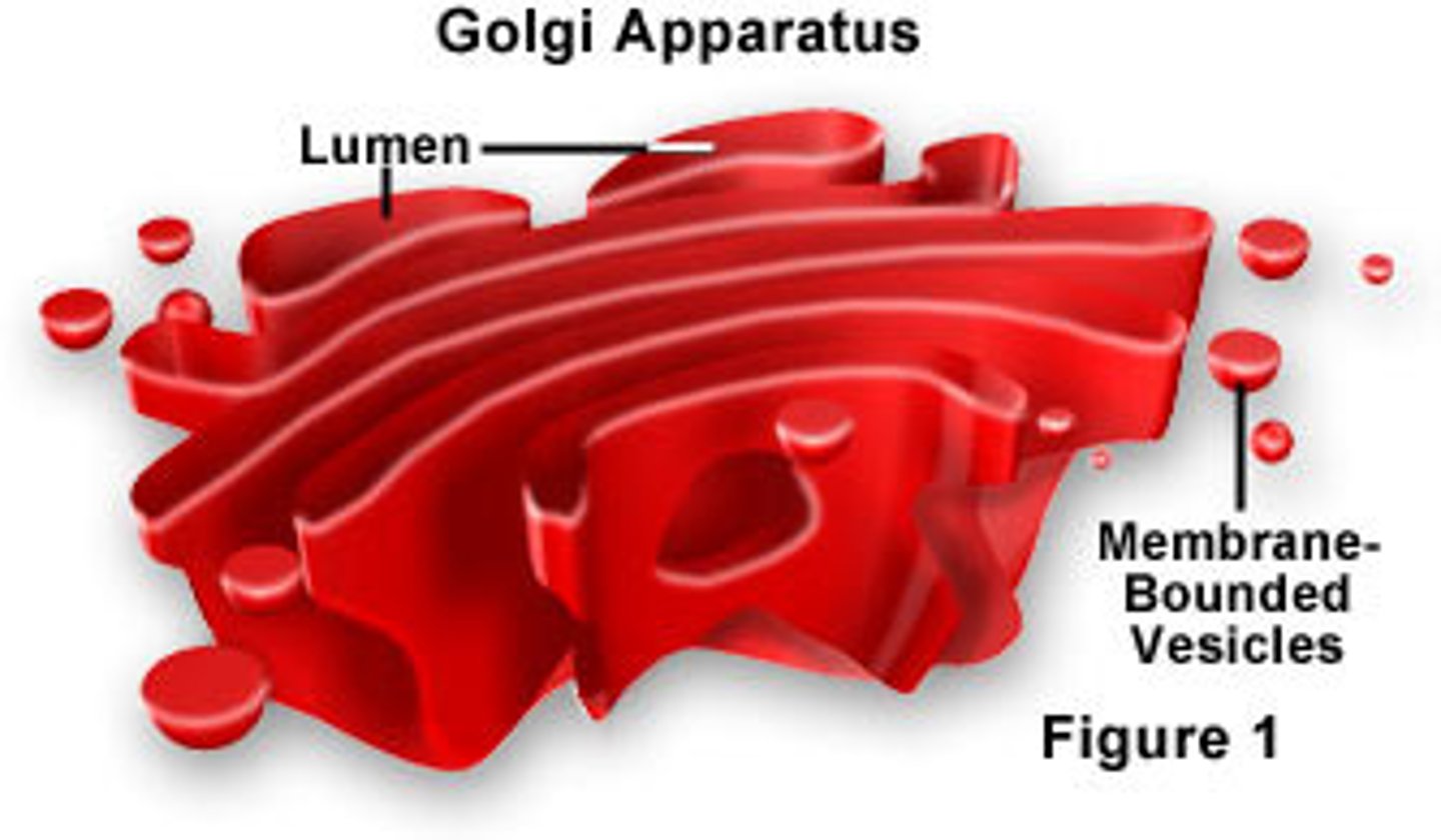
What is the function of the Golgi Apparatus?
It modifies and packages proteins into vesicles for storage or transport
What are Vesicles?
membranous sacs that are used for storage or transport in the cell
What is the structure of Vesicles?
Single membrane with fluid inside
What type of cell are Chloroplast found in?
Plant cells
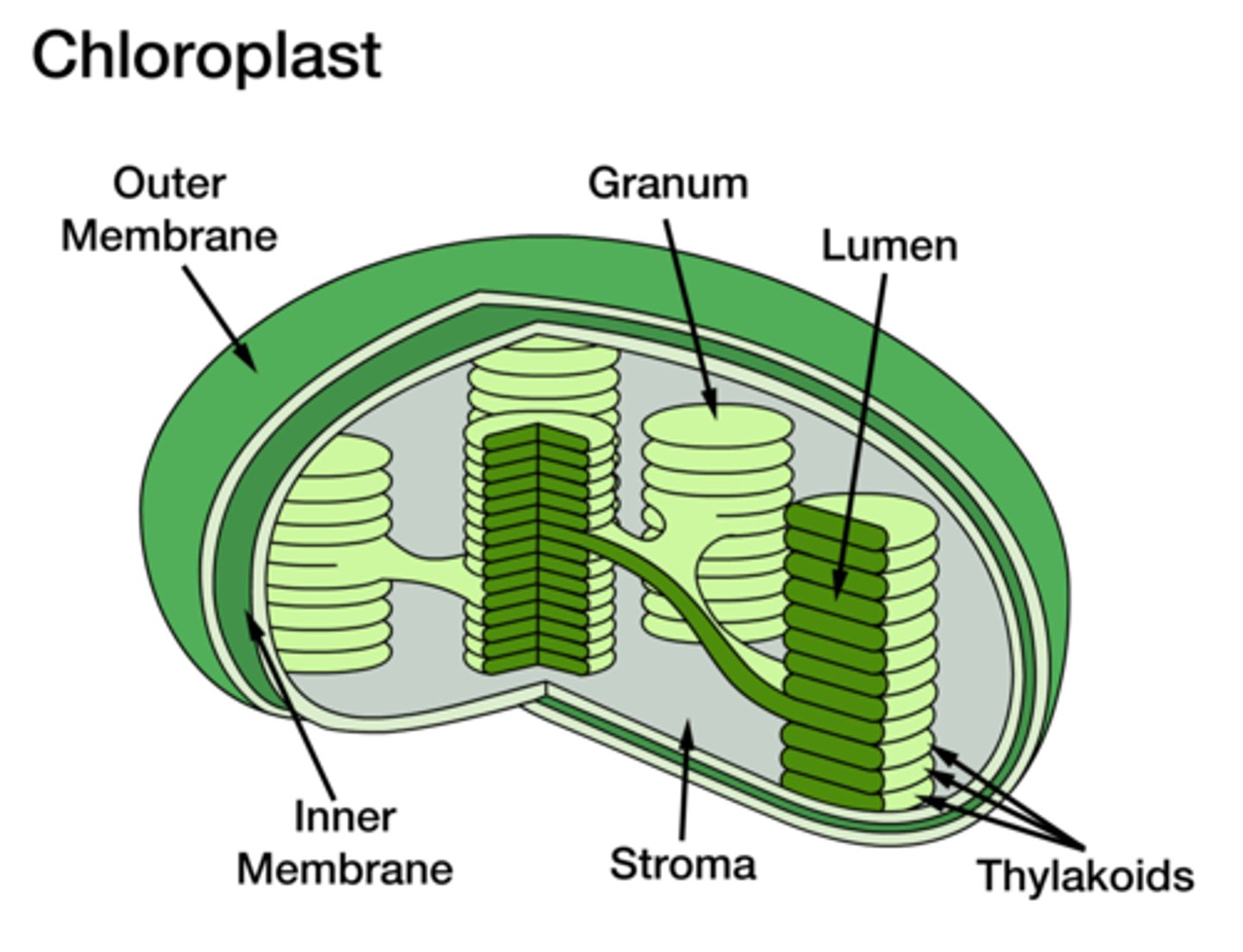
What is the structure of a chloroplast?
A double membrane. The inner membrane is highly folded and embedded with chlorophyll

What is the name of the fluid enclosed in the chloroplast?
Stroma
What are thylakoids?
Internal network of membranes that formed flattened sacs in the chloroplast
What is the name for several thylakoids?
Granum
How are Granas connected
They are connected by membranes called lamellae
Which part of the chloroplast contains chlorophyll?
The grana
What is the function of lysosomes?
They break down waste material in cells , including old organelles. They break down pathogens ingested by phagocytic cells. They also play a role in programmed cell death
What is the structure of lysosomes?
They are specialised vesicles
Do human cells have vacuoles?
They have small transient ones(not permanent)
What is the structure of Vacuoles?
Membrane lined sacs called tonoplast
What is the function of a vacuole?
They keep the cell rigid by pushing against the cell.
Used for storage e.g. Sap
What is the diameter of a ribosome?
20nm
What is the function of ribosomes
The site where amino acids are assembled into proteins
Are ribosomes membrane bound?
No; ribosomes have no membrane.
What type of cells are centrioles found in ?
Animal cells
What is the structure of Centrioles?
2 hollow cylinders made of microtubules positioned at right angles to each other
What is the function of Centrioles?
They form spindle fibres during cell division and are involved in the formation on cilia and flagella
What is the cell wall made from?
Cellulose
What is the function of the cell wall?
It provides mechanical strength to maintain the cell's shape e.g. protects the cell from bursting while maintaining turgor pressure
Are cell walls permeable?
Yes, they are fully permeable
Define Tissue
A group of similar cells that work together to perform a particular function
What is the function of the cytoskeleton?
Provides mechanical strength and establishes the cell shape. Helps with movement of organelles within cells
What is Locomotion?
movement
What is the diameter of Microtubules?
25 nm
What are Microtubules made of?
tubulin protein
What is the function of Microtubules?
To transport organelles and make spindle fibres during cell division
What is the diameter of Microfilaments?
7 nm
What are Microfilaments made of?
actin protein
What is the function of Microfilaments?
Cell movement and cell contraction during cytokinesis
What is the diameter of Intermediate fibres?
8-10nm
What is the function of Intermediate fibres?
Give mechanical strength to cells and help maintain integrity in the cell.
What is the structure of Undulipodia and Cilia?
Made of a cylinder of 9 pairs of microtubules in a circle with 2 more in the middle. It is called a 9+2 arrangement
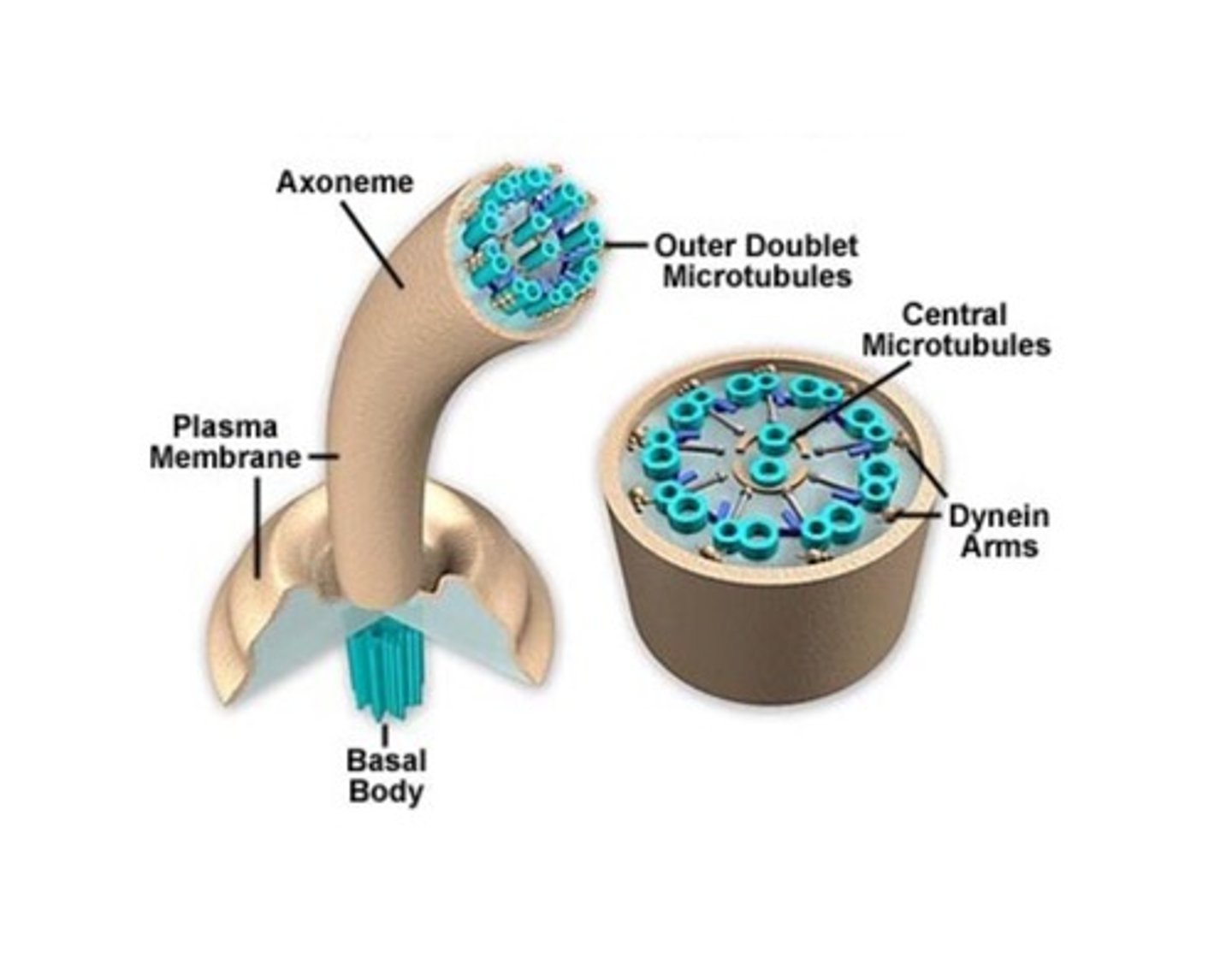
What is the function of Undulipodia and Cilia?
Move liquid past the surface of the cell
How are Undulipodia and flagella different?
Flagella are made of a spiral of flagellin protein attached by a hook to a protein disc that can rotate
What is the cell theory?
All living things are made of cells and new cells are made by the division of pre-existing cells. They contain DNA that act as instructions to growth and this is passed on to new cells
How are permanent mounts prepared?
Dehydrating the specimen and fixing it on wax then slicing it thinly
What are dry mounts used for?
To view the dead specimen whole or in thin slices called sectioning. It is placed on a slide with a coverslip
What are wet mounts used for?
To view live organisms suspended in liquid (Water or immersion oil)
why is a cover slip placed on an angle?
to avoid air bubbles
How are squash slides prepared?
A wet mount is prepared and a coverslip is pressed or a sample is squashed between 2 slides
How are smear slides prepared?
-The edge of a slide is used to smear the sample, creating a thin, even coating on another slide.
-A cover slip is then placed over the sample.
Why are stains used in microscopy?
Stains are used to add contrast as different components in a cell take up stain to different degrees. Certain stains are also used to stain specific cell structures or cell products.
What are the benefits of differential staining?
It can distinguish between 2 types of organisms that would be hard to identify
It can also differentiate between different organelles of a single organism within a tissue sample
What is the magnification equation?
Magnification = image size / actual size
define resolution
The ability to distinguish between two points
What is the order of magnification in microscopes?
TEM > SEM > LM
What image does a SEM microscope produce
Black and white and 3D
How does an SEM microscope work?
Scans the surface of the specimen and the reflected beam makes the image
Why must the specimen be in a vacuum for an SEM and TEM microscope?
To prevent air particles from scattering the electrons
Why is specimen dehydration important for SEM and TEM microscopy?
To ensure the specimen is dead
What is the order of resolution in microscopes?
SEM>TEM>LM
How does a TEM microscope work?
A beam of electrons pass through a very thin slice of the sample, the beam is then focused to produce an image on a screen
What type of image does a TEM microscope produce?
Black and white and 2D
How does a light microscope work?
A beam of light passes through a specimen which travels through the eyepiece lens, allowing the specimen to be observed
What type of image does a light microscope produce?
Black and white and 2D