DCAP Ch 14: Somatic Nervous System
1/103
Earn XP
Description and Tags
Name | Mastery | Learn | Test | Matching | Spaced |
|---|
No study sessions yet.
104 Terms
sensation
the taking in of sensory input
integration
Thinking and deciding what to do next
response
What your muscles actually do (motor output)
Sensation (vs.)
The activation of sensory receptor cells at the level of the stimulus
Perception (vs.)
The central processing of sensory information into a meaningful pattern
Receptors
Cells of structures that detect sensations
What directly changes a receptor?
A stimulus
What detects changes and relay them?
Transmembrane proteins
What opens to relay the changes?
Ion channels
What is generated by transmembrane proteins?
Action potentials
ligand
a chemical that binds to a transmembrane receptor protein
Free nerve ending
dendrites embedded in tissue that would receive a stimulus (ex: pain, temperature)
encapsulated ending
sensory nerve ending is contained in connective tissue that enhances their sensitivity (ex: laminated corpuscles detector pressure and touch)
specialized receptor
has distinct components that intercept a specific type of stimulus (ex: photoreceptors detect light in retina)
exteroceptor
located near a stimulus in the external environment (ex: skin)
interoceptor
interprets stimuli from internal organs and tissues (ex: aorta walls detect bp)
proprioceptor
located near a moving part of the body (ex: muscle)
chemoreceptor
interprets chemical stimuli (ex: taste, smell)
osmoreceptor
responds to concentration of body fluids (ex: hypothalamus detects water balance)
thermoreceptor
reacts to special physical stimuli (heat or cold)
nociceptor
interprets the presence of chemicals from tissue damage (ex: pain, intensity)
mechanoreceptor
interprets physical stimuli (ex: balance, vibration, sound, pressure)
general sense
A type of sensory perception that includes sensations such as touch, temperature, pain, and proprioception, as opposed to special senses like vision and hearing.
proprioception
body position
kinesthesia
body movement
visceral sense
trunk organ sensations like ‘stomach fullness’
somatosensation
group of general senses which includes touch, proprioception, and interoception

Merkel’s discs
mechanoreceptors in the epidermal-dermal junction and mucosal membranes that detect low frequency vibrations

Ruffini’s corpuscle
a type of mechanoreceptor located in the dermis and joint capsules that responds to deep pressure and high frequency vibration; also referred to as a bulbous corpuscle

Pacinian corpuscle
a type of mechanoreceptor located deep in the dermis and subcutaneous tissue that detects high frequency vibrations and pressure; also called a lamellated corpuscle
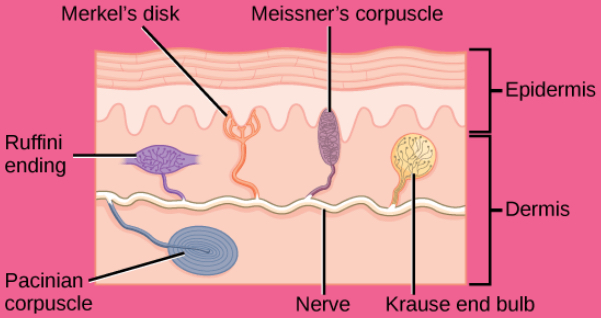
Krause end bulb
a type of mechanoreceptor found in the dermis and mucous membranes that is sensitive to cold temperatures

Golgi tendon organ
Proprioceptor in line with tendons that detects stretchand tension in muscles, helping to prevent excessive force during contraction
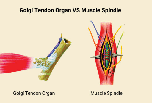
muscle spindle
a type of proprioceptor located within muscles that detects changes in muscle length and the rate of stretch, playing a crucial role in the regulation of muscle tone and reflexes
interoceptors
in walls of visceral organs and vessels to detect internal changes, such as osmoreceptors, humoral receptors, and glucoreceptors that monitor internal conditions and physiological states, providing information about the body's internal environment.
Special senses
sensory modalities that provide information about the environment through specialized organs, including vision, hearing, taste, smell, and balance
Papillae
small projections on the tongue that contain taste buds and are involved in the sensation of taste.
taste buds
sensory receptors located within the papillae of the tongue that detect taste stimuli, allowing the perception of different flavors such as sweet, sour, salty, bitter, and umami
gustatory receptor cells
specialized sensory cells responsible for detecting taste stimuli and transmitting taste information to the brain.
taste nerves
facial, glossopharyngeal, vagus nerves
Olfactory epithelium
Layer the superior nasal cavity and contains bipolar sensory neurons
olfactory sensory neurons
nasal nerves that extend from the olfactory epithelium into the mucus lining
olfactory bulb
region in the brain where axons of the olfactory neurons lead
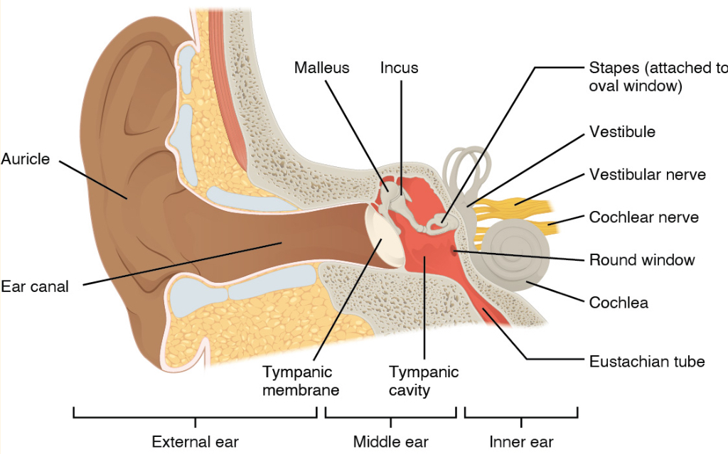
Auricle
the fleshy outer ‘ear’ structure

external acoustic meatus
auditory canal that funnels sound to the eardrum

tympanic membrane
the eardrum; vibrates in response to sound

Eustachian tube
Connects to the pharynx, equilibrates air pressure

ossicles
ear bones; malleus, incus, and stapes

cochlea
spiral-shaped, fluid-filled organ in the inner ear that plays a crucial role in converting sound vibrations into neural signals
spiral ganglia
cluster of sensory neurons in the inner ear that convey auditory information from the cochlea to the brain.
scala vestibuli
fluid-filled tube at the top of the cochlea
cochlear duct
center of the cochlea that contains the spiral ganglia, organs of Corti and the basilar membrane
organs of Corti
Structures in the cochlear duct of the inner ear containing hair cells that convert sound vibrations into electrical signals for the brain.
basilar membrane
A flexible membrane in the cochlea that converts sound vibrations into neural signals, supporting the organ of Corti with sensory hair cells and varying in width to respond to different sound frequencies.
scala tympani
the fluid-filled chamber in the cochlea below the cochlear duct that aids in sound transmission from the oval to the round window
tectorial membrane
A gelatinous structure in the cochlear duct that converts sound vibrations into electrical impulses by displacing hair cells.
stereocilia
tiny hair-like projections on sensory hair cells in the inner ear that convert mechanical sound vibrations into electrical signals, critical for hearing
vestibule
The vestibule is the central part of the inner ear between the cochlea and semicircular canals, involved in balance by detecting linear acceleration and gravitational forces through the utricle and saccule.
otolithic membrane
gelatinous layer located in the inner ear that contains tiny calcium carbonate crystals called otoliths; aids in balance by moving in response to head movements to help inform the brain about the body's position relative to gravity
semicircular canals
three ring-like extensions of the vestibule that sense head movement
ampulla
Enlarged base of each semicircular canal that contains hair cells
cupulla
Upper membrane of semicircular canals that moves stereocilia as the head rotates
vestibular ganglion
pathway for positional sensory information leaving the inner ear
vestibulocochlear nerve
nerve bundle that contains spiral ganglia and vestibular ganglia, transporting inner ear input to the brainstem
palpebral conjuctiva
thin membrane on the inner surface of each eyelid
lacrimal gland
produces tears
lacrimal duct
directs tears to flow over the conjuctiva
optic chasm
point in the visual system at which medial retina fibers cross to the other side of the brain; causes the left field of view of each eye to be processed on the right side of the brain and vice versa
fibrous tunic
outermost tissue layer of the eye, includes the sclera and cornea
vascular tunic
the middle layer of the eye that contains many blood vessels
choroid
highly vascularized connective tissue that supplies blood to the eyeball
lens
focuses light onto the retina
ciliary body
muscular structure attached to the lens by ligaments called Zonule fibers
iris
colored part of the eye, composed of smooth muscle; controls the opening and closing of the pupil
neural tunic
innermost layer of the eye; includes the retina
retina
contains nervous tissue responsible for photoreception
aqueous humor
Water fluid that fills the anterior cavity of the eye
vitreous humor
viscous fluid that fills the posterior cavity of the eye
rods
photoreceptors in the retina that contain rhodopsin; detect light at low levels
cones
contain pigments called opsins that detect red, green, and blue light
retinal ganglion cell
produces action potentials in response to visual stimuli
optic disc
where axons of the retinal ganglion cells connect to the leave eye as the optic nerve
blind spot
region of the retina where the optic nerve leaves the eye; possesses no photoreceptors
macula
exact center of the retina
fovea centralis
region of the retina that contains nothing but photoreceptors where visual acuity is the best
contralateral
descriptive term for the nerves on one side of the body being connected to the opposite side of the brain; applies to the spinal nerves
ipsilateral
descriptive term for the sensory nerves being controlled by the same side of the brain and body; applies to the nerves of the head and neck
ascending pathways
tracts that carry peripheral sensations (from below the neck) to the brain or spinal cord
dorsal column system
associated with fine motor touch and proprioception
nerve pathway of the dorsal column system
Axon 1 enters the dorsal root region and joins the dorsal column in the spinal cord, terminating in the medulla. Axon 2 ascends the brainstem bundled as the medial lemniscus. Axon 3 ends in the post central gyrus of the cerebral cortex.
spinothalamic tract
sensory pathway primarily associated with pain and temperature
nerve pathway of the spinothalamic tract
Axon 1 enters the dorsal root and extends into dorsal horn. Axon 2 starts in the spinal cord and connects to the thalamus. Axon 3 travels from the thalamus to the post central gyrus.
Trigeminal pathway
carries somatosensory information from the face, head, mouth, and nasal cavity
nerves of trigeminal pathway
Axon 1 enters the brainstem at the pons. Axon 2 travels one of three pathways but all end in the thalamus. Axon 3 ends in the primary somatosensory cortex of the cerebrum.
thalamus
serves as an important relay for communication between the cerebrum and the rest of the nervous system; required transfer point for sensory input except smell
sensory homunculus
a visual representation of the regions of the cerebral cortex that correlate to their somatosensory signals
executive functions
cognitive functions that lead to goal-directed behavior
working memory
higher cognitive processing that helps to organize and represent information that is not in the immediate environment
premotor cortex
aids in controlling movements of core muscles to maintain posture during movement
supplemental motor area
a region of the brain located in the frontal cortex that plans and coordinates movements
Betz cells
neurons located in the primary motor cortex that descend and synapse with lower motor neurons in the brainstem or spinal cord