Ch.11 - Muscles
1/83
Earn XP
Description and Tags
95 to remember! Assume Agonist if not stated otherwise. L = Location, A= Action, O = origin, I = Insertion, PM = Prime Mover (more specific than agonist)
Name | Mastery | Learn | Test | Matching | Spaced | Call with Kai |
|---|
No analytics yet
Send a link to your students to track their progress
84 Terms
How does reversing muscle action work?
Muscles can’t undo their own actions, only other muscles can. Muscles on opposite sides will oft have opposite functions. Likewise, if muscles are in different compartments, they’ll usually have different functions.
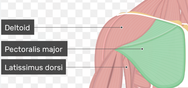
Ex: Pectoral Major Dorsi will flex, Latissimus Dorsi will extend
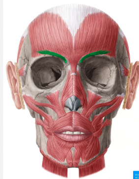
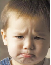
Corrugator Supercilii
L = Above eyebrows, deep to orbicularis oculi
A = draws eyebrows in, frowning
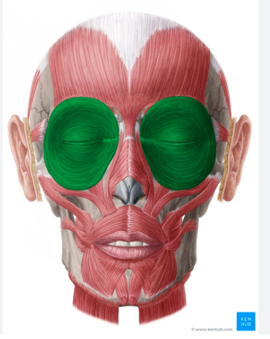
Orbicularis Oculi
L = full circle in orbital region
A = closes eyes
Zygomaticus (major + minor)
L = from zygomatic bone to mouth
A= corners of lips UP and LATERAL, :)
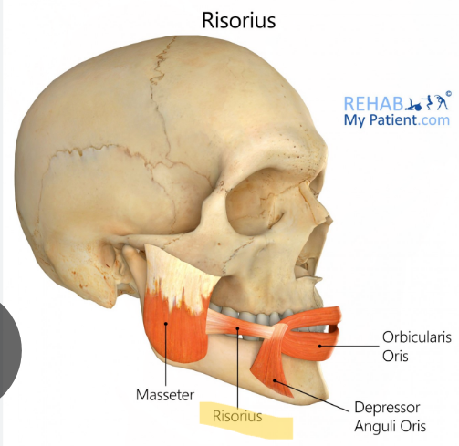
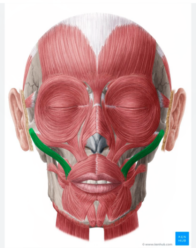
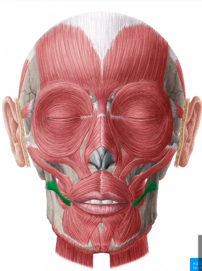
Risorius
L = looks like small muscle going from masseter → oris
A = synergist to zygomaticus :)
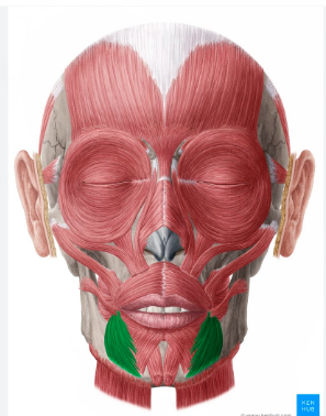
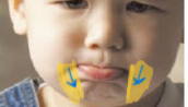
Depressor Anguli Oris
L = below mouth, on the sides of chin
A = antagonist to zygomaticus, draws lips laterally, :(
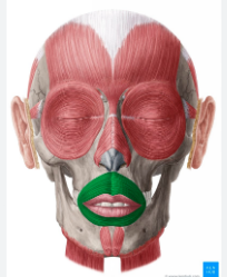
Orbicular Oris
L =surrounds oral cavity
A = closes mouth

Oris
relating to the mouth

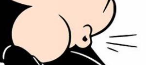
Buccinator
L = wall of the cheek
A= compresses cheeks, to whistle or suckle
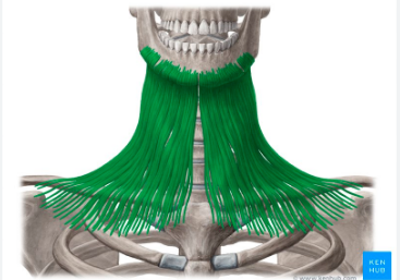
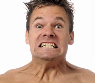
Platysma
L = spans from mandible to clavicle
A = tenses neck muscles
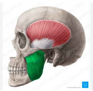
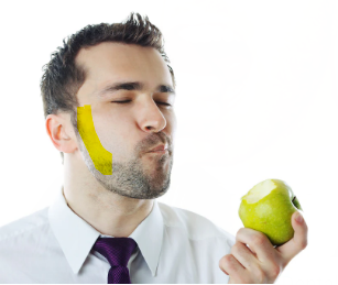
Masseter
O = Zygomatic Bone + Arch
I = Mandible
A = PM ; Jaw closure, mastication
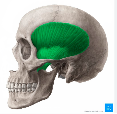
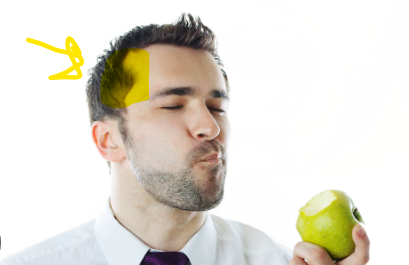
Temporalis
O = = temporal fossa
I = Mandible
A = jaw closure, mastication
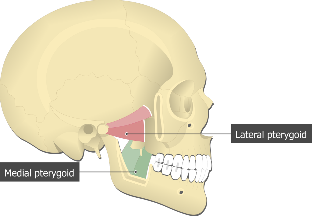
Medial/Lateral Pterygoid:
L = deep, part of sphenoid bone
A = grinding food, side-to-side chewing
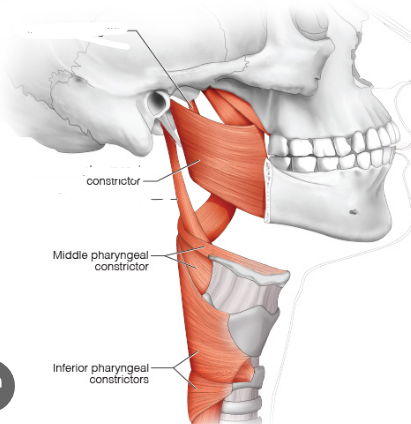
Pharyngeal* constrictor muscles
*Pharynx = throat
A = contract sequentially downward to swallow

Muscles of Mastication (ID 5):
Masseter, Temporalis, Medial Pterygoid, Lateral Pterygoid, Pharyngeal Constrictor
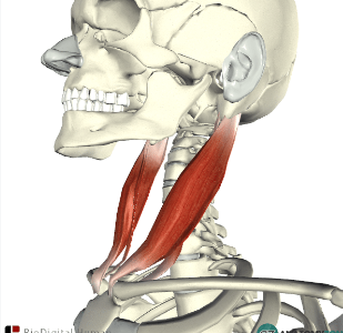
Sternocleidomastoid
*Sternum, Clavicle, Mastoid (zygomatic bone)
O = manubrium + clavicle
I = mastoid process + occipital bone
A = flexes laterally, PM: head flexion
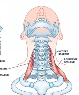
Scalenes
O = vertebrae
I = ribs 1+2
A = Synergist for inhalation/inspiration, helps w breathing
Intrinsic Muscles of the back [ID 5]:
L = deep in back
A = extend trunk, vertebrae/spine
Includes: [Iliocostalis, Longissimus, Spinalis], Splenius, Semispinalis (deep)
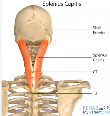
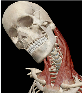
Splenius
L = deeper than the trapezius
A = antagonist to sternocleidomastoid, extends head
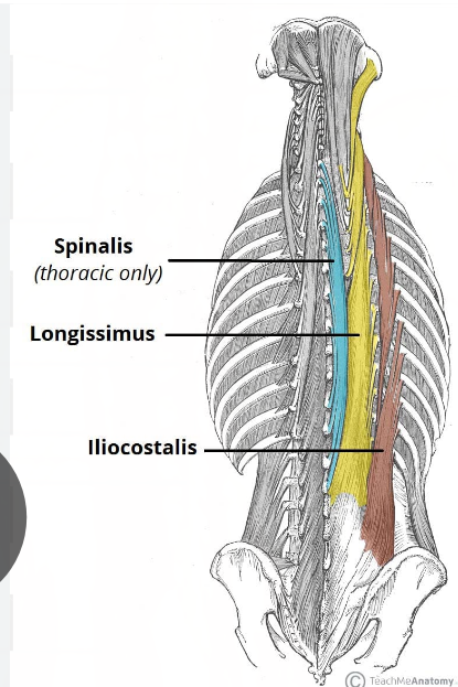
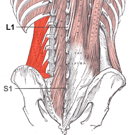
Erector Spinae:
L = L → M, Iliocostalis, Longissimus, Spinalis
A = same as intrinsic muscles of the back
I love spaghetti!
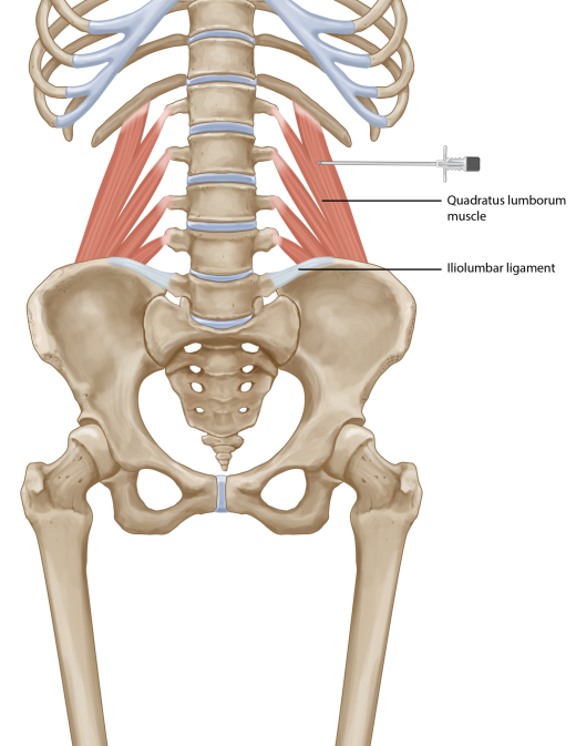
Quadratus Lumborum
L = deep in posterior wall
A= flex vertebral column
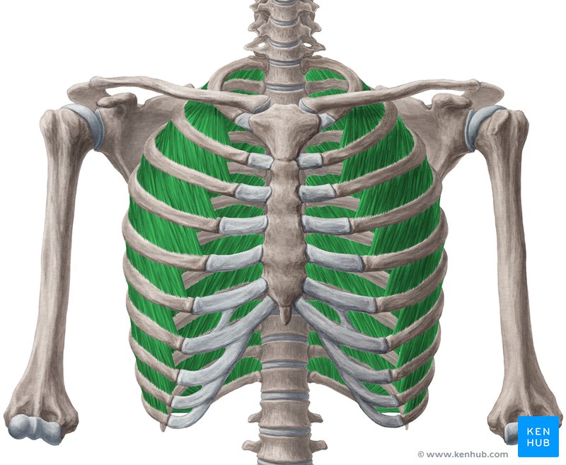
External Intercostals:
L: w/in rib cage and most superficial; going diff. direction than other intercostals
A = synergist to diaphragm; lifts ribcage for inhalation
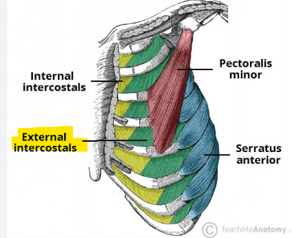
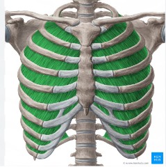
Internal Intercostals
L = w/in rib cage and deeper; going diff. direction than other intercostals
A = synergist to inner intercostals, aids expiration
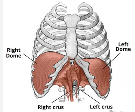
Diaphragm
L = between thoracic vertebrae and top of lumbar vertebrae
A = PM for inspiration
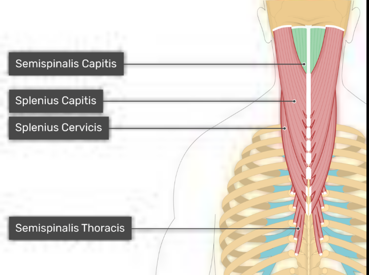
Semispinalis
L = NOT part of the Erector Spinae, deep neck muscle
A = extends trunk, vertebrae/spine
Deep muscles of the thorax (ID 3):
External Intercostals, Internal Intercostals, Diaphragm
Muscles of the Abdominal Wall (4):
External Oblique, Internal Oblique, Transversus abdominis, Rectus Abdominis
*No bony attachment
Muscles of the face (ID 8):
Corrugator Supercilii, Orbicularis Oculi, Zygomaticus, Risorius, Depressor Anguli Oris, Orbicularis Oris, Buccinator, Platysma
Anterolateral Neck Muscles (ID 2):
Sternocleidomastoid and Scalenes
Rotator Cuff:
teres minor
supraspinatus
infraspinatus
subscapularis
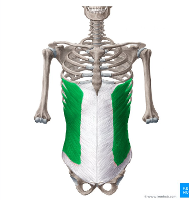
External Oblique:
A = antagonist to vertebral spine; flex vertebral column + compress wall
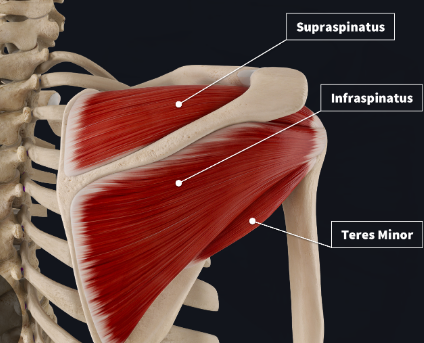
Posterior View of RC:
Infraspinatus:
O - infraspinous fossa
I = greater tubercle
Supraspinatus:
O - supraspinous fossa
I = lesser tubercle
Teres Minor
O - pectoral girdle
I = humerus
(!) All subscapularis shares I w/ supraspinatus
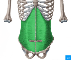
Transversus Abdominis
L = goes transversus abdominis → internal oblique → external oblique
A= antagonist to vertebral spine, compress abdominal wall
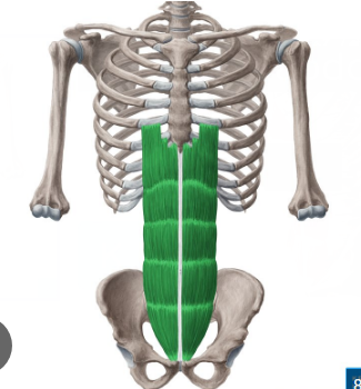
Rectus Abdominis
A = flex/rotate lumbar region
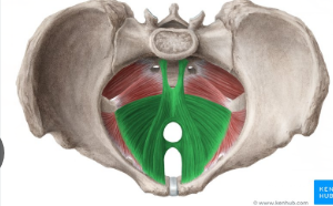
Levator Ani
A = elevating anus, moving fecus along trac
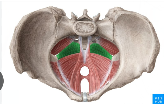
Coccygeus
A = seals bony pelvis to keep abd. organs in place
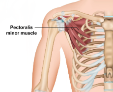
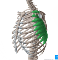
Pec. Minor
O = ribs
I = scapula
A = PM, shrugging (scapula forward and down)
Serratus Anterior
O = Ribs
I = Scapula
A = abduction/protraction, upward rotation
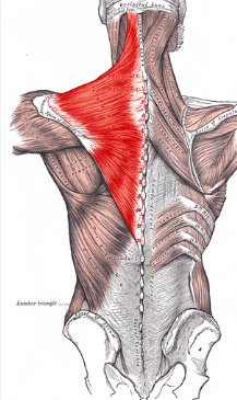
Trapezius
O = occipital + vertebrae
I = sternum + clavicle
A = shrug/depress shoulder + elevator shoulder + adduction + rotation
(!) 3 in 1

Levator Scapuale:
O = vertebrae
I = scapula
A = PM, elevate shoulder
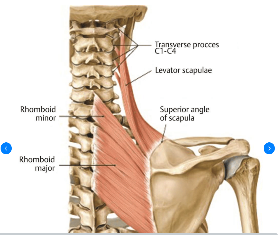
Rhomboids (major + minor)
O = vertebrae
I = scapula
A = elevate shoulder, downward rotation
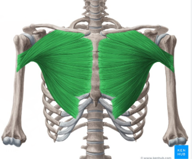
Pecs. Major
O = Axial Skeleton
I = greater tubercule on humerus
A = PM, adduction
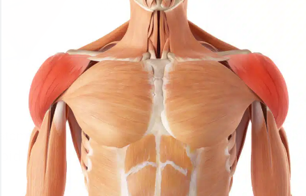
Deltoids
O = pectoral girdle
I = deltoid tuberosity
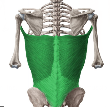
Lats. Dorsi
O = Axial Skeleton
I = humerus
A = PM, extension
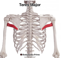
Teres Major
O = pectoral girdle
I = humerus
A= extension/adduction
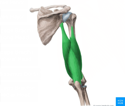
Triceps Brachii
O = scapula + humerus
I = olecranon humerus
A= extend forearm
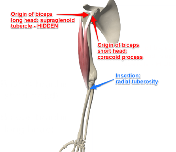
Biceps Brachii
O = Scapula
I = radial tuberiosity
A = PM, supinates forearm to flex
(!) 2nd strongest
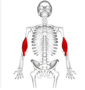
Brachialis
O = humerus
I = coronoid process of ulna
A = PM, flexes forearm
(!) strongest of 3 forearm flexers
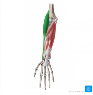
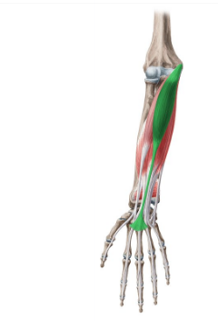
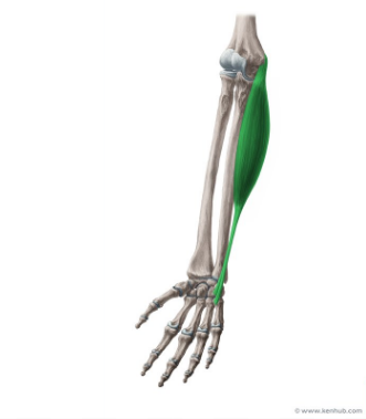
Brachioradialis
O = humerus
I = radial/lateral styloid process
A = synergist, flexes forearm
(!) 3rd weakest

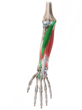
Pronator Teres:
L→ M (1, 2, 3,4)
A = synergist, pronation
Flexor Carpi Radialis
L → M (1, 2, 3,4)
A = flexes wrist
Palmaris Longus
L → M (1,2,3,4)
A = tenses skin
Flexor Carpi Ulnaris
L → M (1,2,3,4)
A = flexes wrist
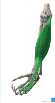
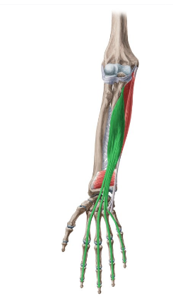
Flexor Digitorum Superficialis
L = deep to other 4 superficial anterior muscles
A = flexes wrist + fingers
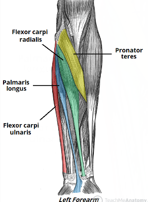
Anterior Superior Muscles of the Upper Limb
M → L
Flexor Carpi Ulnaris → Palmaris Longus → Flexor Carpi radialis → Pronator Teres
Flexor Digitorum Profundus
L = deep
A = flex fingers
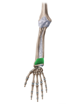
Pronator quadratus
L = deep anterior muscles
A = PM, pronate forearm
Posterior Superior Muscles of the Upper Limb (L →M)
Extensor carpi radialis longus and brevis → Extensor Digitorum → extensor carpi ulnaris
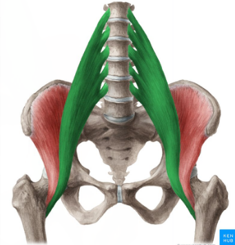
Iliopsoas
O = pelvis + ventral column
I = femur
A = PM, flexion
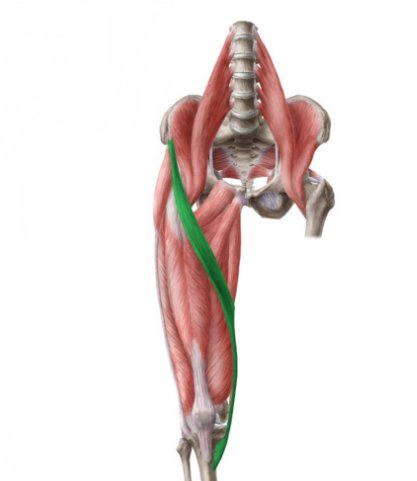
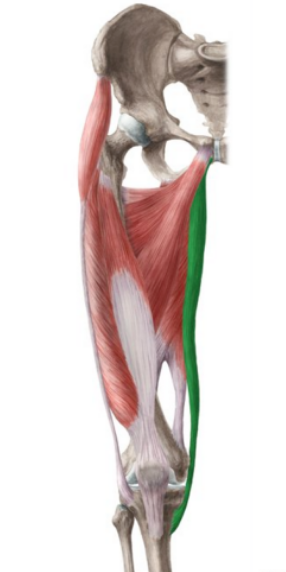
Sartorius
O = Anterior Superior Iliac Spine
I = Tibia
A = Synergist for thigh abduction
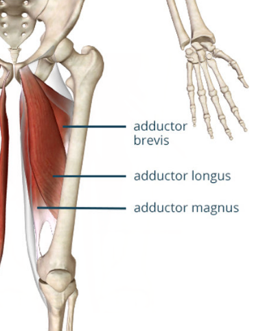
Adductor Longus + Magnus
O = Coxal bone
I = femur
A= adduction (duh!) + medially rotates thigh
Gracilis
O = coxal bone
I = tibia
A= adduction
(!) Most medial in lower limb
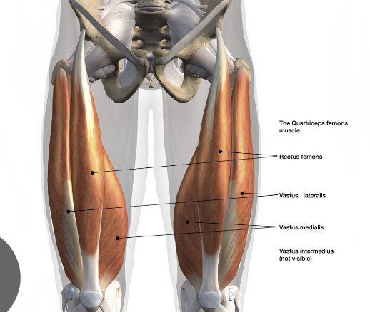
Quadriceps Femoris:
Rectus Femoris, Vastus Lateralis, Vastus Medialis, Vastus Intermedialis
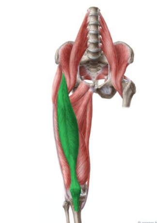
Rectus Femoris
O = Coxal Bone
I = patella + tibial tuberosity
A = synergist that flexes thigh and extends knee
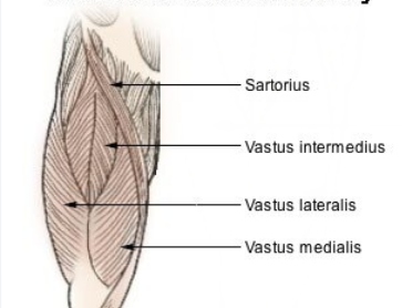
Vastus Lateralis/Medialis/Intermedius
O = femur
I = patella + tibial tuberosity
A = extends knee
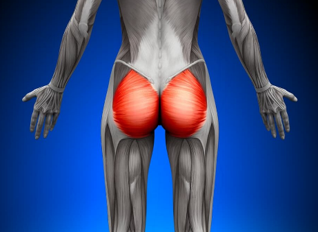
Gluteus Maximus
O = Pelvis
I = femur
A= PM, extension
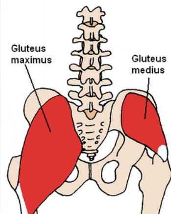
Gluteus Medius
O = Ilium
I = femur
A = PM, abduction + medial rotatio
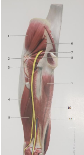
Piriformis
O = Sacrum
I = Femur
A= lateral rotation
(!) lateral rotators w/ same I and different O
Quadratus Femoris
O = Ischium
I = femur
A = lateral rotation
(!) Same I as Piriformis but different O
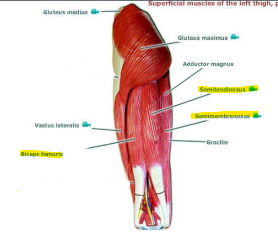
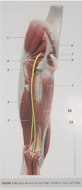
Hamstrings
All muscles have at least Ischium as their O and at least Tibia as their I. Biceps Femoris (lateral) to the Semitendinosus’ (medial). Semimembranosus is the deepest, having both the Tibia and Femur as its’ I.
Biceps Femoris
O = Ischium + Femur
I = Tibia + Femur
(!) part of Hamstrings, most lateral
Semitendinosus
O = Ischium
I = Tibia
(!) pt. Hamstrings, most medial and is simplest
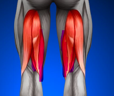
Semimembranosus
O = Ischium + Femur
I = Tibia + Femur
(!) deepest pt. of Hamstrings + medial
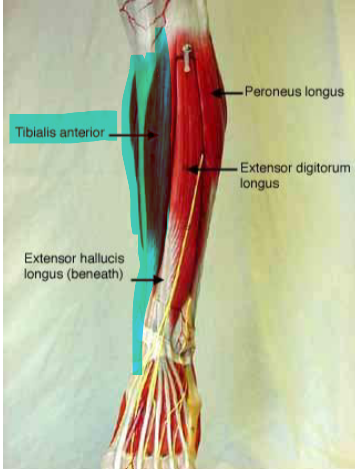
Tibialis Anterior
O = Tibia
I = metatarsal 1 + medial cuneiform
A = PM, dorsiflexion
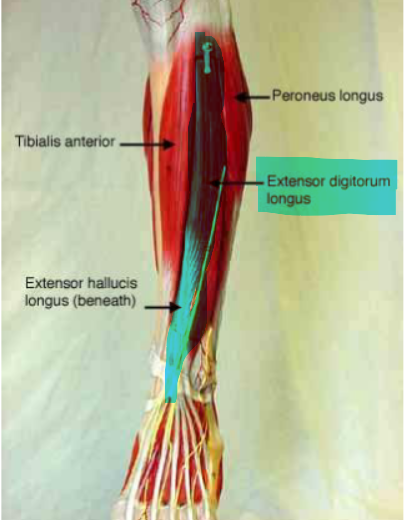
Extensor Digitorum Longus
O = Tibia + Fibula
I = Phalanges
A = PM, Extend digits
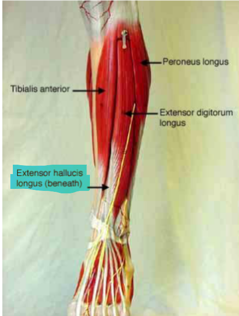
Extensor Hallucius Longus
O = Fibula
I = hallux
A = extend hallux
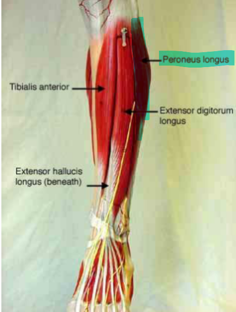
Fibularis/Peroneus Longus
O = Fibula
I = metatarsal 1 + medial cuneiform
A = plantar flexion + foot eversion
(!) Most lateral
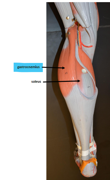
Gastrocnemius
O = Femur
I = calcaneus
(!) pt. of Triceps Surae, shared I
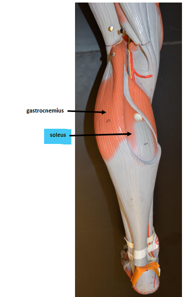
Soleus
O = tibia + fibula
I = calcaneus
A = plantar flexion
(!) Triceps Surae, same I
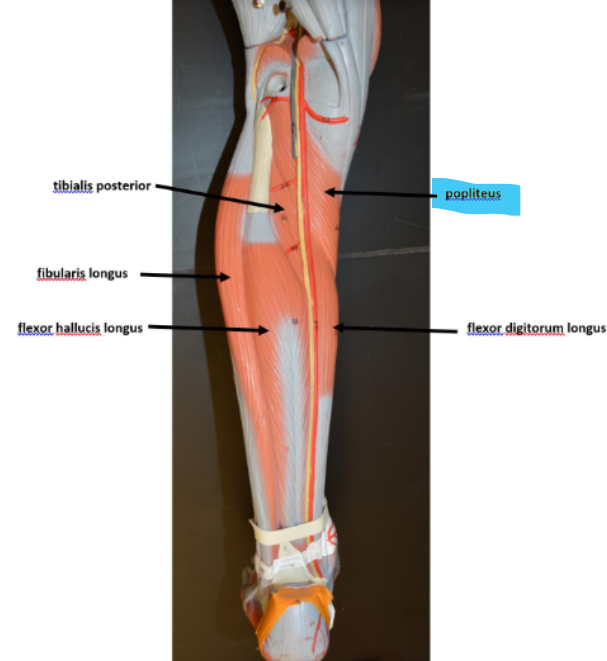
Popliteus
L = behind the knee, wraps around the Tibia to the femur
A = knee flexion

Flexor Digitorum Longus
L = runs beneath the popliteus
A = flexes digits
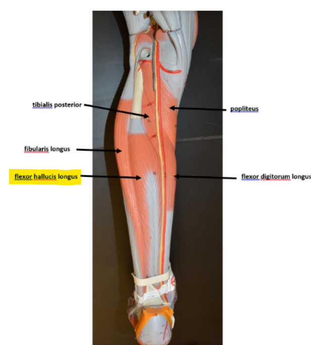
Flexor Hallucius Longus
A = flexes hallux
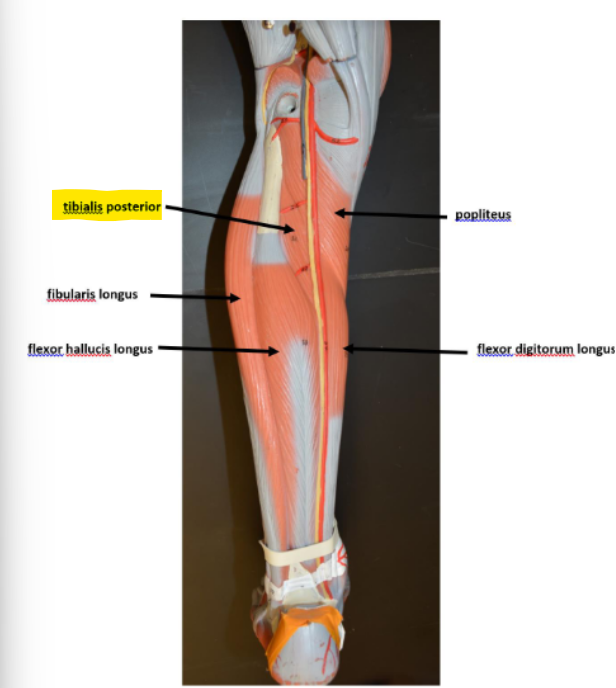
Tibialis Posterior
L = most medial within the leg
A = foot inversion
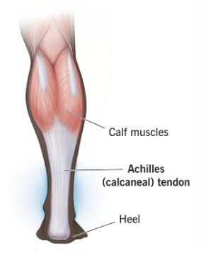
Archilles/Calcaneal Tendon
the largest and the strongest tendon