BCM.34 - BACTERIAL CELL WALLS
1/38
There's no tags or description
Looks like no tags are added yet.
Name | Mastery | Learn | Test | Matching | Spaced | Call with Kai |
|---|
No analytics yet
Send a link to your students to track their progress
39 Terms
Name the essential and inessential structures of a bacteria

General features of a bacterium
-Most have 1 circular, covalently closed chromosome.
-Around 0.25-3mm
- 0.8 - 8 x10^6 base pairs
- 1000-9000 genes
strain with the smallest genome
Mycoplasma
Plasmids in bacteria
Extra chromosomal circular DNA
- 1x10^3 – 1x10^5 BP
- Not required by bacteria in all growth conditions
Eg F-Plasmid that is transmissible via conjugation
- may have antibiotic resistance genes
Eg TOL plasmid in p.putida ( encodes genes for metabolic pathway needed to degrade benzene )
Pilli
- Pilli are found on gram -ve ( range of pilli that carry out a range of functions )
- 3-25nm diameter, 12 micrometers in length.
- Some pilli are involved in adhesion to surfaces via adhesins and electrostatic intercations.
- F-Pilli are involved in conjugation and provides a route for horizontal gene transfer.
- Type 4 pilli confer motility via twitching allowing bacteria to move across solid surfaces.
How do bacteria move using pilli
Type 4 pilli
Adhesin at end of the pilli attaches to surface ahead of cell ( polymerisation ) and then the pilli contracts and pulls the bacteria closer to the bound adhesin via depolymerisation ( depolymerisation requires energy )
Flagella
- Enables motility in liquid environments
- 20micrometers long, 20 nm wide
- Polar arrangement ( single flagella at one pole of the cell, eg vibrio cholerae )
- Attached to bacteria via a basal body ( around 20 different proteins ) and is the only example in nature of a electrical motor powered by the proton motive force/ protonic potential. Charged movements releases free energy that is transferred into electrical energy.
- Other bacteria have a peritrichaus arrangement where multiple flagella are attached to the sides of bacteria ( eg E.Coli ) and they form a flagella bundle that forms behind the bacteria cell.
- Flagella rotate.
- Not visible with light microscope but are with electron microscopes.
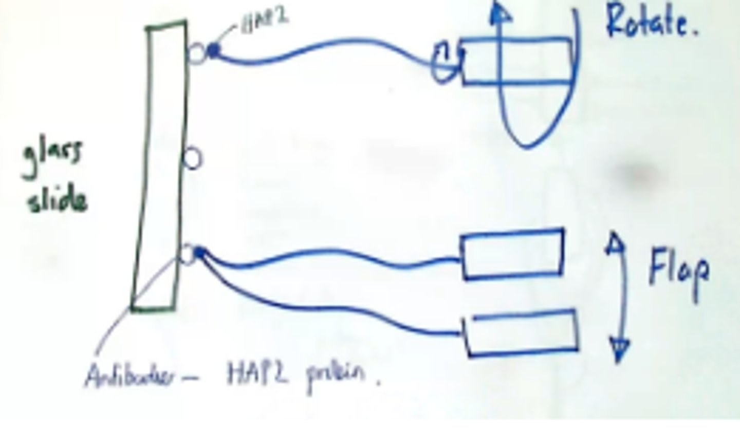
How did we find out that the flagella rotate bacteria not flap them?
The tethered cell experiment:
Glass slide in aqueous environment with bacteria ( E coli ).
Glass slide covered with antibodies, that are antibodies to a protein at the tip of the bacterial flagellum ( HAP 2 ).
Bacterial flagella bind, bacteria become stuck.
They try to rotate their flagella but instead rotate the whole cell, so we see bacteria rotating if this has happened correctly.
( Another suggestion was that flagella flap rather than rotate, so we should see bacteria flapping up and down, which we do not )
Bacterial cell walls
They help:
- Maintain shape
- Prevent osmotic lysis
Freely permeable.
Gram stains reveal +/- and effects the ability of some bacteria to uptake a dye called cystal violet.
Gram + retain dye, gram - do not.
Both have Peptidoglycan
Gram -ve cell wall structure
outer membrane made from lipopolysaccharide /LPS.
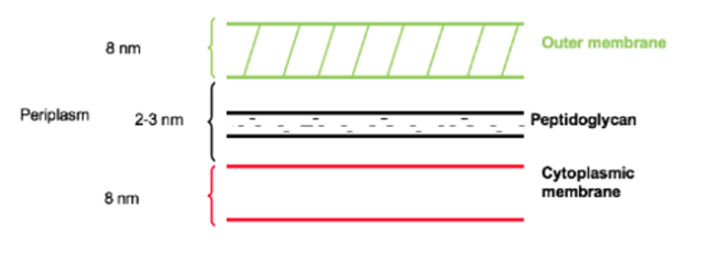
Periplasm
The gel-like material that fills the region between the cytoplasmic membrane and the outer membrane of Gram-negative bacteria.
What does the outer membrane of the gram -ve cell look like
provides another permeability layer from toxic compounds like bile salts and antibiotics.
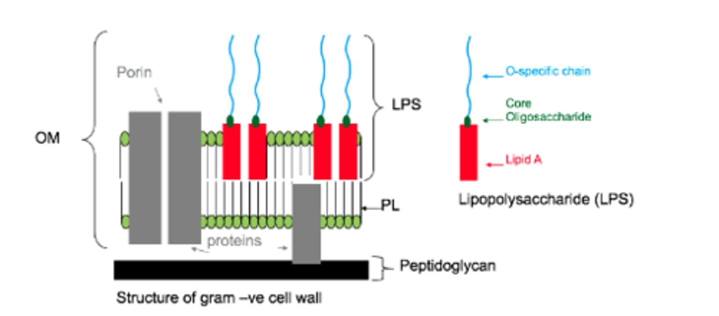
Structure of LPS
O specific chain is also an oligosaccheride and may be called O-specific antigen. It is highly specific to species.

Function of LPS
1) They confer a negetive charge to the outer surface and this plays roles in eg phagocytosis
2) The ability for bacteria to cause disease resides in the ability of the O specific chain to vary in structure. When in the body, antibodies attack the o-specific chain. If it can be varied it avoids detection. There is a highly selective pressure for disease causing bacteria to develop new structual varients of this chain to help evade immune system detection.
- Important with enteric bacteria eg salmonella enterica and shigella flexneri.
The LPS is also know as..
eg why?
The endotoxin ( as its toxic to us ). N.menintidis ( gram –ve ) causes meningitis, makes masses of LPS more than the cell needs, and is partitioned off of the cell and spread about the body an causes capilleries to become leaky.
Gram +ve cell wall
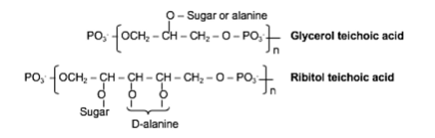
Teichoic acid
40-90% peptidoglycan. Teichoic acid is cross linked into the PDG, it confers a negetive charge but its actual role is unknown.
May petrude from surfaces
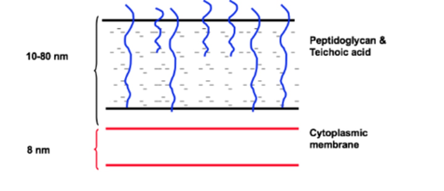
What does techoic acid look like
What do you think are the advantages an and limitations of a cell having 1-2 layers of PDG (Gram negative) versus several layers (Gram positive)?
+ More layers = more defensive, harder to get through
- Some antibacterial drugs such as penicillin interfere with the production of peptidoglycan so less layers = more destruction What
Compare what gram -ve and gram +ve bacterial cells have
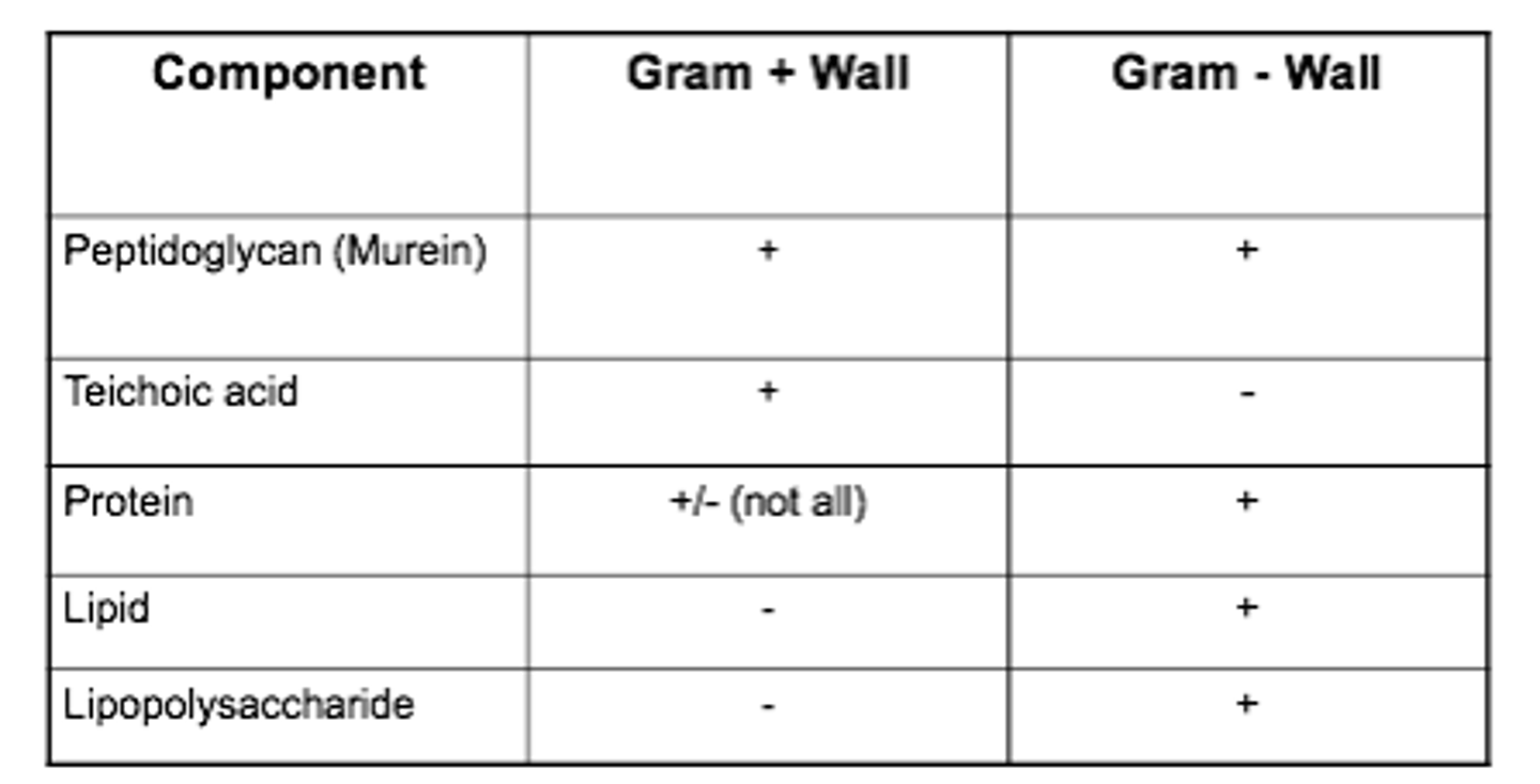
Peptidoglycan and lipopolysacheride are
unique to bacterial cell walls
Structure of peptidoglycan
Polysaccharide chain made up of two sugars NAG and NAM linked through glycosidic bonds and then cross-linked by peptide bridges to produce a strong lattice structure
Polymer 10-65 repeats long. G-M-G repeats - 3 in a strand. 5-15% of GN weight is peptidoglycan.
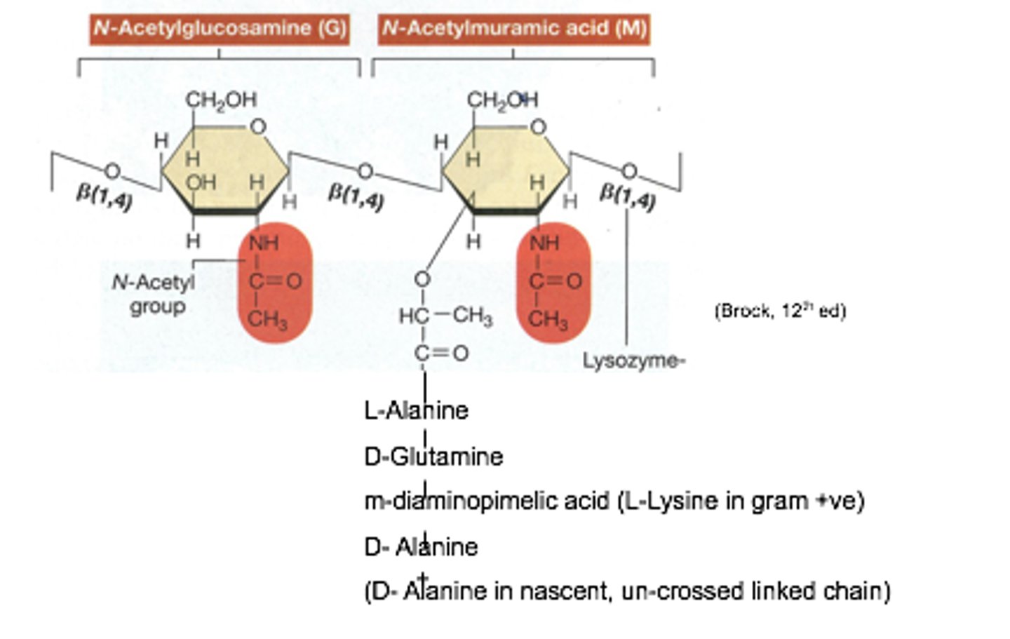
The third amino acid in the peptidoglycan crosslinking chain is
Role?
Catalysed by
either diaminopimilic acid (DAP) ( GRAM -VE ) or L-lysine ( GRAM +VE )
1 Is used in a peptide bond, the other is free and allowing cross linking to take place in adjacent molecules via joing to adjacent D-alanine
Transpeptidase
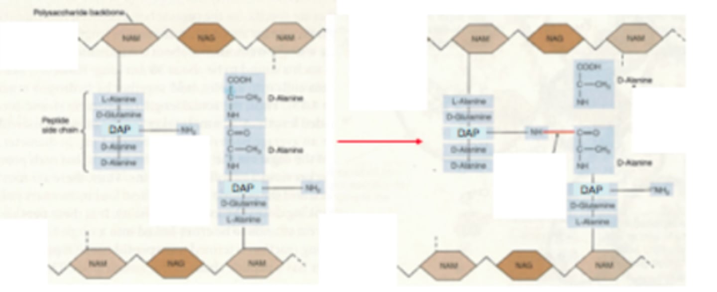
Transpeptidase
a bacterial enzyme that cross-links the peptidoglycan chains to form rigid cell walls
Lysozyme and penicillin are natural compounds and
attack the cell wall.
Action of lysozyme
Lysozyme is a compound found in egg whites and tears.
It cleaves the beta-1-4-glycosidic bonds between NAG and NAM.
Most effective on gram –ve as theres less PDG so it can weaken easier, causes the cell to lyse from inability to withstand pressure.
Action of penicillin
- Penicillin works better on gram positive bacteria; it inhibits transpeptidase.
- When a bacteria is growing in preparation for binary fission, it makes more space in the cell wall by cleaving cross links in PG cell wall.
- Autolysins cleaves these cross links for new molecules to be inserted.
- Transpeptidase and Autolysin activity must therefore be balanced.
-If penicillin is applied, autolysins continue to work but no cross links are MADE as transpeptidae is not working, therefore the cell wall progressivly weakens and turgor pressure cannot be maintained so cells lyse.
A patient presents at hospital complaining of a non-productive cough that has prescribed for 6 days. The doctor prescribed a 7 –day course of amoxicillin (a B –lactam antibiotic). After day 7 the patient is no better that before she started the treatment. Laboratory tests showed that the infection was caused by Mycoplasma pneumoniae. Explain why the antibiotic did not work.
Penicillin only works on
actively growing bacteria
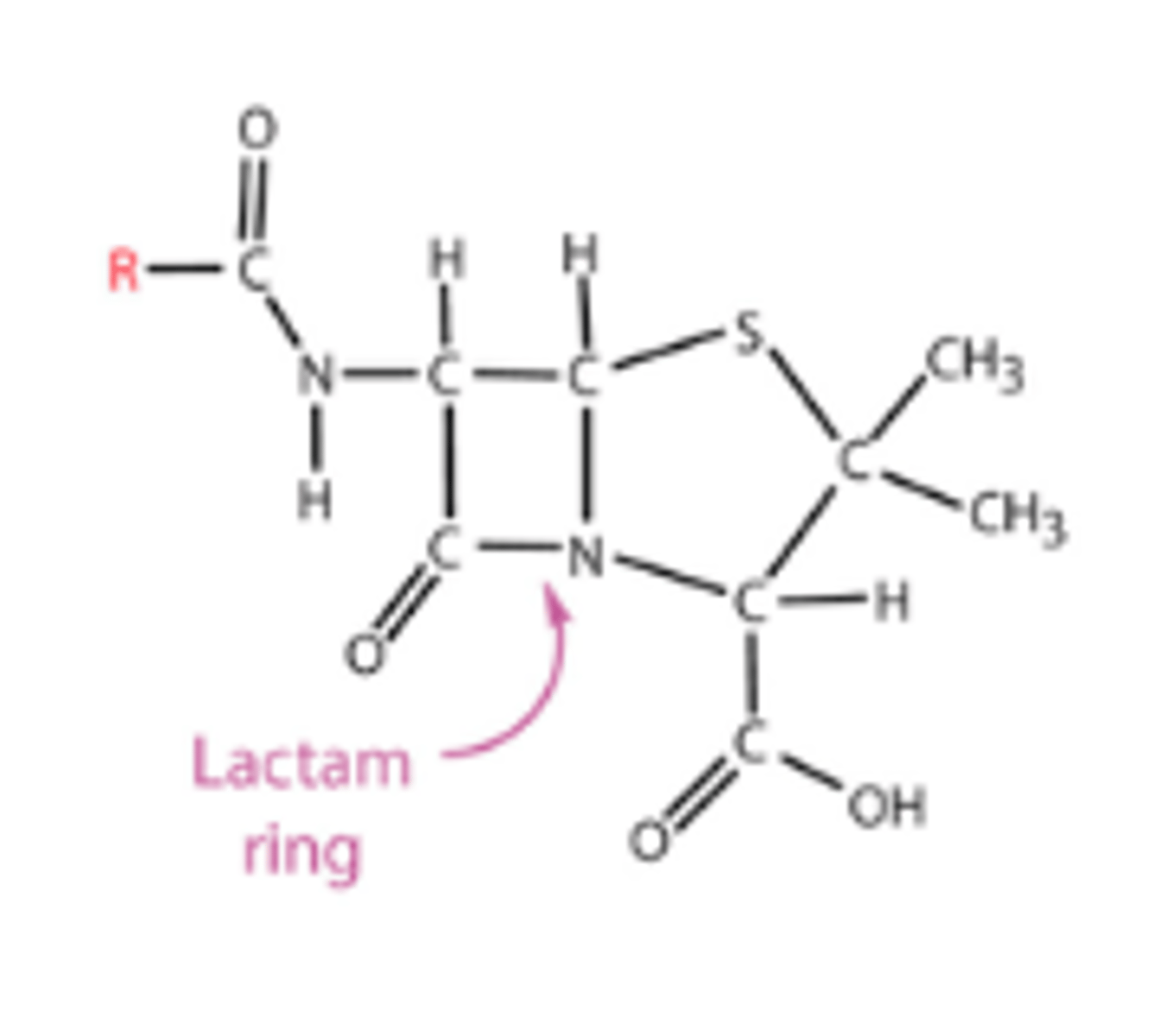
Penecillin structure
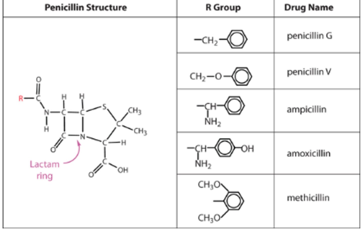
Different types of penecillin
Penicillin is a beta-lactam antibiotic.
Different penicillins have different R groups.
Pen G is only effective on gram + as it cannot get across the outer membrane of gram -ve bacteria.
When R group is modified that allow it to move across the outer membrane eg amplicillin and amoxicillin and are called broad spectrum antibiotics ( these are semi synthetics ).
Penecillin resistance
Beta lactam resistance
- Bacteria Inherit a gene encoding beta lactamase. These cleve the beta lactam ring and destroy the antibiotic.
- Found in both gram + and -.
What is reststant to beta lactamase?
- Methicillin is resistant to beta lactamases as it’s a poor substrate for the enzyme due to R groups,
- MRSA developed other ways to combat it.
- Eg New NDM-1 is carried on plasmids and confers resistance to bacteria against methicillin
How can we combat MRSA
- Vancomycin is not a beta lactam, it’s a glycopeptide and is last line of defense eg used to create MRSA and was taken from a. orientalis.
- Vancomycin binds to terminal d-alanines on peptodidoglycan so it cannot cross link.
- However we now have VRSA which has van genes make a PTG structure with d-lac-d-ala instead of d-ala-d-ala so vancomycin cant bind.
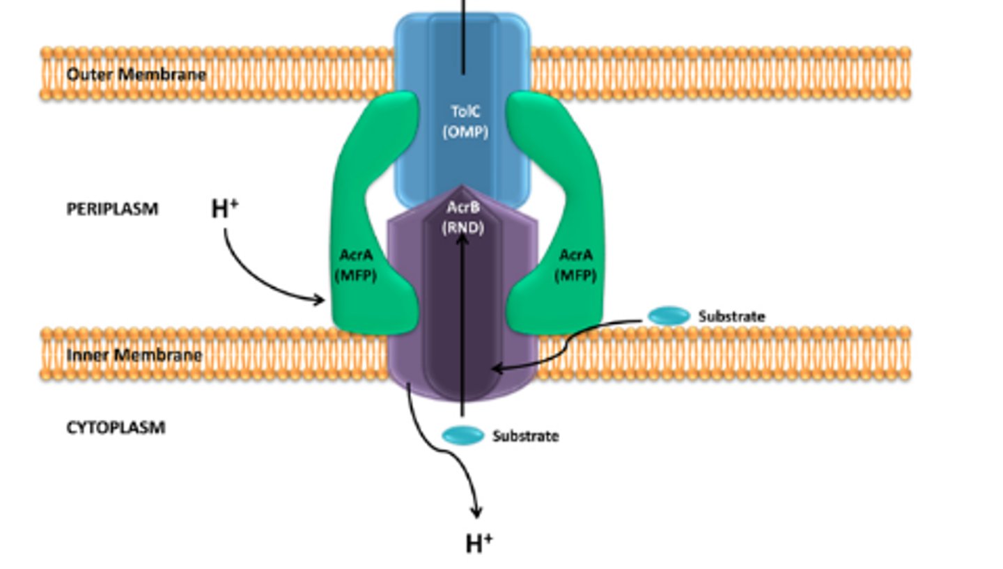
Efflux pumps
- Complex proteins that sit in the cell wall/ membrane.
- They span the whole envelope and pick up the molecule up in the cytoplasm and chuck it out of the cell using PMF, transferring H+ in. - They have very high specificity.
Eg multidrug ArcAB Tol C pump in e coli. Export antibiotics like ciprofloxarin and a dye called acridine orange and hexane. These pumps can transfer out antibiotics.
MexT
Bacteria mutate so mexT is expressed at high levels all the time so many efflux pumps are created. Act on many antibiotics.
Common cause of infections in hospital.
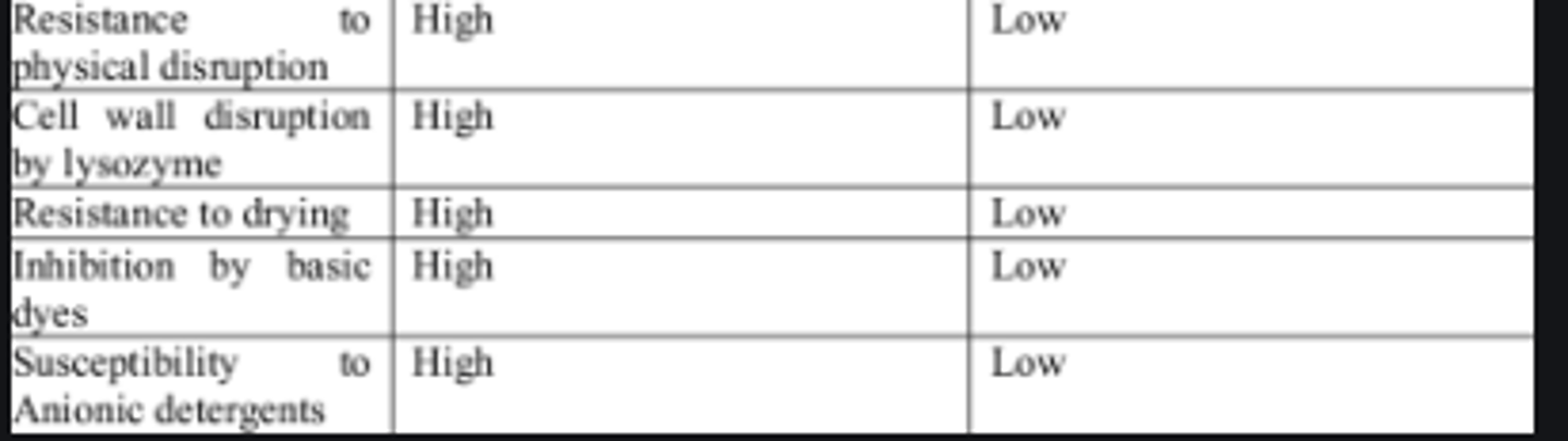
Advantages and disadvantages of gram +ve vs gram -ve
Not all bacteria are gram + or -,
there are other cell wall types.
- Mycobacteria - high lipid rich cell wall
-Some gram +ve have an extra layer outside cell wall called the S layer which is a protein coat
- Archae have unique cell walls that are diverse and different.
- Mycoplasma ( smallest bacteria ) are obigate intracellular pathogens, and live in a constant homeostatic environment. No cell wall and are pleomorphic and take on many shapes.
A patient presents at hospital complaining of a non-productive cough that has prescribed for 6 days. The doctor prescribed a 7 -day course of amoxicillin (a B -lactam antibiotic). After day 7 the patient is no better that before she started the treatment. Laboratory tests showed that the infection was caused by Mycoplasma pneumoniae. Explain why the antibiotic did not work.
- no PTG layer
- nothing for the antibiotic to act to to lyse the cell
Chelating agents
are organic compounds capable of linking together metal ions to form complex ring-like structures called chelates.
chelating agents
molecules that attract or bind with other molecules and are therefore useful in either preventing or promoting movement of substances from place to place