neurons and action potentials
1/24
There's no tags or description
Looks like no tags are added yet.
Name | Mastery | Learn | Test | Matching | Spaced |
|---|
No study sessions yet.
25 Terms
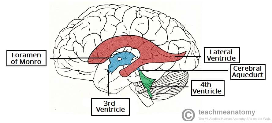
ventricles
four spaces filled by CSF, interconnected
constant flow of CSF, providing cushioning/buoyancy
flow of nutrients to and out of brain
brain-blood barrier
semi-permeable separation between blood and brain
pathogen protection in sensitive brain tissue
endothelial cells, astrocytes, pericytes
passes: water, gas, hydrophobic, glucose, amino acids
blocks: large, hydroPHILIC, bacteria
challenge for drug delivery
human vs rodent brain
similarities
genes, neurons, proteins, structures, connectivity patterns
differences
size and neuron no.
brain structural proportions, small organizational diffs
smooth rodent brain cortex
glia cells (50% of cells)
do not fire AP, support
3 types
astrocytes: touch neurons/BV cells; anchoring, balance chem. concentration outside neurons, injury repair
microglia: immune defense by destroying foreign
oligodendrocytes: wrap neurons with myelin to increase electrical signaling
neurons
Ramon y Cajal’s Neuron Doctrine that says the brain is made up of small, discrete intersecting bodies
soma (cell body), dendrites, axon terminals, terminal branches
parts of the neuron
dendrites: branching projections that collect info
soma: contains the nucleus and integrates info
axon: conducts neural signal across a distance (long distances such as SC to toe)
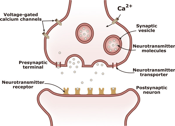
synapse stages
presynaptic: sending side
postsynaptic: receiver
voltage
difference in electrical potential between 2 points
voltage at resting: -70 mV
higher negative charge inside the cell
Na/K pump
high K+ and low Na+ inside b/c NA/K pump ejects 3 Na+ and pulls 2 K+, using 1 ATP
overall negative charge inside the neuron
inactivation of neuron voltage
equal concentrations inside and out —> death, no electrical activity
Ouabin: african arrow poison that stops Na/K pump
action potential
spikes to +30 mV at peak AP
action potential graph
time (ms) vs voltage (mV)
depolarization
membrane potential becomes less negative
hyperpolarization
membrane potential becomes more negative
all or nothing principle
an action potential is initiated after threshold, -55mV, is met
Na+/K+ pump brings back to baseline during weak stimulations
strong stimulation (depolarization) at +30mV
steps in action potental
depolarization: above -55mV —> opens voltage dependent Na+ channel and Na+ rushes in due to low concentration in the cell
at peak voltage of 30mV, Na+ channels close
stops depolarization (transitioning to repolarization) and channels cannot open for 1 ms
high voltage opens voltage dependent K+ channels
repolarization and K+ move out
Hyperpolarization closes K+ channels, resulting in afterpolarization
Inactive Na+ channels cause a refractory period where no new action potentials occur and Na+/K+ pump continuously brings back to baseline
afterpolarization
being less than -70mV of baseline following an action potential
tetrodotoxin (TTX)
caused by puffer fish in eyes
inactivates Na+ channels by blocking them
contribute to paralysis
lidocaine/novocaine
occuring in pain-sensitive neurons where Na+ channels do not cause signaling
beginning of an action potential, location
begins at the axon hillock, traveling along the axon to axon terminals
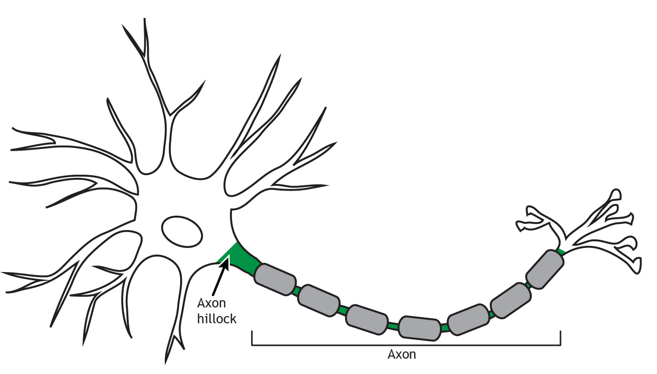
axon hillock
rich in Na+ channels, causing volatile movement
depolarizing formed by Na+ influx flows down the neuron, depolarizing the next section
Na+ channels close prior, then travel, preventing A.P. from moving backward
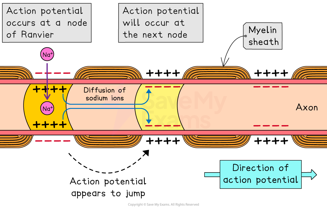
saltatory conduction
use of myelin sheaths (fatty substance) surrounding the axon
in CNS: oligodendrocytes
in PNS: schwann cells
speeds up AP from 2 m/s to 120 m/s
multiple schlerosis
breakdown of myelin sheath
slows down neural communication, impairing vision, muscle weakness, movement/coordination issues, and cognitive impairments
neurotransmission steps
general: higher Ca2+ extracellularly
Action potential opens up Ca2+ channels and they rush in the axon terminal
vesicles get fused to the extracellular membrane and open up at the synaptic cleft
neurotransmitters bind to the receptors on dendrite of postsynaptic neuron
what determines neurotransmission
types of neurotransmitters, type of receptors (ionotropic vs metabotropic), types of ion channels opening