Kines 202 Lab
1/121
There's no tags or description
Looks like no tags are added yet.
Name | Mastery | Learn | Test | Matching | Spaced | Call with Kai |
|---|
No analytics yet
Send a link to your students to track their progress
122 Terms
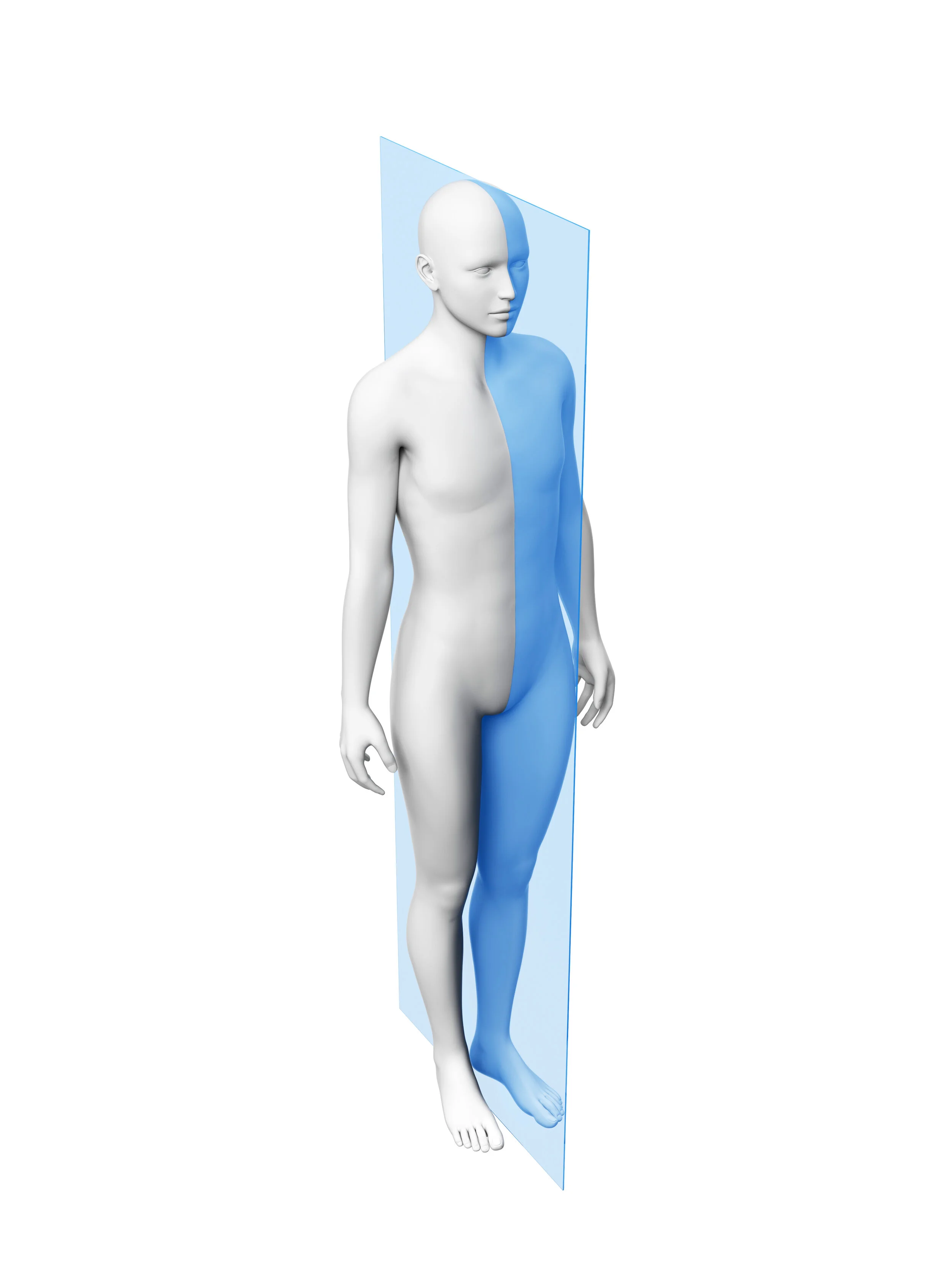
What plane cut is this?
Sagittal Plane
What does the Sagittal Plane split the body up?
left and right
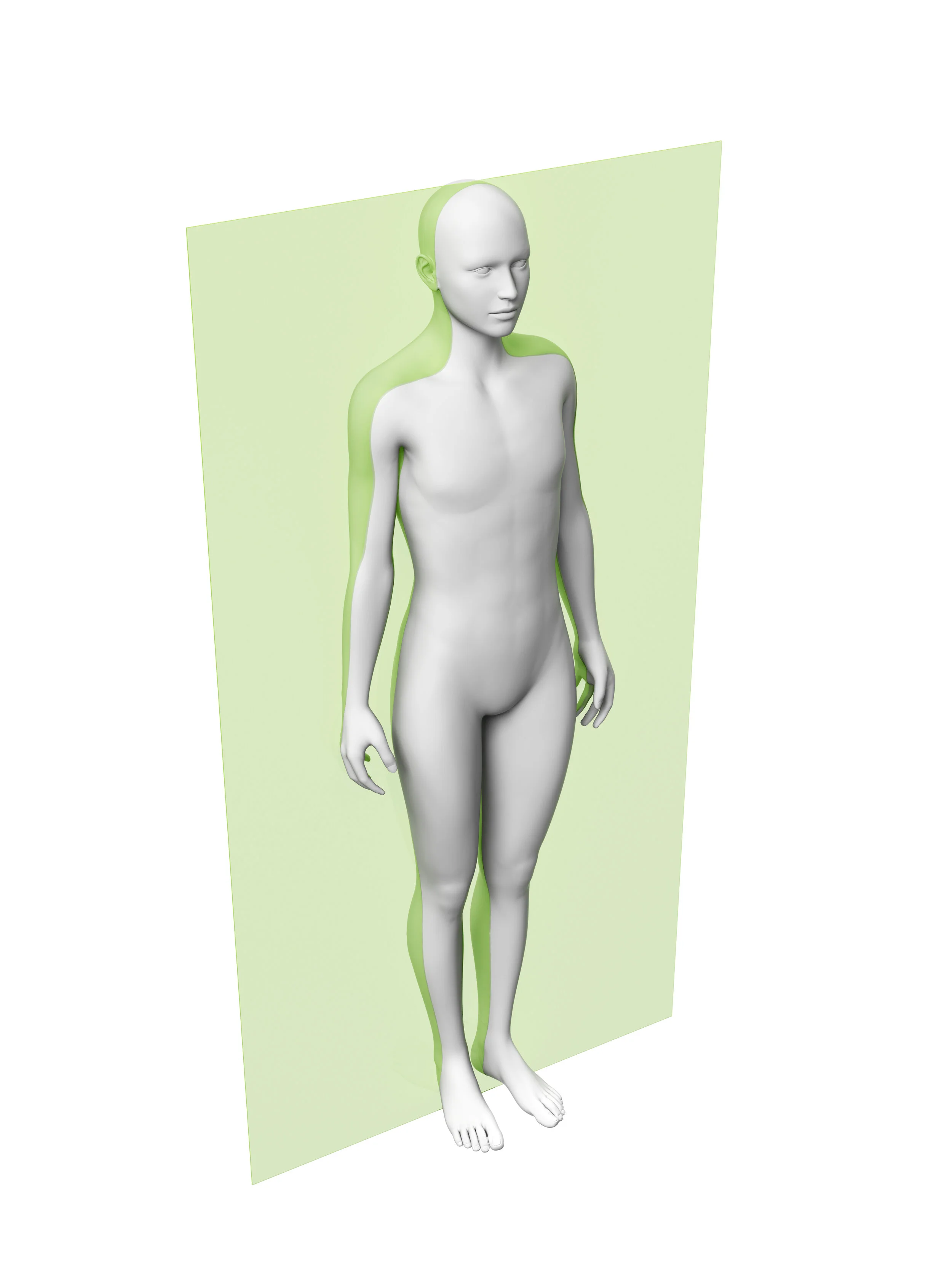
What plane cut is this?
Frontal
What does the Frontal plane cut?
Anterior and Posterior views
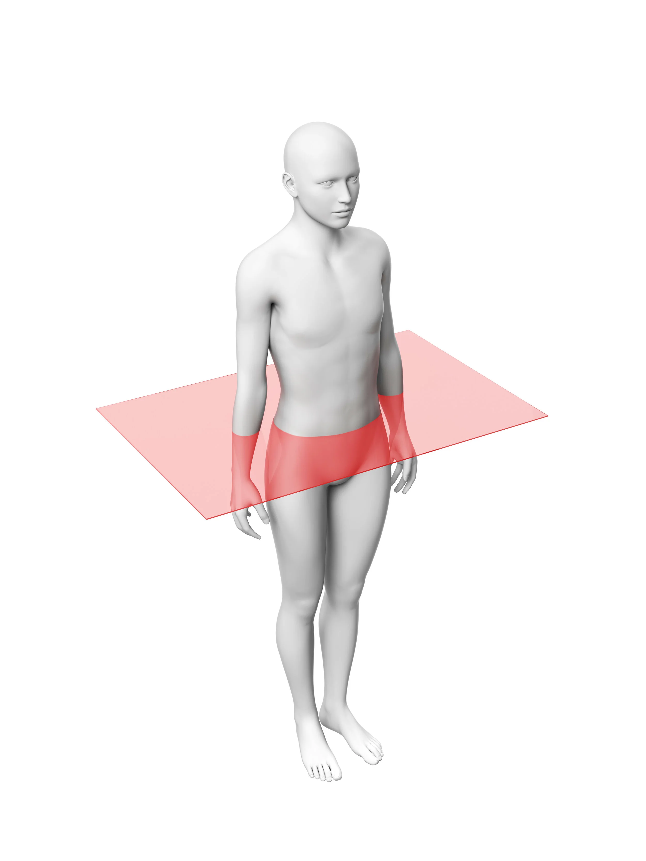
What plane cut is this?
Transversal?
What does the Transverse cut the body into? What movement can the body do?
Superior and Inferior; axial rotation of the head
What axis does the body rotate on the Sagittal plane? What movement can the body do?
Mediolateral axis; flexion and extenstion
What axis does the body rotate on the frontal plane? What movement can the body do?
Anteroposterior axis; adduction and abduction
What axis does the body rotate in the transverse plane?
Longitudinal axis
What movement is this?
flexion
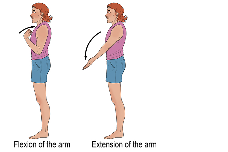
What movement is this?
Extension
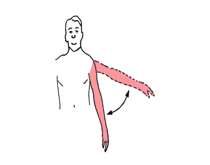
What movement is this?
abduction of shoulder and adduction of shoulder
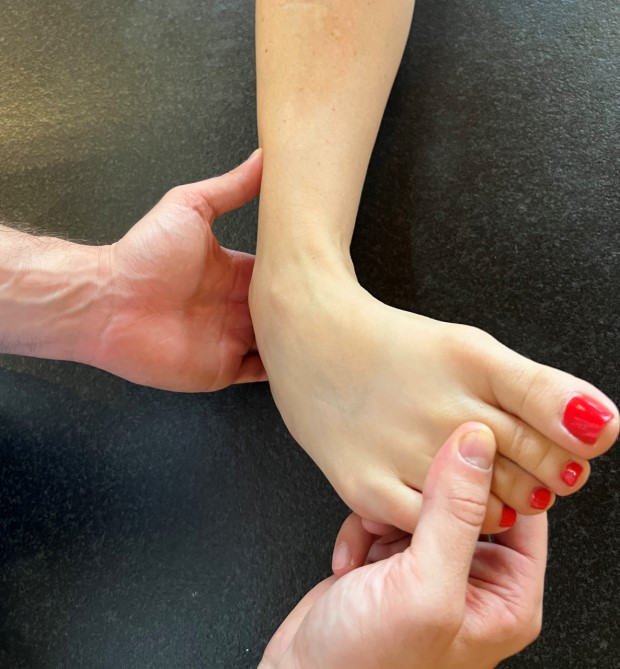
What movement is this?
Inversion of ankle
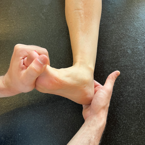
What movement is this?
Eversion of ankle
What movement is this?
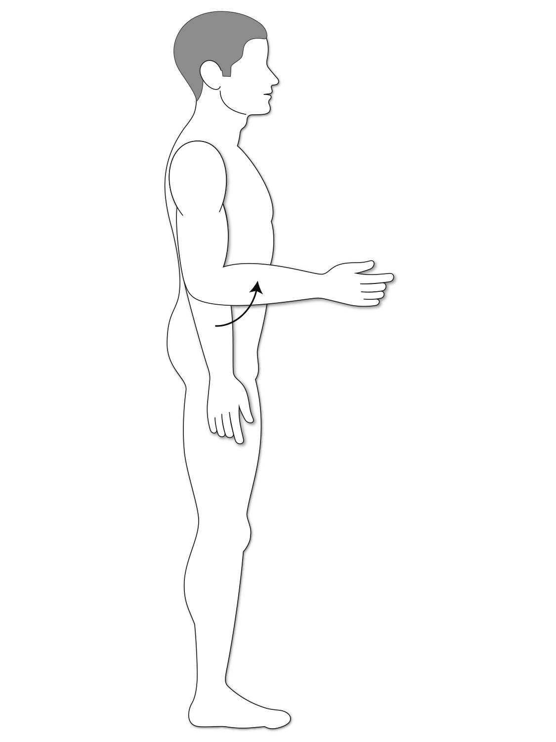
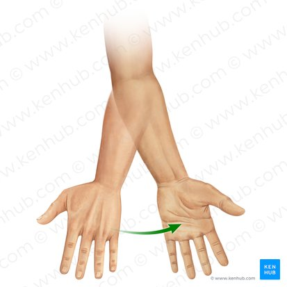
Supination of forearm
What movement is this?
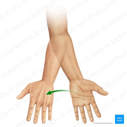
Pronation?
List all the types of Synovial Joints?
Planar joint
Hinge joint
Pivot joint
Ellipsoid joint
Saddle joint
Ball-and-socket joint
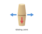
What movement does the Planar joint allow? give an example
sliding of bone across flat surface/ fossa
example: intercarpal and intertarsal joints
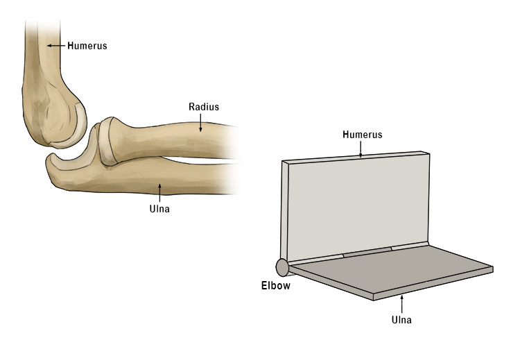
What movement does the Hinge joint allow? give an example
flexion and extension
example: elbow flexion and extension
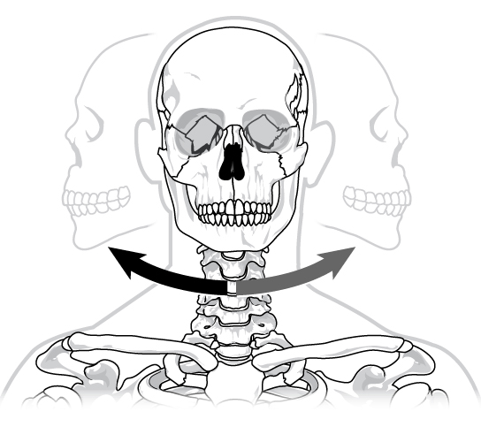
What movement does the Pivot joint allow? give an example
Medial and Lateral rotation
example: atlas/axis of the cervical vertebrae
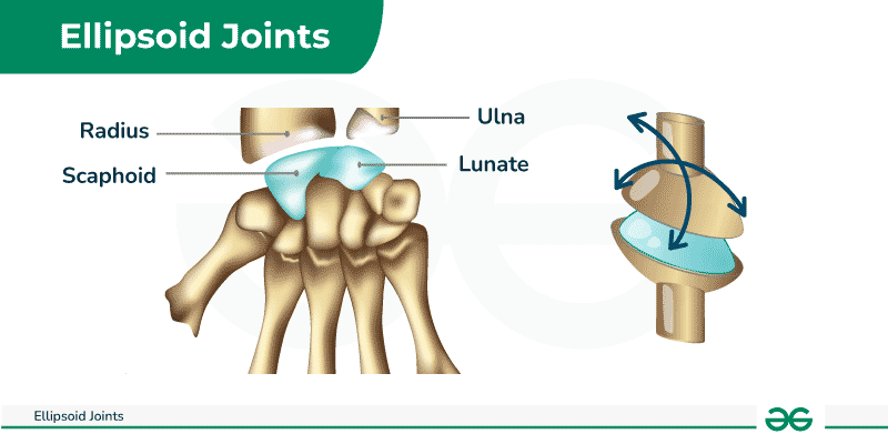
What movement does the Ellipsoid joint allow? give an example
allows for adduction/abduction, flexion/extension, circumduction
example: wrist or knuckles
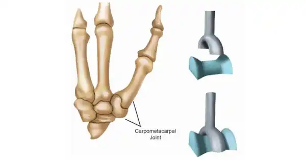
What movement does the Saddle joint allow? give an example
flexion and extension
example: thumb flexion and extension
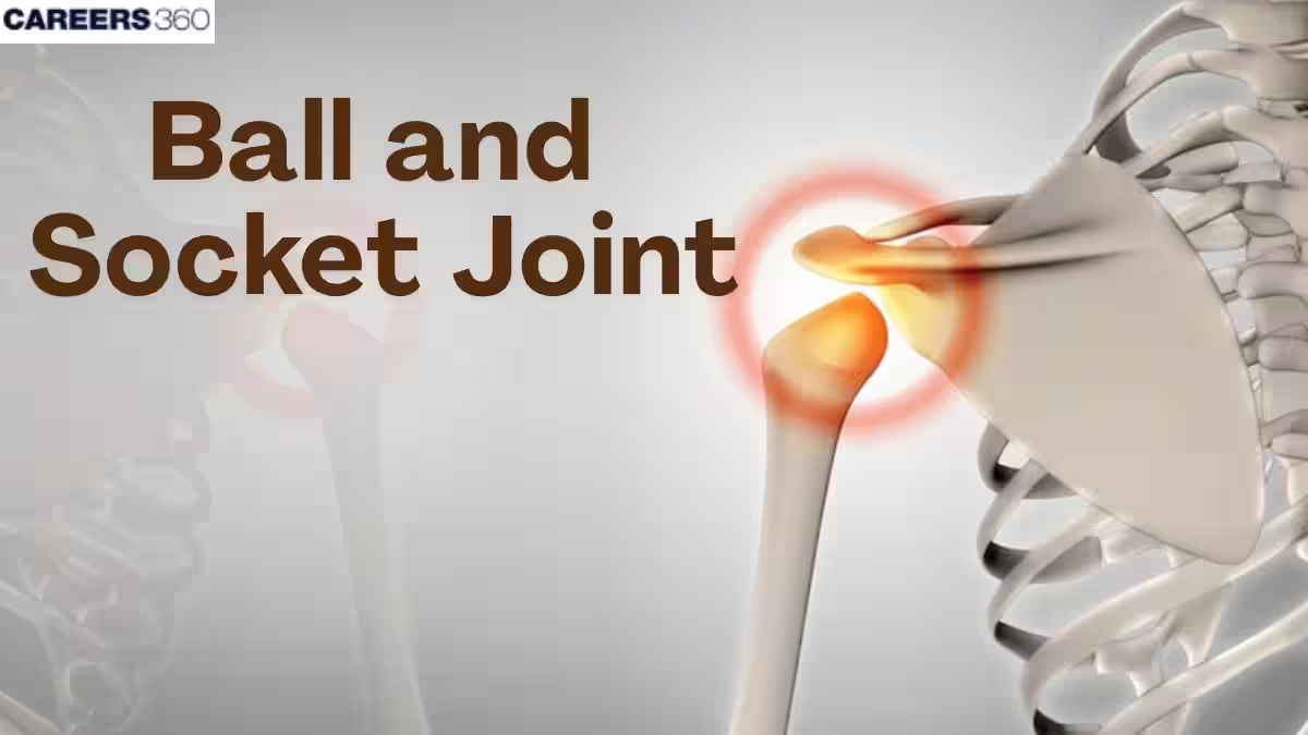
What movement does the Ball-and Socket joint allow? give an example
medial and lateral rotation, flexion/extension, abduction/adduction
example: shoulder joint
What does Palpation mean?
the process of using one’s hands to examine a body
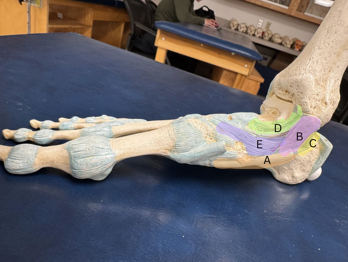
Label each ligament from A-D
A. Plantar Calcaneonavicular (Spring) Ligament
B.Tibiocalcaneal Ligament
C. Posterior Tibiotalar Ligament
D. Anterior Tibiotalar Ligament
E. Tibionavicular Ligament
What ligament is on the bottom of the foot?
Long Plantar ligament, from the Calcaneus to the 2nd-5th Metatarsals
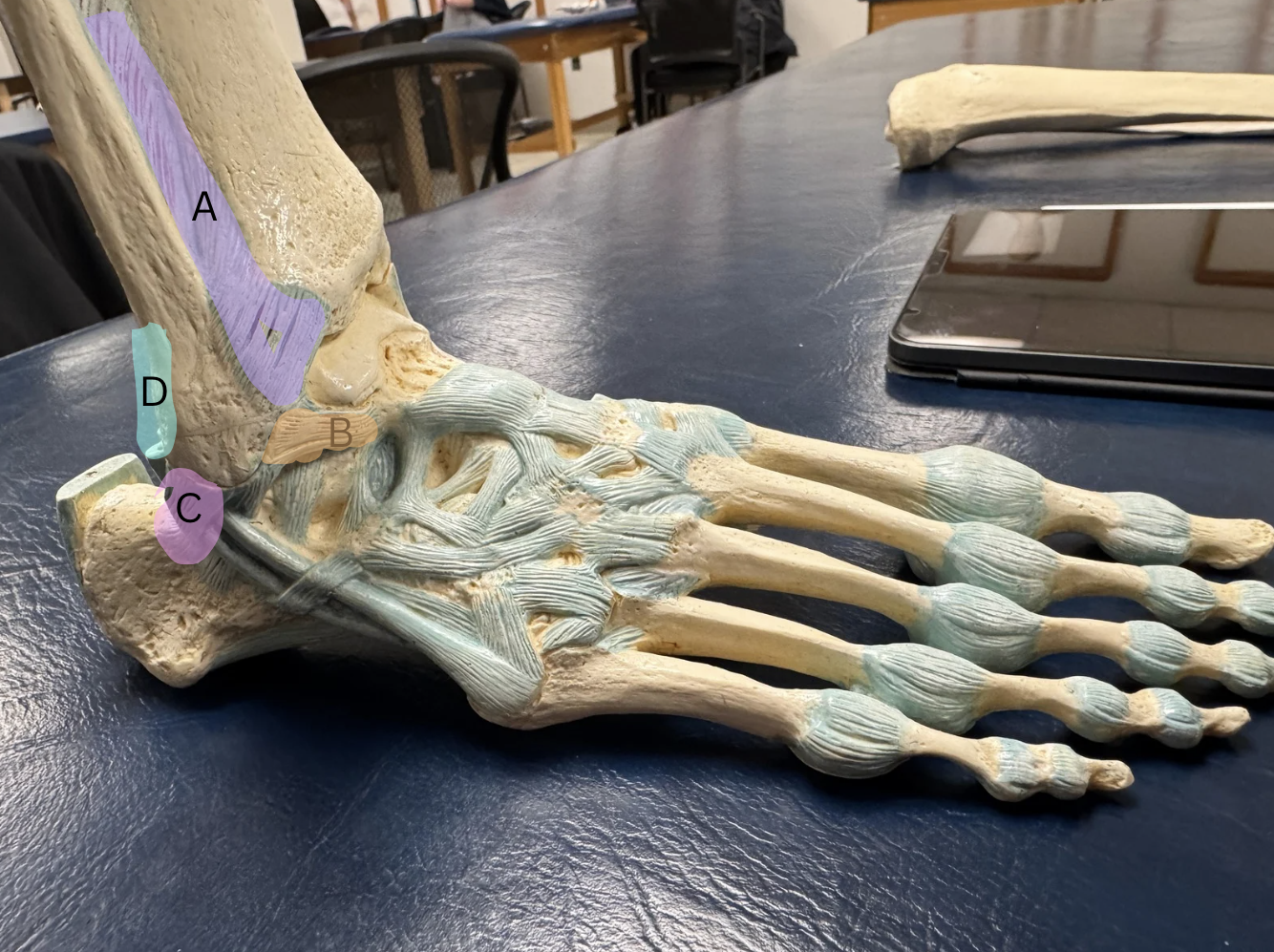
Label the ligaments from A-D
A. Anterior Tibiofibular Ligament
B. Anterior Talofibular Ligament
C. Calcaneofibular Ligament
D. Posterior Tibiofibular Ligament
What is the function of 3 arched in our foot?
Shock absorption due to elasticity and support by ligaments, muscles, and other fascia.
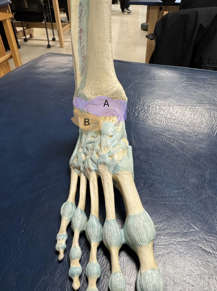
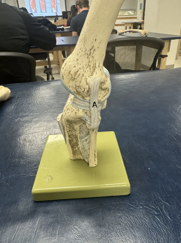
Label the two joints A and B, and what movements they do.
A. Talocrural Joint, Plantar Flexion and Dorsiflexion
B. Subtalar Joint, Ankle Eversion and Inversion
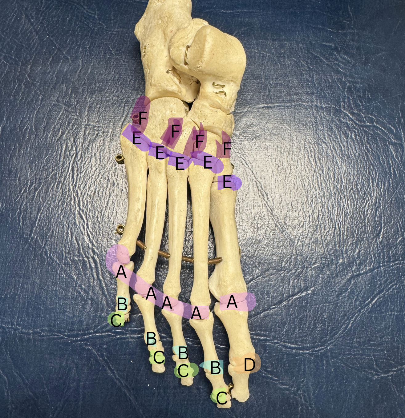
Label these joints A-F
A. Metatarsophalangeal joint
B. Proximal Interphalangeal Joint
C. Distal Interphalangeal Joint
D. Interphalangeal Joint
E. Tarsometatarsal Joint
F. Intermetatarsal Joint
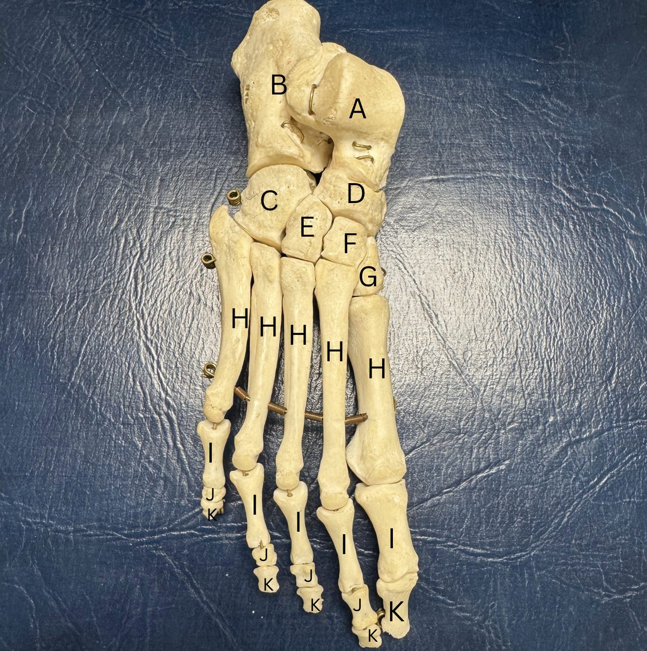
Label all the bones of the foot A-K
A. Talus
B. Calcaneus
C.Cuboid
D. Navicular
E.Lateral cuneiform
F. Intermediate cuneiform
G. Medial cuneiform
H. Metatarsals (1-5)
I. Proximal Phalange (1-5)
J. Middle Phalange (2-5)
K. Distal Phalange (1-5)
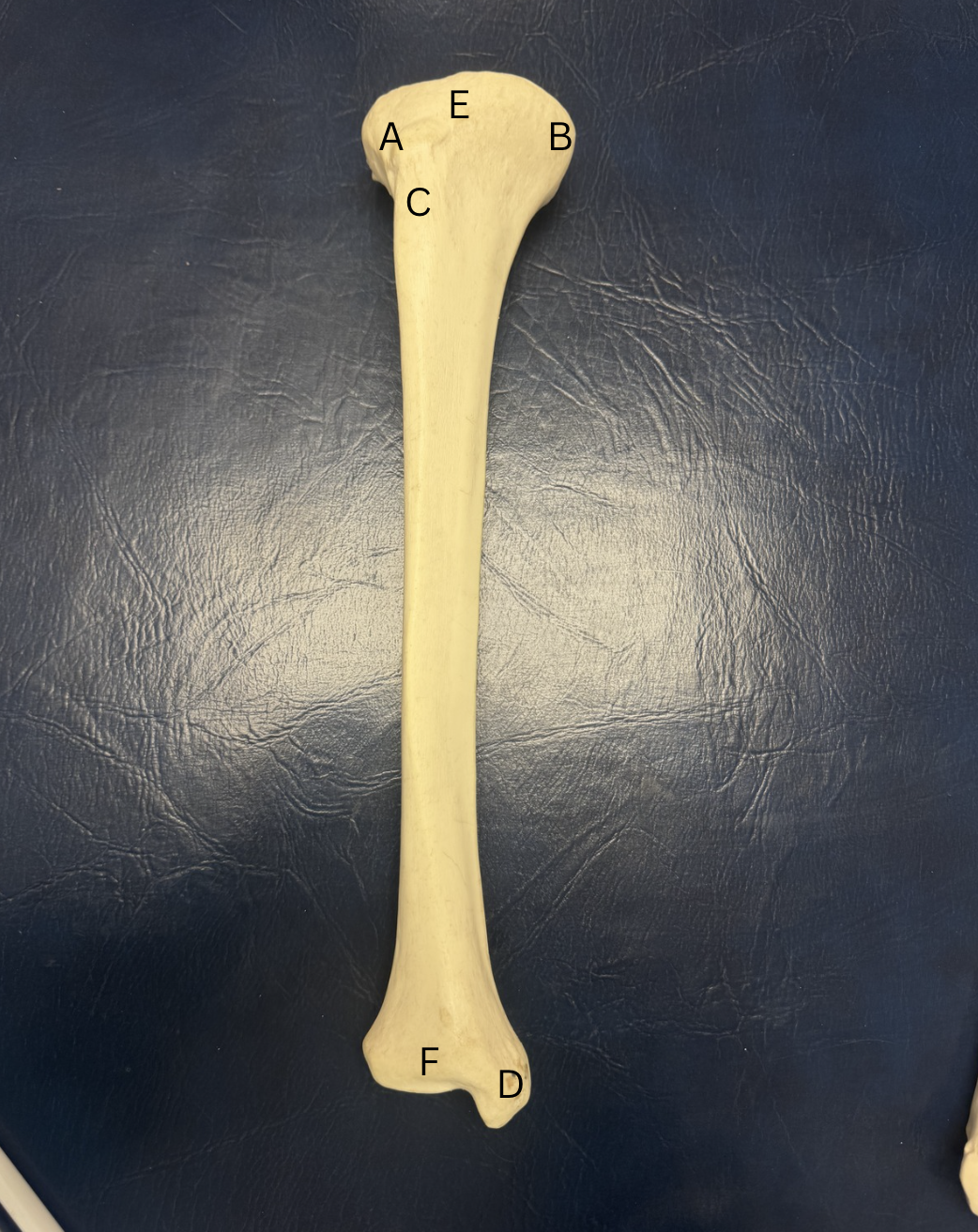
Label A-F of this bone
A. Lateral Condyle
B.Medial Condyle
C.Tibial Tuberosity
D.Medial Malleolus
E. Intercondylar Eminence
F. Articular Surface
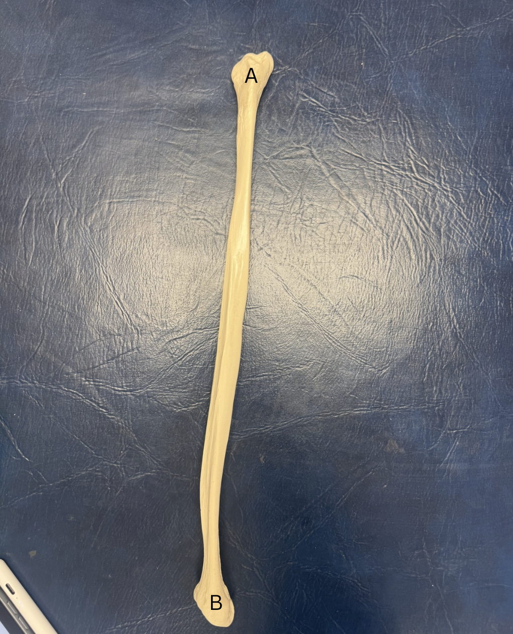
Label A and B of this bone
A. Head of Fibula
B. Lateral Malleolus
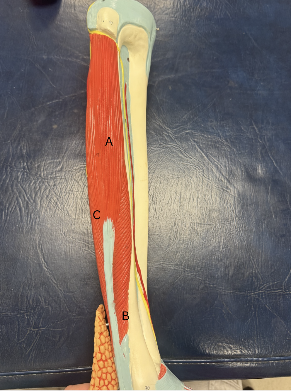
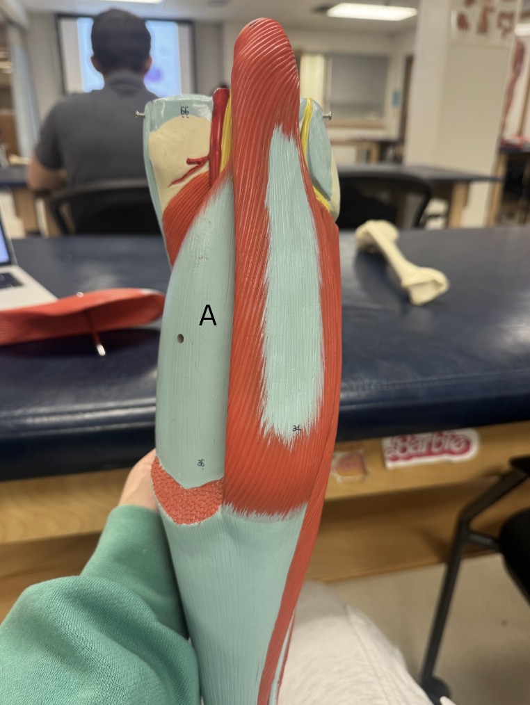
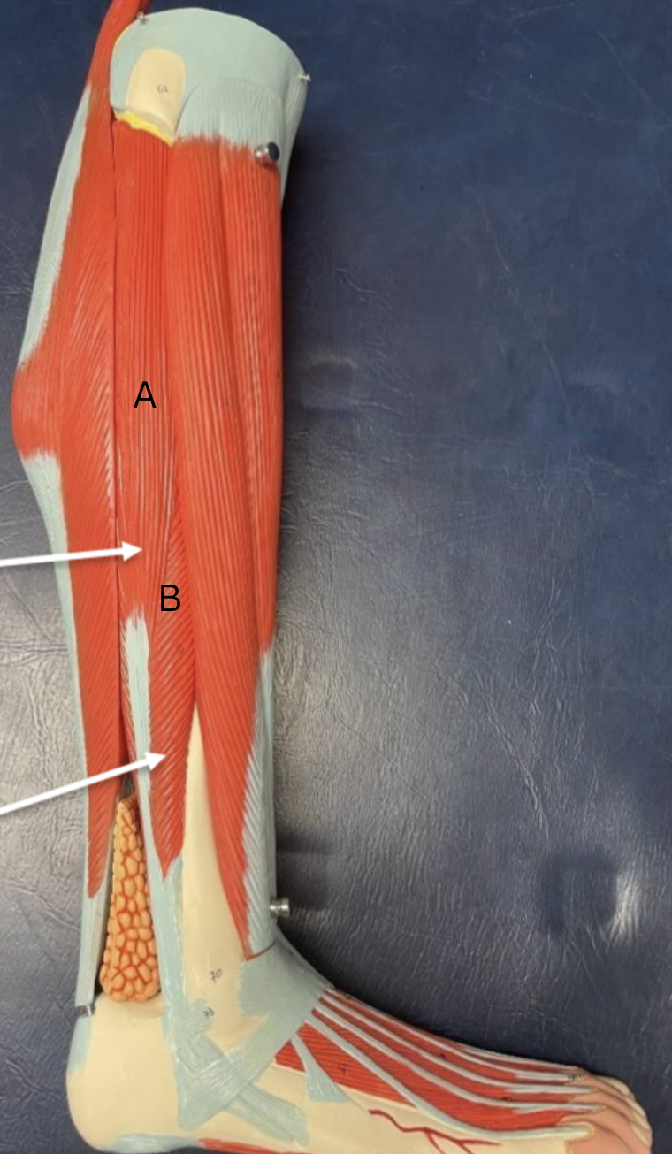
Label this muscle from A-C
A. Tibialis Anterior
B. Extensor Hallucis Longus
C. Extensor Digitorum Longus
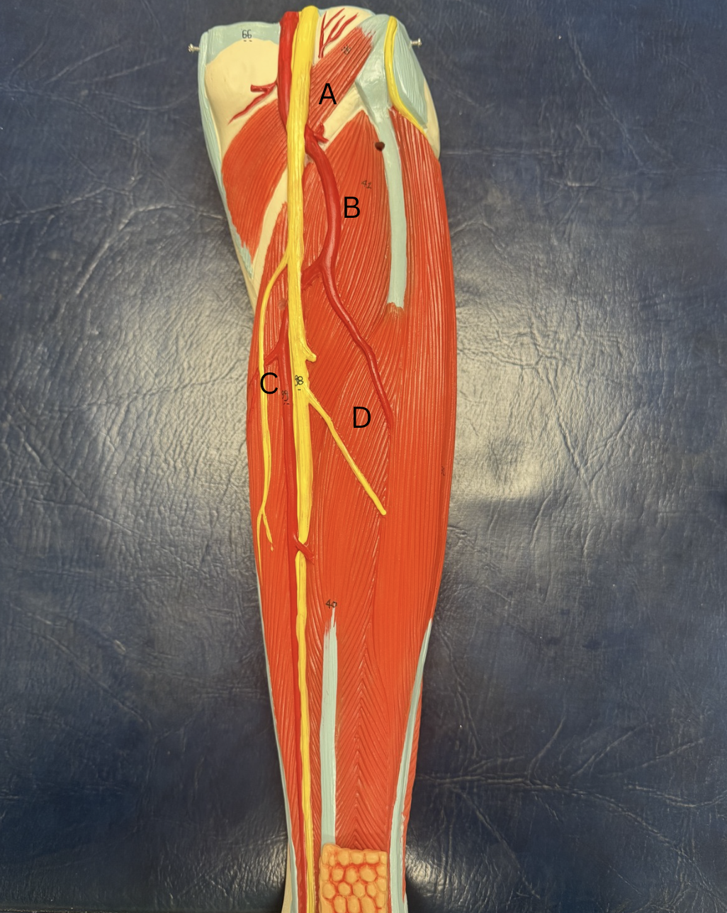
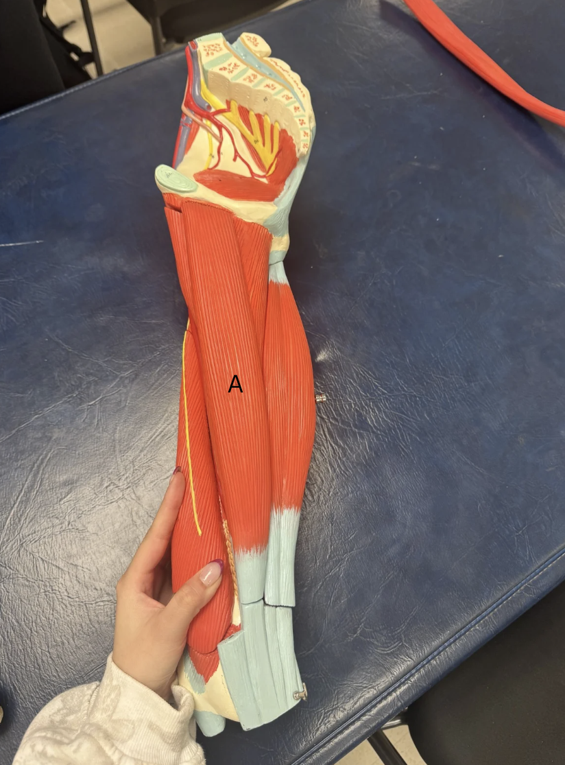
Label the muscles from A-D
A. Popliteus
B. Tibialis Posterior
C. Flexor Digitorum Longus
D. Flexor Hallucis Longus
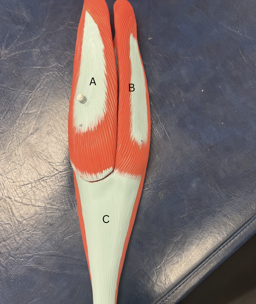
Label the muscles A-B and the tendon C
A. Medial Gastrocnemius
B. Lateral Gastrocnemius
C. Achilles Tendon
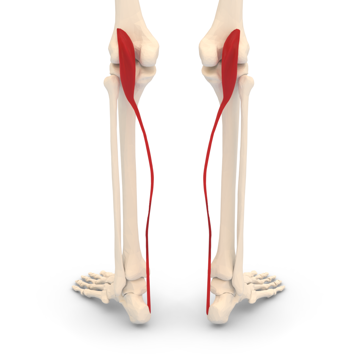
Label the muscle A
A. Soleus
OIF of Gastrocnemius
O:Medial head/above Medial Femoral Condyle
I: Achilles Tendon
F:Plantar Flexion
OIF of Soleus
O:Posterior surface of Tibia (soleal line) and posterior surface of the head of Fibula
I: Achilles Tendon
F: Plantar Flexion
Name this muscle
Plantaris
OIF of Popliteus
O: Lateral Epicondyle of Femur
I: Posterior Surface of the Tibia
F: Plantar Flexion
List all the muscles that the Tibial Nerve innervates
Gastrocnemius
Soleus
Plantaris
Popliteus
Posterior Tibialis
Flexor Digitorum Longus
Flexor Hallucis Longus
OIF of Posterior Tibialis
O: Posterior Surface of Upper Intake interosseous membrane
I: Lower surface of navicular and cuneiform
F: Plantar Flexion
OIF of Flexor Digitorum Longus
O: Middle 1/3rd of Posterior Tibia
I:Base of distal phalanges
F: 4 digit plantar flexion
OIF of Flexor Hallucis Longus
O: Posterior Fibula
I: Base of the distal phalanx of the big toe
F: plantar flexion of big toe
OIF of Anterior Tibialis
O: Lateral Tibial condyle
I: Medial Cuneiform and 1st Metatarsal
F:Dorsiflexion
OIF of Extensor Digitorum Longus
O: Lateral Tibia and head of Fibula
I: Dorsal (bottom side) o f 2-5 Phalanges
F: Dorsiflexion of 4 digits
OIF of Extensor Hallucis Longus
O: Anterior surface of Fibula
I: Dorsal side (bottom side) of 1st Phalange
F: Dorsiflexion of big toes (1st metatarsal)
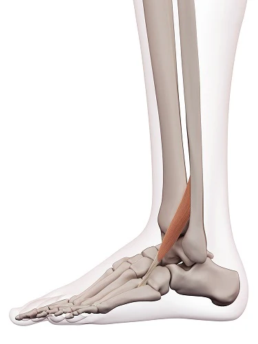
What muscle is this?
Peroneus Tertius
OIF of Peroneus Tertius
O: 2/3rd of Lateral Fibula
I: Long Medial Cuneiform/base of 1st Metatarsal
F: Dorsi Flexion
Label A and B muscles
A. Peroneus Longus
B. Peroneus Brevis
OIF of Peroneus Longus
O: Head of Superior and 2/3 of Fibula
I: Medial Cuneiform base of 1st Metatarsal
F: Eversion of Ankle
OIF of Peroneus Brevis
O: Inferior 2/3 of lateral Fibula
I: base of 5th metatarsal
F: Eversion of Ankle
What muscles does the Deep Peroneal Nerve innervate
Anterior Tibialis
Extensor Digitorum Longus
Extensor Digitorum Longus
What muscles does the Superficial Peroneal Nerve innervate
Peroneus Tertius
Peroneus Longus
Peroneus Brevis
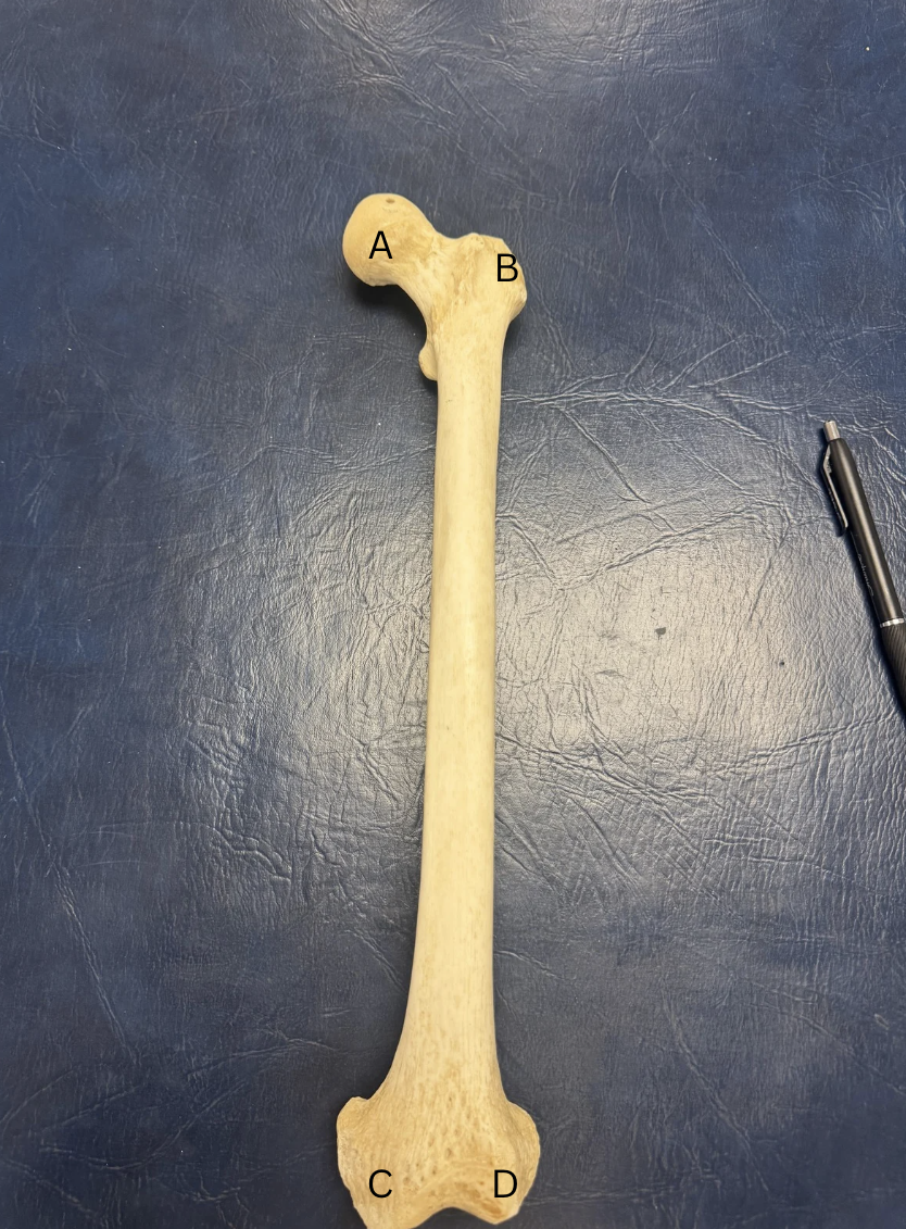
Label the Femur
inside head of Femur is Fovea Capitis
A.Head of Femur
B. Greater Trochanter
C. Medial Condyle (above is epicondyle)
D. Lateral Condyle ( above is epicondyle)
E. (Not labeled) lesser Trochanter
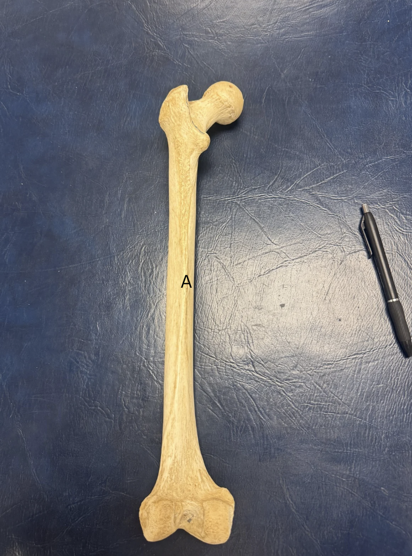
Label A
A. Linea Aspera
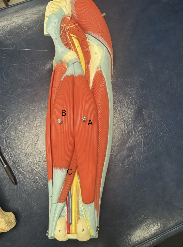
Label the quad muscles
A. Vastus Intermedius
B. Vastus Medialis
C. Vastus Lateralis
Label this muscle
Gracilis
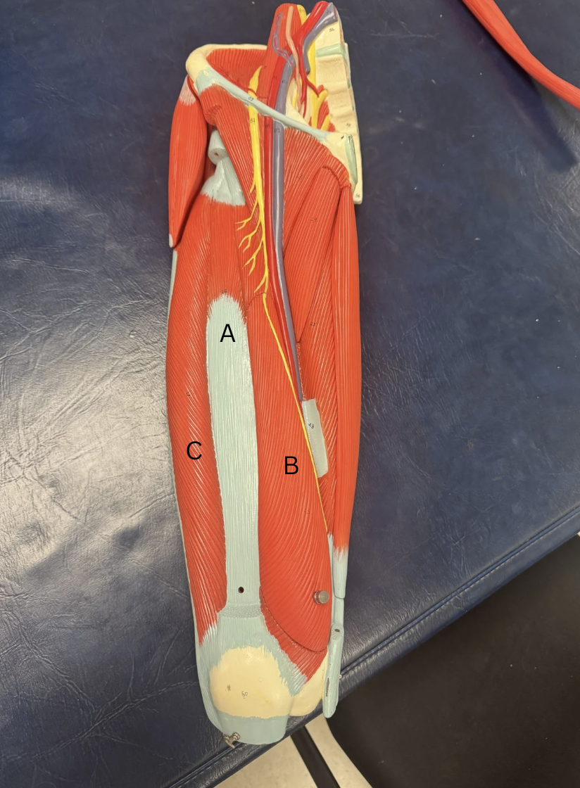
Label these muscles
A.Biceps Femoris
B.Semitendinosus
C.Semimembranosus
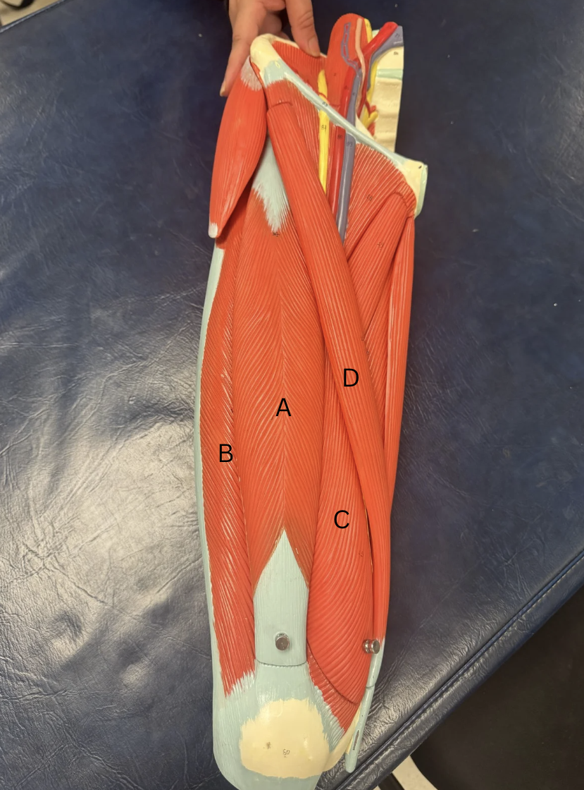
Label the quad muscles
A. Rectus Femoris
B. Vastus Lateralis
C. Vastus Medialis
D. Sartorius
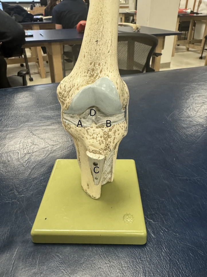
Label the menisci and ligaments
A.Medial Menisci
B.Lateral Menisci
C.Infrapatellar tendon
D. Anterior Cruciate Ligament
Label this ligament
A. Posterior Cruciate Ligament
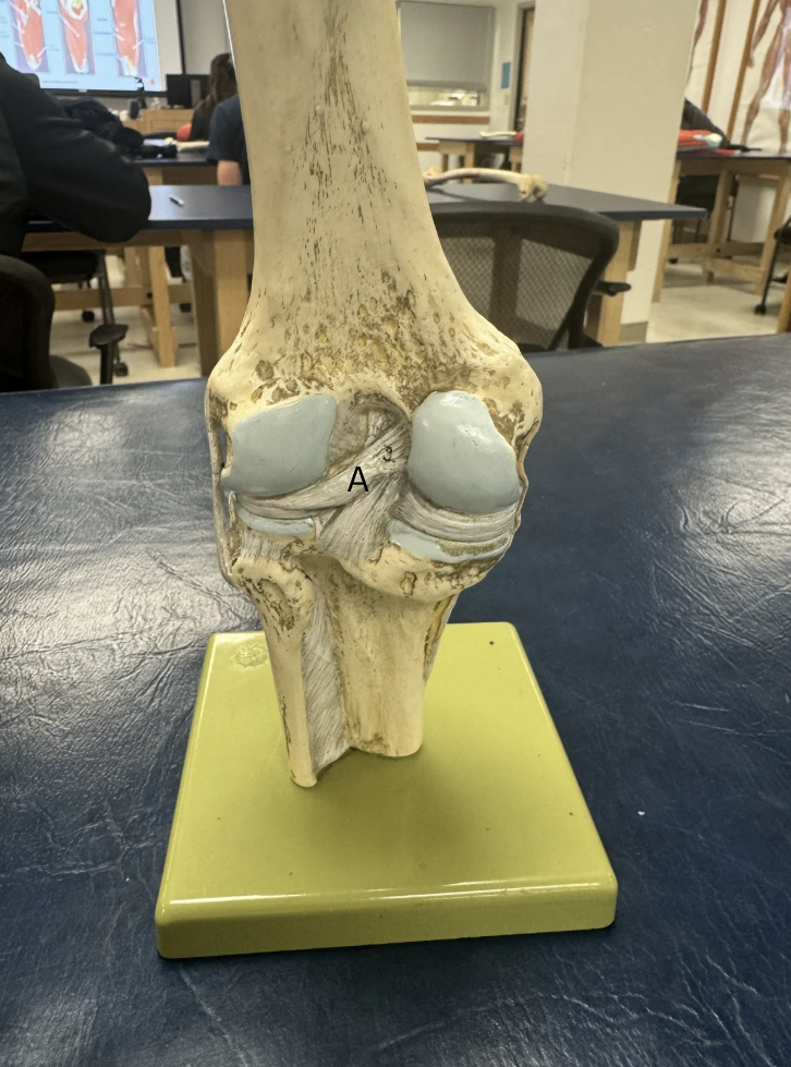
Label this ligament
A. Medial Collateral Ligament
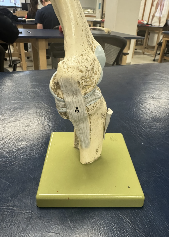
Label this Ligament
A. Lateral Collateral Ligament
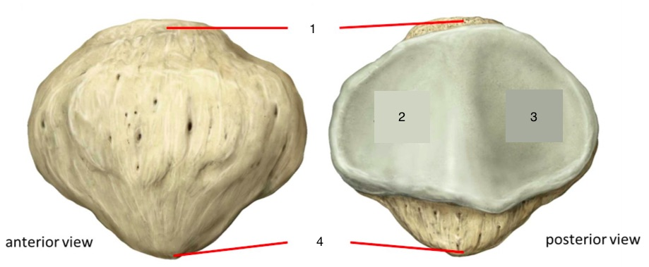
Label this bone
Patella bone
Base
Lateral Facet
Medial Facet
Apex of Patella
What are the joints of the knee?
Tibiofemoral joint (epicondyles and condyles)
Patellofemoral joint
Proximal Tibiofibular joint (head of Tibia and Fibula)
What is the terrible triad?
an injury to the ACL, MCL, and medial meniscus at the same time
What tendon do all the quad muscles insert into?
Quadriceps tendon inserts into the Suprapatellar tendon
What tendon does the Gracilis, Sartorius, and Semitendinosus insert into?
The Pes Anserine Tendon (AKA Goose foot) medially
What nerve innervates the quad muscles and Sartorius?
Femoral Nerve
What nerve innervates the hamstring muscles?
The Tibial portion of the Sciatic nerve
What nerve innervates the Gracilus?
Obturator Nerve
OIF of Rectus Femoris
O: Anterior of iliac spine
I: Tibial Tuberosity
F: Extension of knee
OIF of Vastus Medialis
O: Linea Aspera of Femur
I: Tibial Tuberosity
F: Extension of knee
OIF of Vastus Lateralis
O: Greater Trochanter
I: Tibial Tuberosity
F: Extension of Knee
OIF of Vastus Intermedius
O: Intertrochanteric line of anterior proximal femur
I: Tibial Tuberosity
F: Extension of Knee
OIF of Semimembranosus
O: Ischial Tuberosity
I: Medial condyle of Tibia
F: Knee Flexion
OIF of Semitendinosus
O: Ischial Tuberosity
I: Medial Condyle of Tibia
F: Knee Flexion
OIF of Biceps Femoris
O: Ischial Tuberosity
I: Head of Fibula
F: Knee Flexion
OIF of Sartorius
O: Anterior Superior Iliac spine
I: Anteromedial surface of upper Tibia
F: Knee Flexion
OIF of Gracilis
O: Body and inferior ramus of pubis
I: Medial Tibial condyle
F: Slight knee flexion
What is the “Screw-Home-Method”?
Popliteal muscle allows for full extension and flexion, to lock knee in for stability
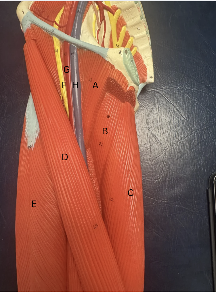
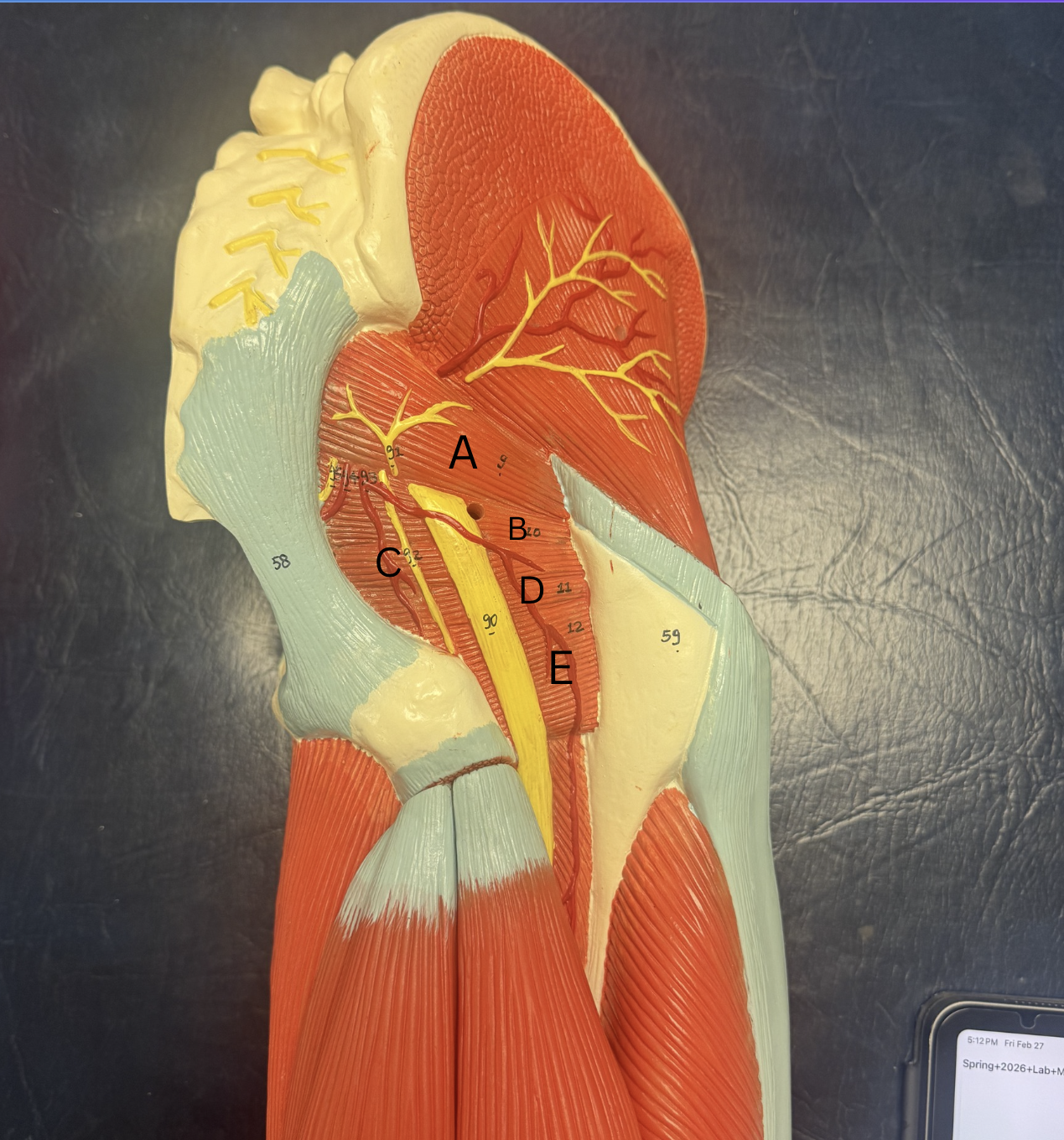
Label the proximal anterior muscles of the thigh
A. Pectineus
B. Adductor Magnus is under Adductor Longus
C. Vastus Lateralis
D. Sartorius
E. Vastus Lateralis
F. Femoral Nerve
G. Femoral Artery
H. Femoral Vein

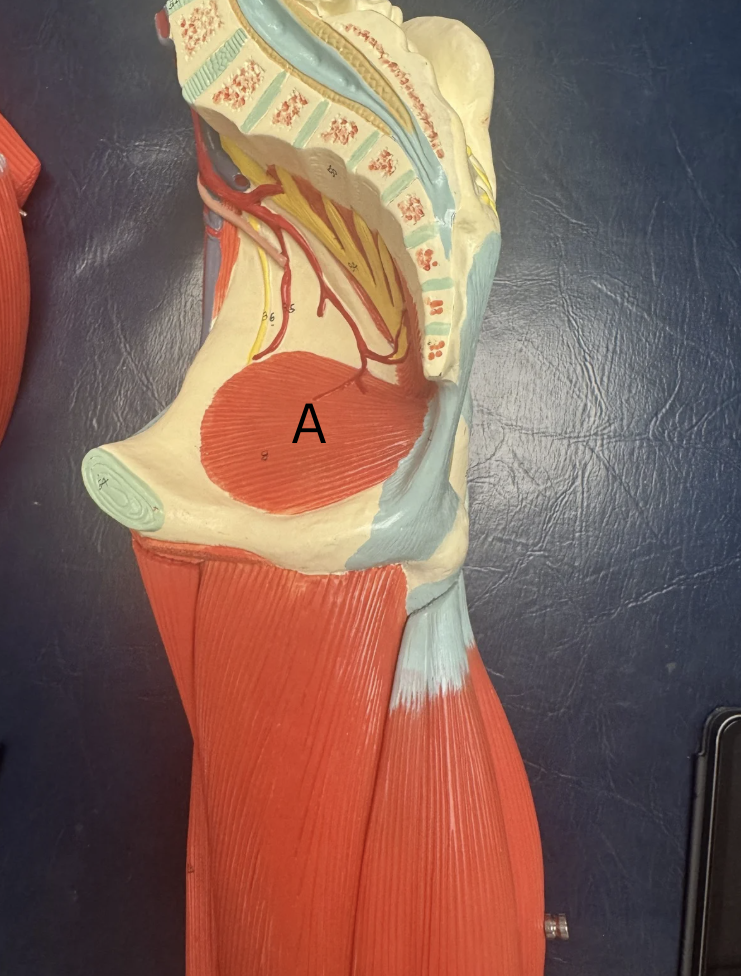
Label these muscles
A. Iliacus
B. Psoas Major
entire muscle group is iliopsoas
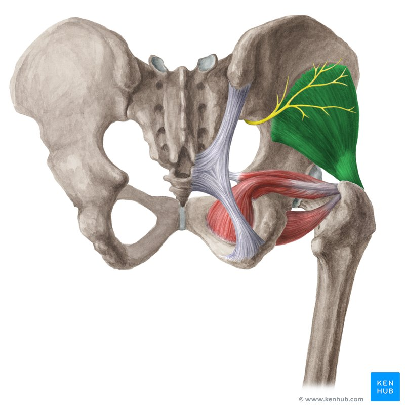
Label this muscle
A. Obturator Internus
Label these muscles
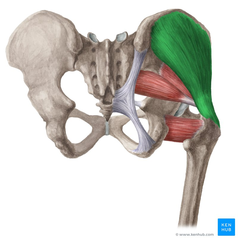
A. Piriformis
B. Gemellus Superior
C. Obturator Internus
D. Gemellus Inferior
E. Quadratus Femoris
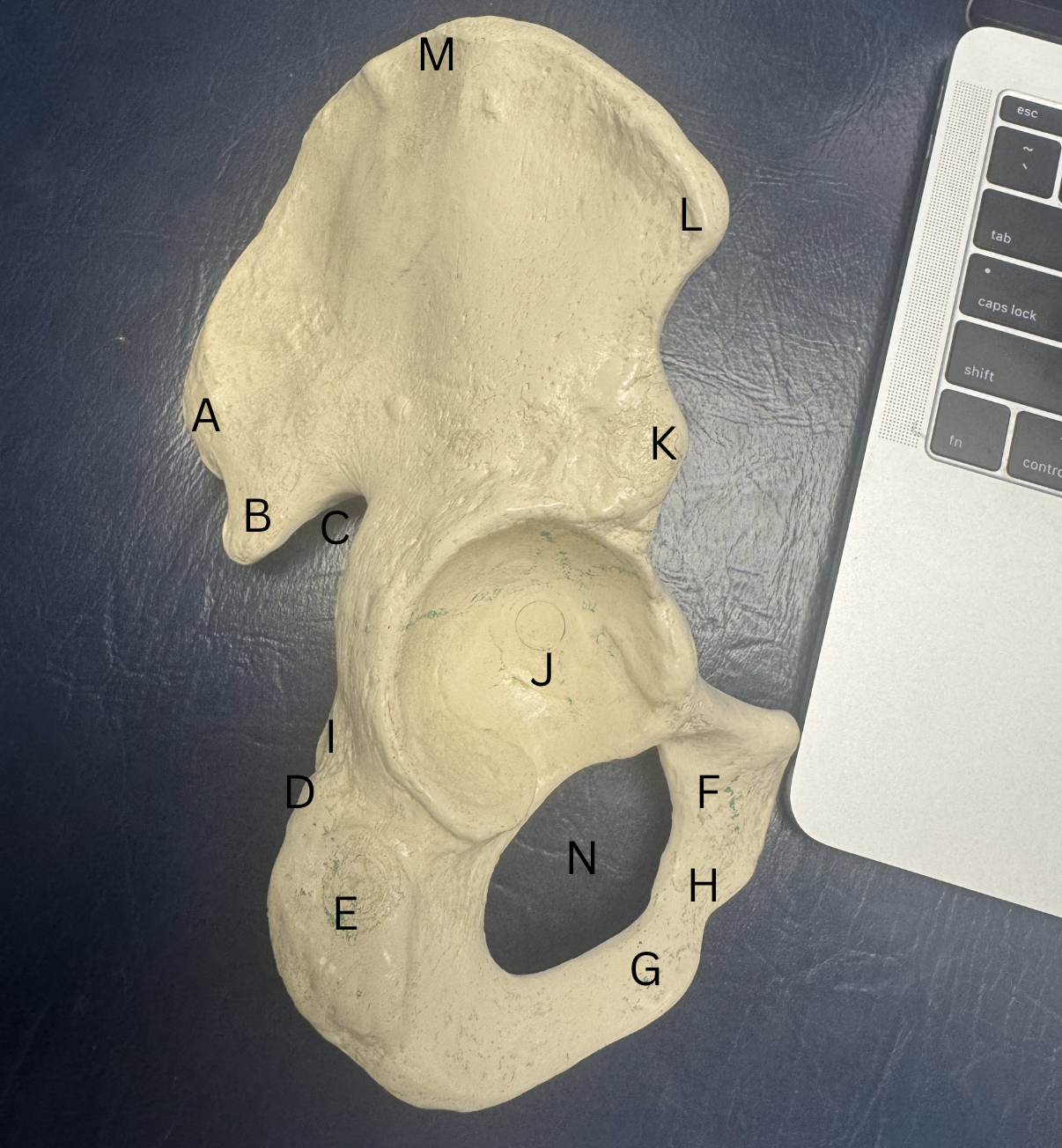
Label these structures from the Innominate bone
A. Posterior Superior Iliac Spine
B. Posterior Inferior Iliac Spine
C. Greater Sciatic Notch
D. Lesser Sciatic Notch
E. Ischial Tuberosity
F. Superior Pubic Ramus
G. Inferior Pubic Ramus
H. Pubic Body
I. Ischial Spine
J. Acetabulum
K. Anterior Inferior Iliac Spine
L. Anterior Superior Iliac Spine
M. Iliac Spine
N. Obturator Foramen
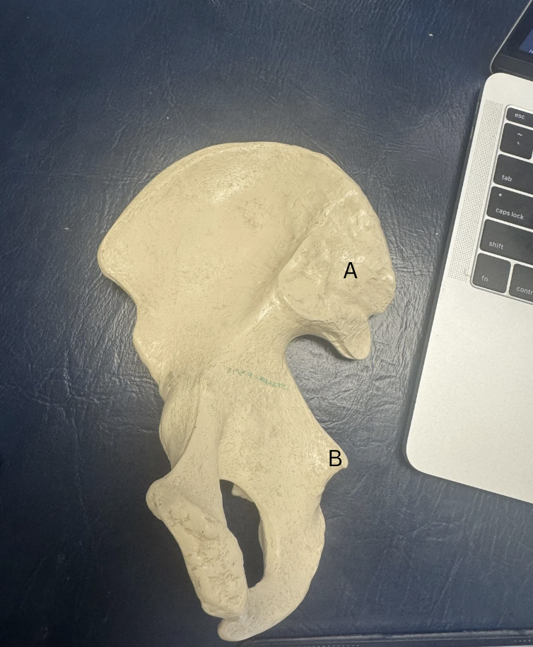
Label these two structures
A. Articular Surface (attaches Sacrum to Pelvis)
B. Ischia; Spine
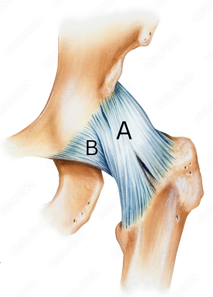
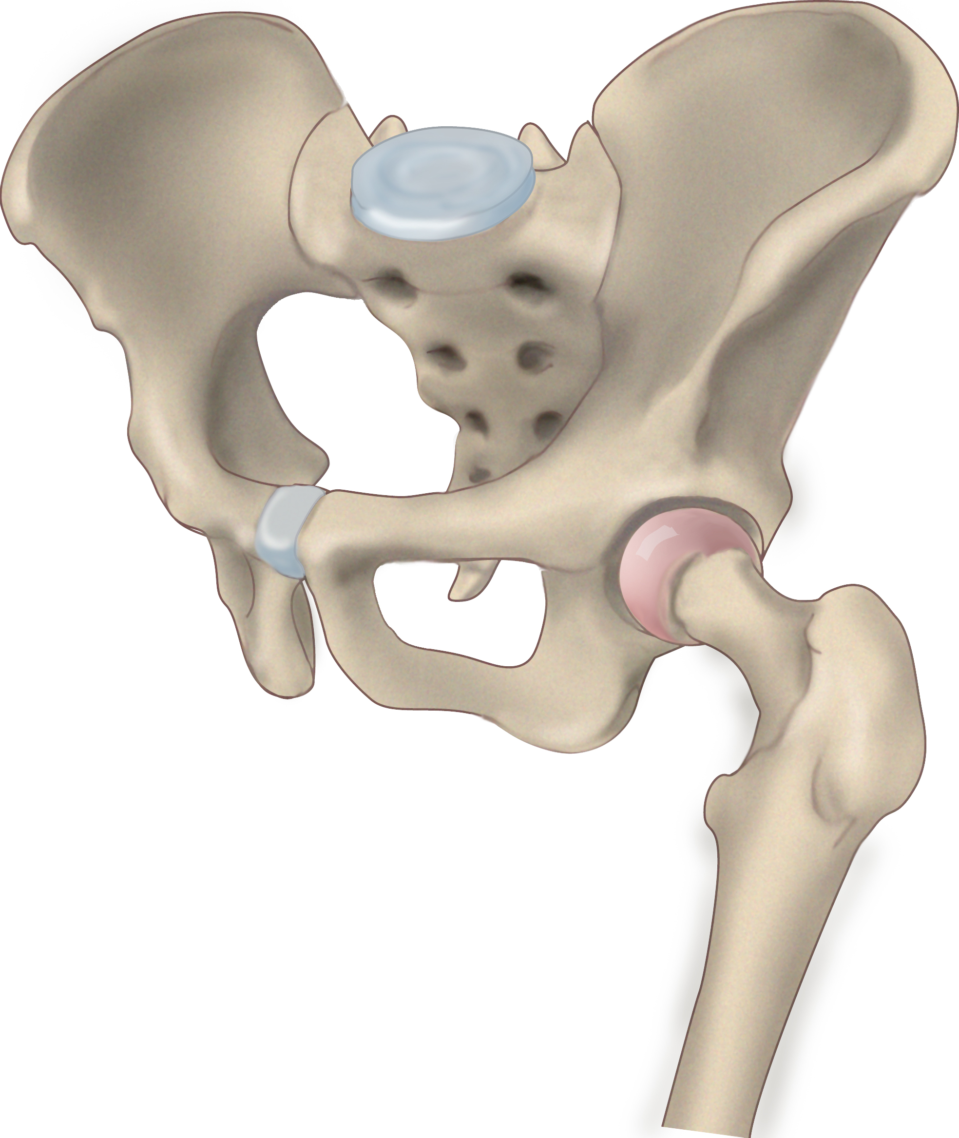
Label these two hip ligaments
A. Iliofemoral Ligament
B. Pubofemoral Ligament
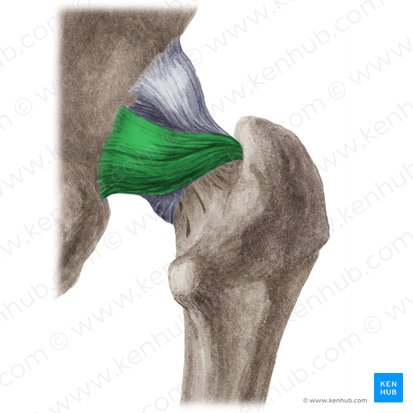
Label this ligament
Ischiofemoral Ligament
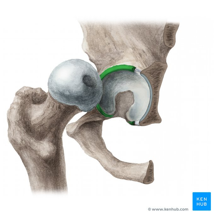
What is this specific tissue called that deepens the socket of the Acetabulofemoral Joint
Acetabular Labrum
Label these joints
Acetabulofemoral joint
Sacrococcygeal joint
Sacroiliac joint
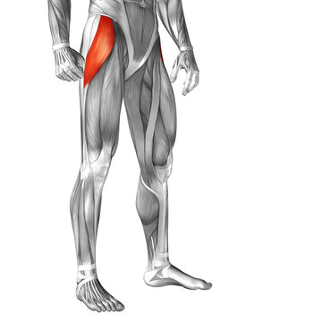
Label this muscle, what tendon does this muscle insert into
Tensor Fasciae Latae; Iliotibial tract
Label this muscle
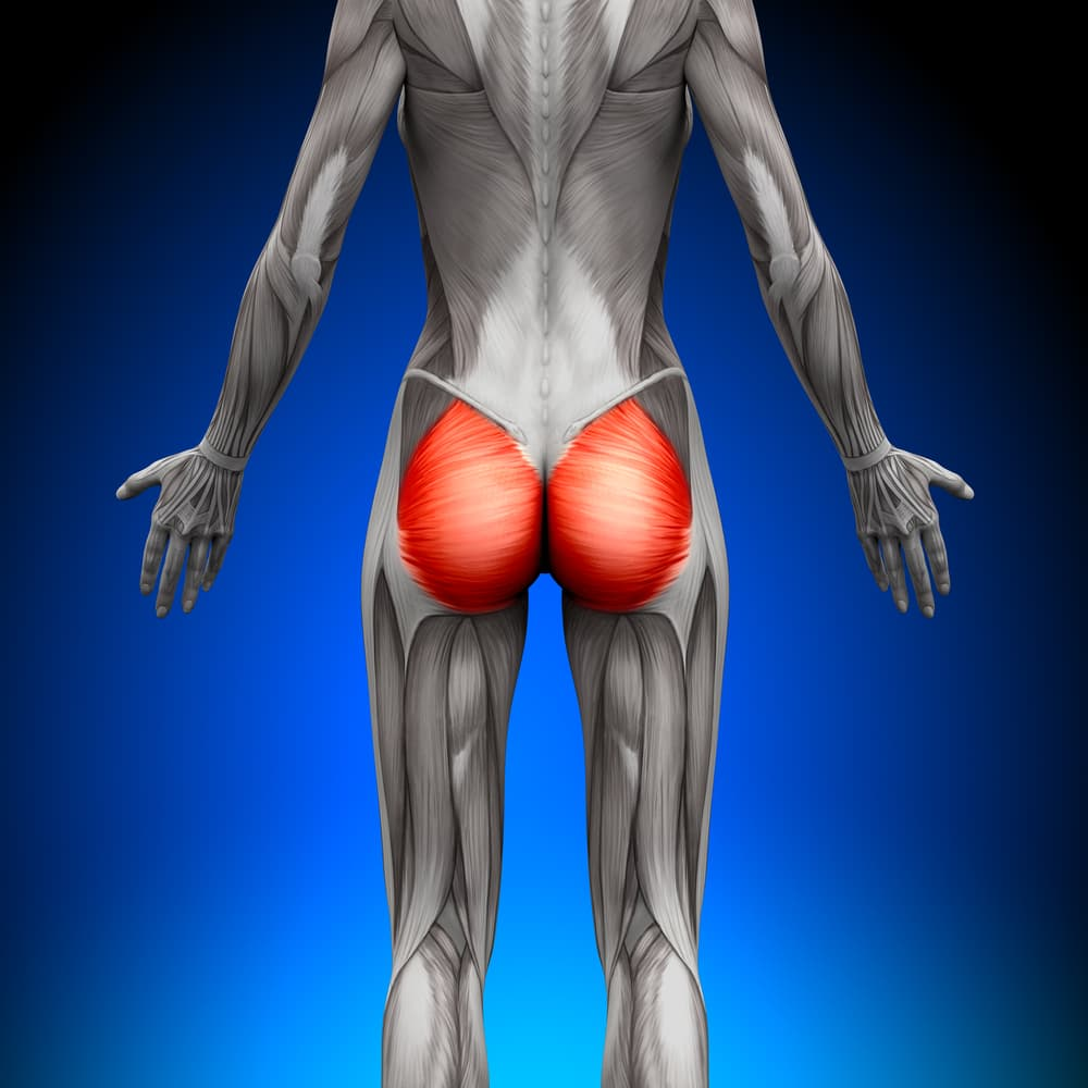
Gluteus Maximus
Label this muscle
Gluteus Medius
Label this muscle
Gluteus Minimus
What does the Iliofemoral ligament help prevent/limit?
prevents hyperextension
What does the Pubofemoral ligament help prevent/limit?
limits hyperabduction