Histology: dentin and pulp
1/31
There's no tags or description
Looks like no tags are added yet.
Name | Mastery | Learn | Test | Matching | Spaced |
|---|
No study sessions yet.
32 Terms
General aspects
Dentin and pulp come from dental papilla of tooth bud. Dentin is mineralised component of complex while pulp is the unmineralised component.
Physical properties
Pale yellowish color. As enamel is translucent, dentin will be responsible for the color of the crown.
Harder than bone and cementum, but softer and less fragile than enamel.
Very permeable.
Chemical compositionIt is a hard connective tissue:
• 70% Mineral component: Hydroxyapatite crystallites
• 20% Organic component: Type I Collagen, Proteins…
• 10% Water
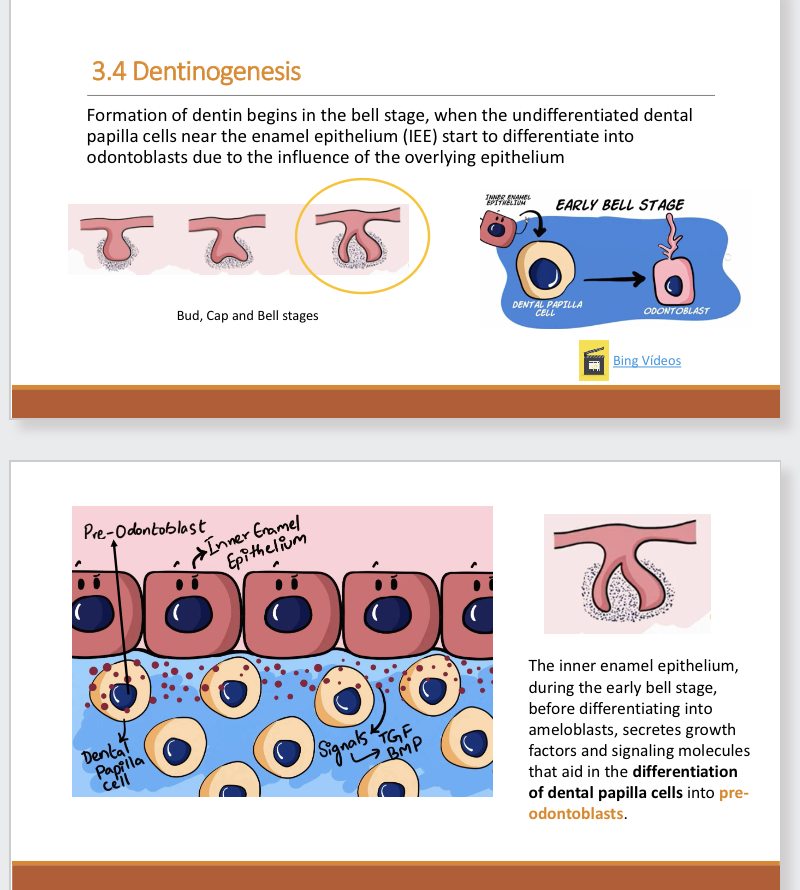
Dentinogenesis
Formation of dentin begins in bell stage, where undifferentiated dental papilla cells near enamel epithelium start to differentiate into odontoblasts due to influence of overlying epithelium.
Dentinogenesis 2
Dentinogenesis 3
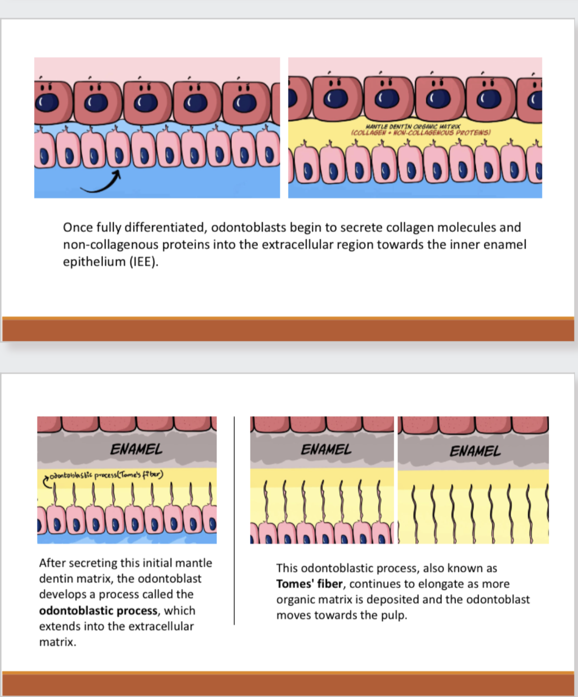
Tomes process and tomes fiber
•Tomes' process is the distal extension of an ameloblast, responsible for the secretion of rod and interrod enamel.
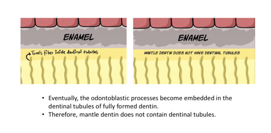
•Tomes' fiber refers to the odontoblastic process of the odontoblast, which is present within the dentinal tubules of fully formed dentin.
Dentinogenesis 3
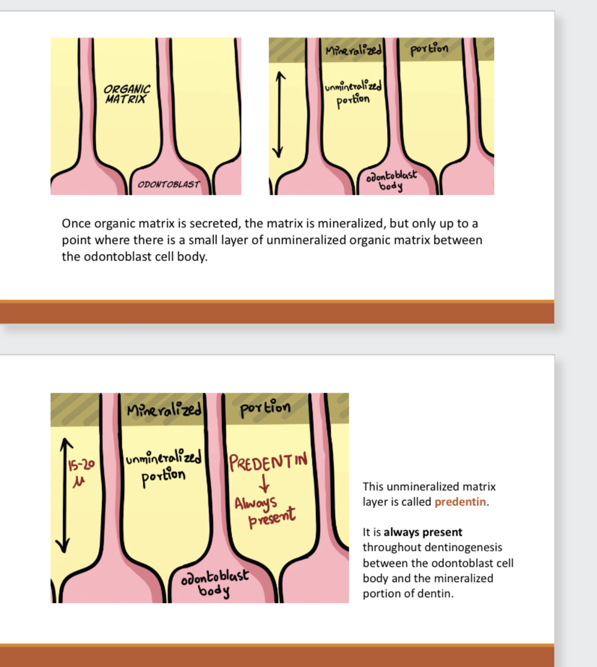
Once organic matrix is secreted, the matrix is mineralized, but only up to a
point where there is a small layer of unmineralized organic matrix between
the odontoblast cell body.
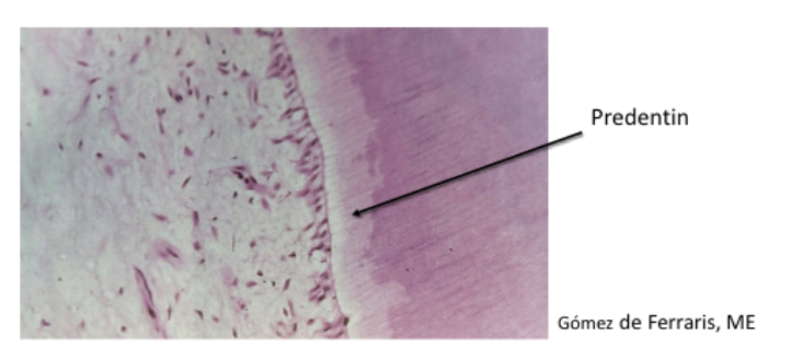
This unmineralized matrix layer is called predentin. It is always present throughout dentinogenesis between the odontoblast cell body and the mineralized portion of dentin.
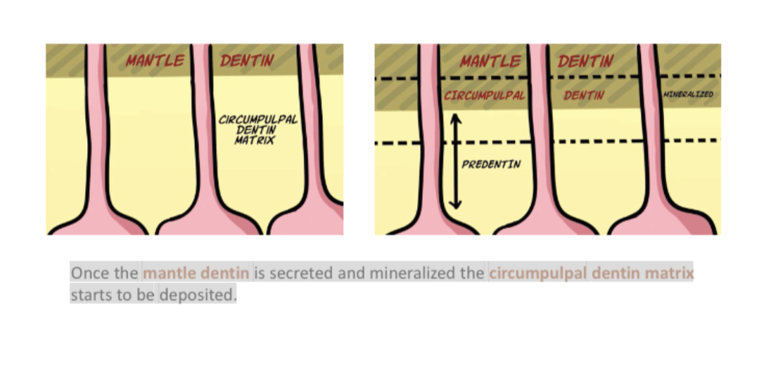
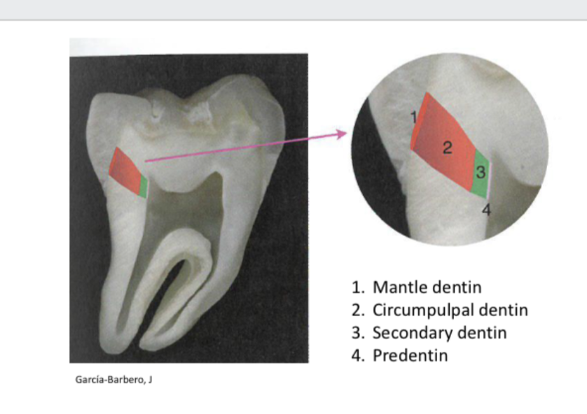
Once the mantle dentin is secreted and mineralized the circumpulpal dentin matrix
starts to be deposited.
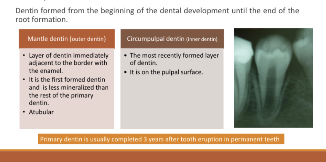
Primary dentin
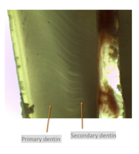
Secondary dentin
It is formed once the root formation is completed.
• Similar structure to primary dentin although less
regular and mineralized.
• A slight change in the direction of the dentinal
tubules occurs when secondary dentin
formation begins.
• A rest line can be observed between primary
and secondary dentin.
Secondary dentin
Secondary dentin forms on all internal aspects of the pulp cavity, but in the pulp
chamber it tends to be thicker on the roof and floor than on the side walls.
Predentin
• It is the unmineralized zone of dentin immediately next to the pulp.
• 30 μm thick
• Odontoblasts initially form predentin, which undergoes mineralization and becomes
dentin.
Dentin tubules
• Small canals that extend through the entire width of
dentin, from the pulp to the DEJ.
• Their course is a slight S-curve.
• The end of the tubules are perpendicular to the DEJ.
And how are they formed?
As each odontoblast deposits dentin matrix and retreats toward the central pulp, it extends
an elongated process. Mineralization of dentin matrix around the odontoblast process
creates a tubule within the dentin
• Filled with dentinal fluid
• Each tubule contains the cytoplasmic cell process of an odontoblast.
• The odontoblastic cell body lies at the border between dentin and dental pulp
• There are also nerve fibers that come from the pulp. 40% of tubules around
pulp horns are innervated.
◦ The number of tubules per unit area increases toward the pulp (45,000 tubules
per mm2 against 20,000).
◦ The lumen of the tubules is also bigger close to the pulp (2.5-4μ) than to the DEJ
(1μ).
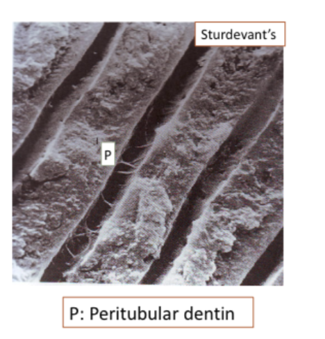
Pertubular dentin
• Formed on the walls of dentinal tubules
diminishing their lumen progressively.
• Very mineralized. Poor in collagen fibers.
• Its formation is influenced by
environmental and pathologic factors.
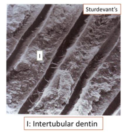
Interbular dentin
Dentin between peritubular dentin
Represents most of the tooth
Less calcified.
Composed of apatite crystallites
embedded in collagen matrix between
tubules.
Dental pulp
The dental pulp is the soft connective tissue that
forms the inner core of each tooth.
It is divided into the coronal part and the
radicular part depending on its position.
It is highly vascularized and innervated: At the
root tip, blood vessels and nerves enter the
tooth.
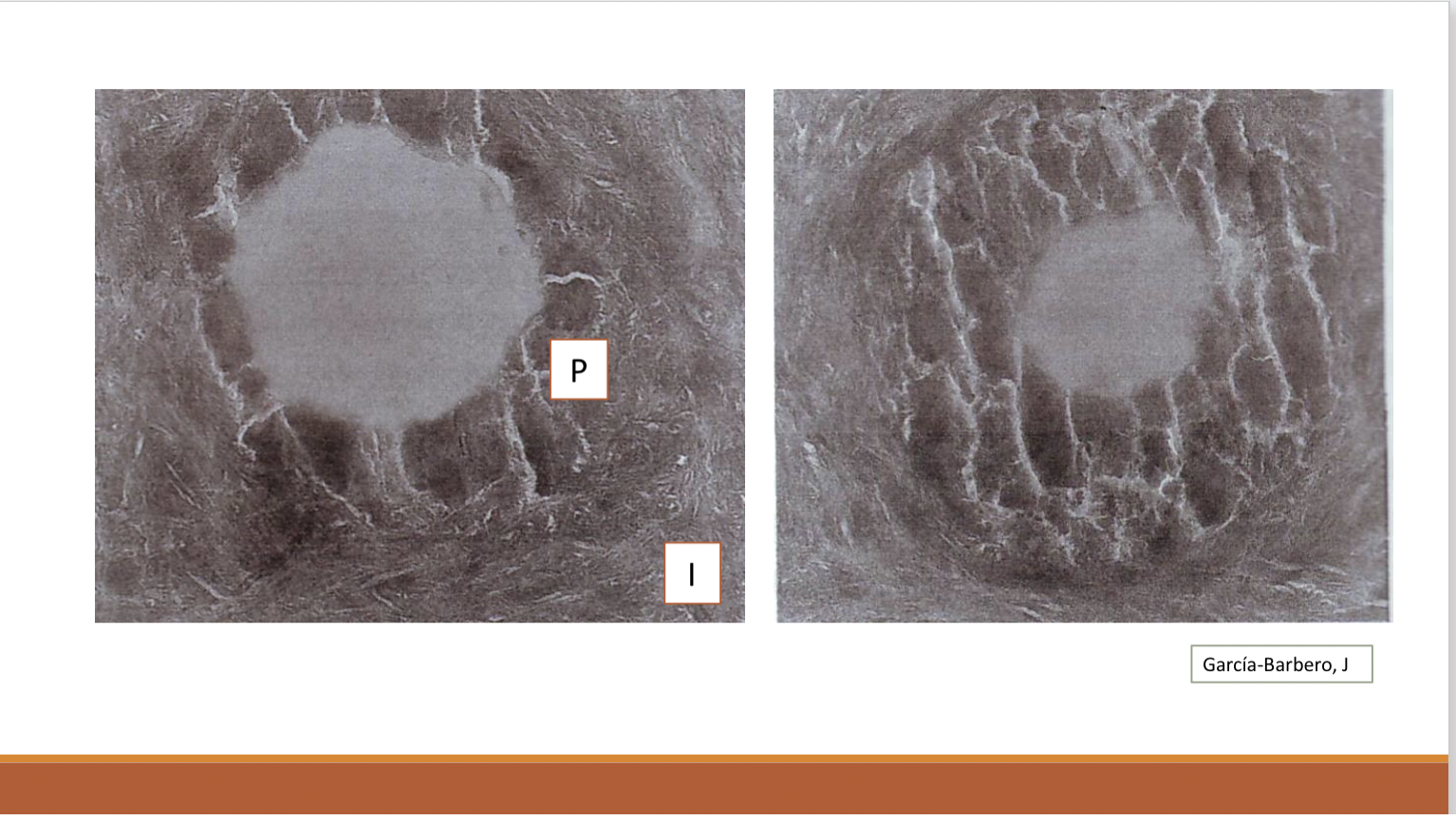
Zones of pulp
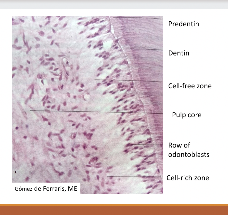
Components of pulp: odontoblast
• They are dentin forming cells.
• Have their cell bodies in the pulp cavity,
but their long cytoplasmic cell
processes extend into the tubules in the
mineralized dentin.
• They can reach the DEJ.
• They are arranged in a single layer.
Fibroblast
• Predominant cell type of pulp.
• Function: to form and maintain the matrix that
consists of collagen, fiber and ground substance
throughout the pulp.
• Cell-rich zone.
Gómez de Ferraris, ME
Undifferentiated mesenchymal cells :
• Under adequate stimulus they may differentiate into
odontoblasts, fibroblasts or macrophages.
• They are present in the cell-rich zone and in the pulp
core.
• In older pulp their number and ability to differentiate
decreases, which implies a reduction in the
regenerative potential of the pulp.
Defense cells:
• They play a major role in local inflammation and immunity.
• They are recruited from the blood stream and remain as transient
inhabitants in the pulp
• Macrophages, Lympocytes…
Collagen Fibers:
• Type I (60%) y Type III.
• Its content increases with age.
Ground substance:
• It is a structureless mass. It makes up the bulk of the pulp.
• It consists of complex proteins, carbohydrate and water.
Pulp vascularisation
• The pulp is a microcirculatory system because it lacks true arteries and veins.
• The arterioles on entering the pulp show a reduction in the thickness of vessel wall musculature and therefore the lumen size increases.
• Pulpal blood flow is more rapid than in most areas of the body.
• Pulp pressure is the highest of body tissues.
Process of pulp vascularisation
1. Arterioles enter into the pulp through the apical
foramen and at times through accessory
foramens, and ascend through the radicular pulp
of the root canal.
2. Once they reach the pulp chamber in the crown
they branch out peripherally to form a dense
capillary network immediately under - and
sometimes extending up into - the odontoblast
layer.
3. Small venules drain the capillary bed and
eventually leave as veins via the apical foramen.
Innervations of pulp
• Nerve fibers enter the tooth through the apical foramen
together with blood vessels.
• After entering the foramen, they branch peripherally and
coronally.
• The majority of the nerve bundles reach the coronal dentin
where they fan out to form the nerve plexus of Raschkow.
• There, they terminate as free nerve endings that synapse
into the odontoblast cell layer and the odontoblastic cell
processes (approximately 100–200 μm deep in the dentinal
tubules).
Cementum
• Cementum is a hard connective tissue that covers the root of the
tooth to furnish a medium into which the main fibers of the
periodontal ligament are inserted.
• It is neither vascularized nor innervated.
• Its thickness changes being:
o Maximum in the apexes and in the interradicular area of multirooted
teeth (50-200 μm)
o Minimum in the cervical site (10-50μm).
Chemical composition of cementum
• 65% inorganic content: calcium phosphate in the form of hydroxyapatite
crystals.
• 23% organic content (collagen fiber type I)
• 12% water
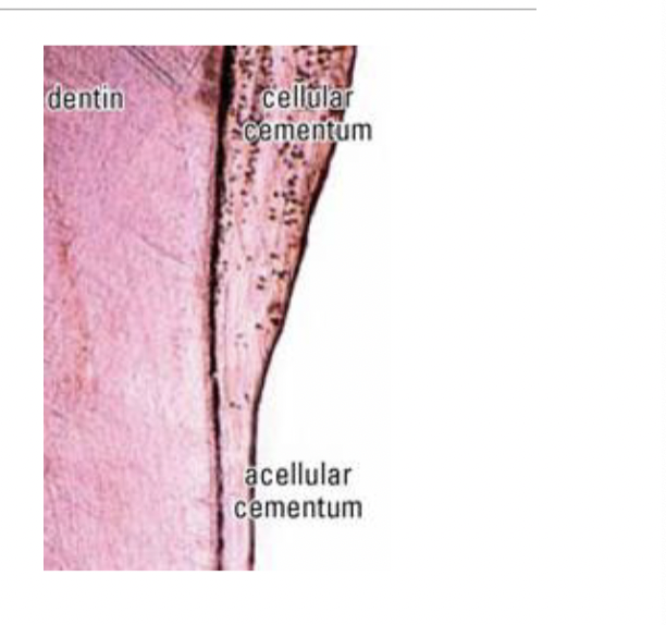
Acellular cementum
Covers root adjacent to cementum
Cellular cementum:
• it is in the apical third of the root, overlying
acellular cementum.
• Its cells are cementoblasts and cementocytes