human anatomy & physio - module 4
1/37
Earn XP
Description and Tags
respiratory system
Name | Mastery | Learn | Test | Matching | Spaced |
|---|
No study sessions yet.
38 Terms
structural classifications of the respiratory system
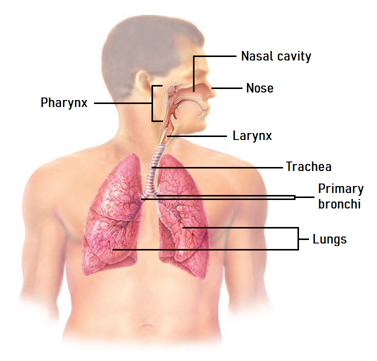
Structurally, the respiratory system can be categorised into the upper respiratory system (also called the upper respiratory tract) and the lower respiratory system (also called the lower respiratory tract)
The upper respiratory system includes the:
Nose
Nasal cavity
Pharynx
Larynx
The lower respiratory system includes the:
Trachea
Left and right primary bronchi
Lungs
lower respiratory system
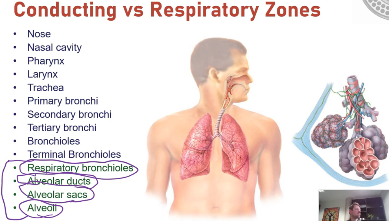
functional classification of respiratory system
Respiratory zone
The airways of the respiratory zone are the areas where gases can move between the air and bloodstream (a process called gas exchange). They include the respiratory bronchioles, alveolar ducts and alveoli
Conducting zone
The tubes that make up the conducting zone provide a passage into and out of the respiratory zone. They function to filter, warm and moisten air however gas exchange cannot occur here. Included in the conducting zone are the nose, nasal cavity, pharynx, trachea, bronchi, bronchioles and terminal bronchioles
nose & nasal cavity
Provide an airway for respiration. They moisten, warm and filter air. They also play a role in speech, smell and reflexes such as sneezing
The floor of the nasal cavity is formed by the palate which separates the nasal cavity from the oral cavity. The nasal cavity is also divided into a right and a left protein by the midline nasal septum
There are two types of mucous membranes that line the nasal cavity. The olfactory mucosa contains receptors for the sense of smell. The respiratory mucosa contains pseudostratified ciliated columnar epithelium with goblet cells
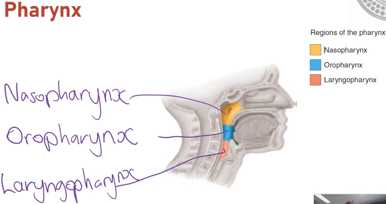
pharynx
The pharynx has 3 parts:
The nasopharynx sits posterior to the nasal cavity
The oropharynx sits posterior to the oral cavity
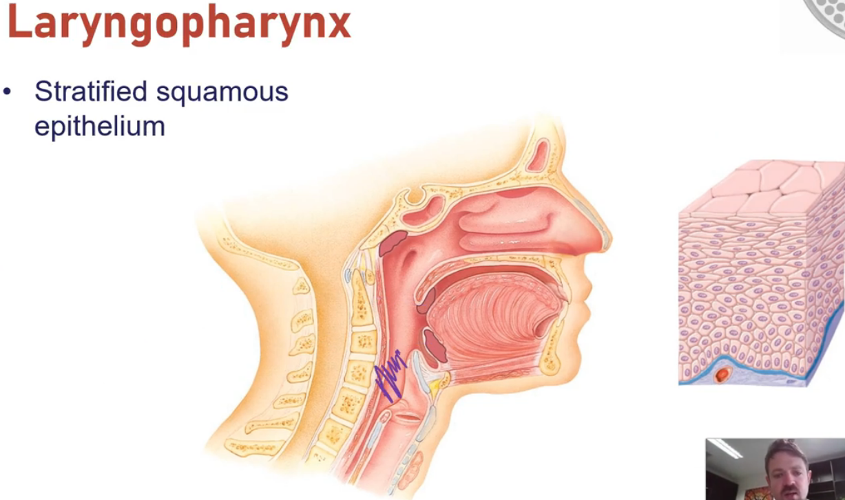
The laryngopharynx sits inferior to the oropharynx, posterior to the larynx
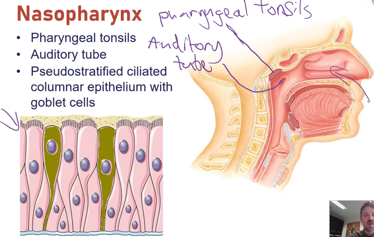
nasopharynx
Serves only as an airway. It is lined with pseudostratified ciliated columnar epithelium with goblet cells and it contains the pharyngeal tonsil on its posterior wall
Tonsils are masses of immune tissue that trap and destroy foreign pathogens
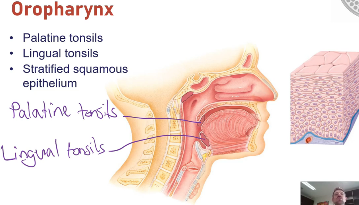
oropharynx
Serves as a passage for both food and air. There are two kinds of tonsils in the oropharynx; the palatine tonsils and the lingual tonsils
The oropharynx is lined with stratified squamous epithelium which provides a high level of physical protection as well as protection against chemical and thermal trauma
laryngopharynx
Common passageway for both food and air. Due to this, it is also lined with stratified squamous epithelium for protective purposes. There are no tonsils in the laryngopharynx
larynx
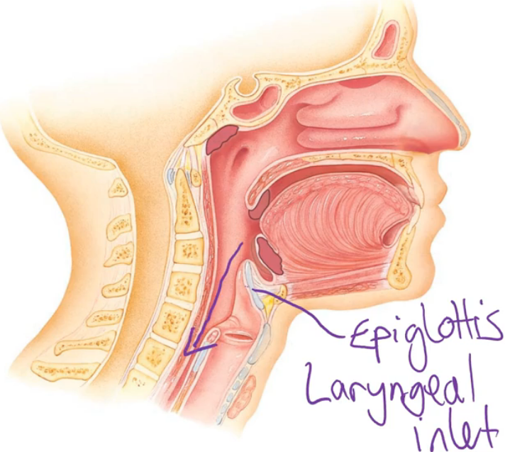
The last part of the upper respiratory system. It contains the epiglottis, which are a flap of cartilage that covers the laryngeal inlet during swallowing. This directs food down the oesophagus and ensures that food/drink does not enter your lower respiratory system, which you swallow
The larynx also contains the voice box. The voice box contains the vocal chords and is responsible for voice production. It also contains a prominence known as the laryngeal prominence
Voice production
Involves the intermittent release of expired air through the constricted opening of the voice box (also called the glottis). Within the glottis are mucous membrane-covered ligaments called vocal chords
The muscles of the larynx can manipulate the length and tension of these vocal chords into a position where they vibrate as air passes between them. This vibration produces a sound wave. The pitch of sound varies depending on the length & tension of the vocal chords, as well as the width of the glottis
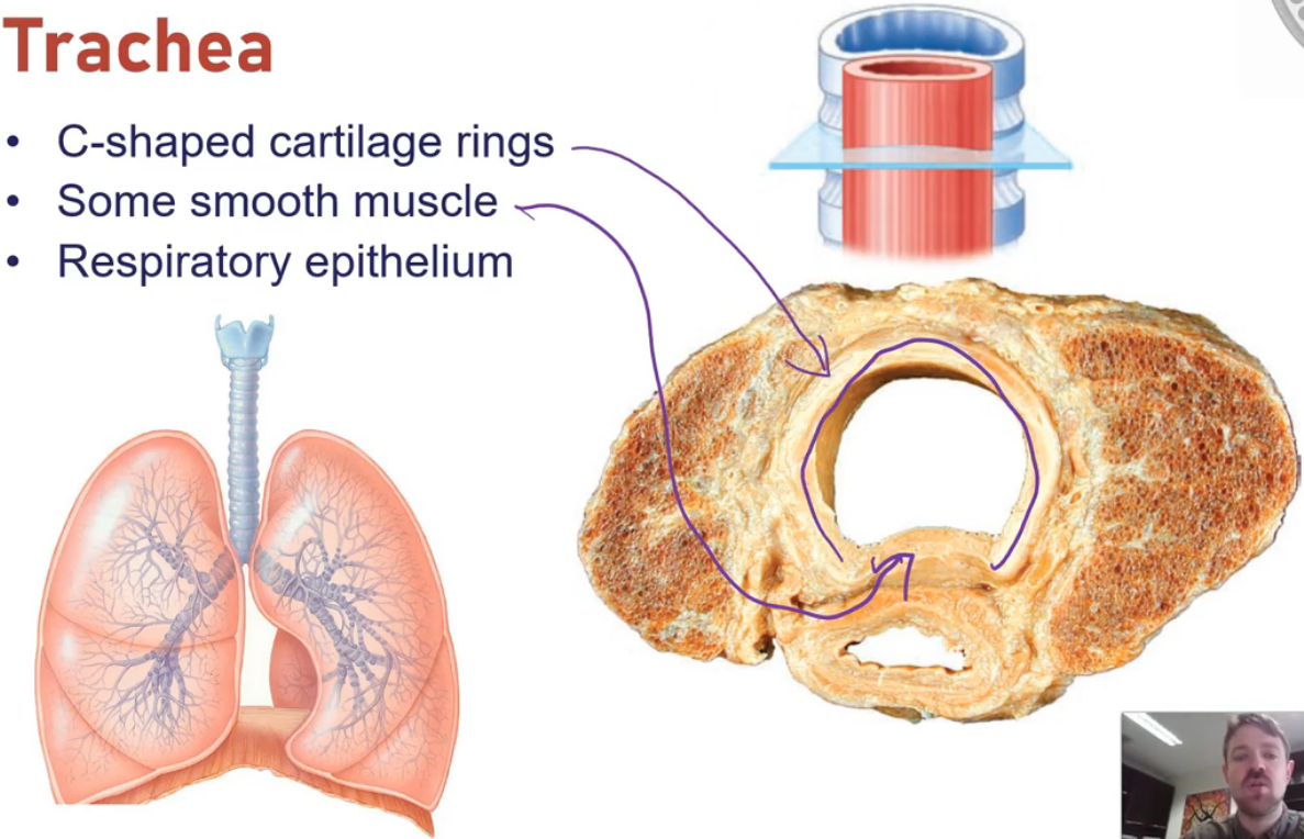
Trachea
A long flexible tube that runs from the larynx in your neck to the bronchi in your chest. It consists of C-shaped cartilages (rings) that prevent it from collapsing
The internal surface is lined with pseudostratified ciliated columnar epithelium with goblet cells which traps foreign particles to ensure they do not enter the deeper parts of your lungs
This is important as foreign matter could interfere with gas exchange if it reached the alveoli
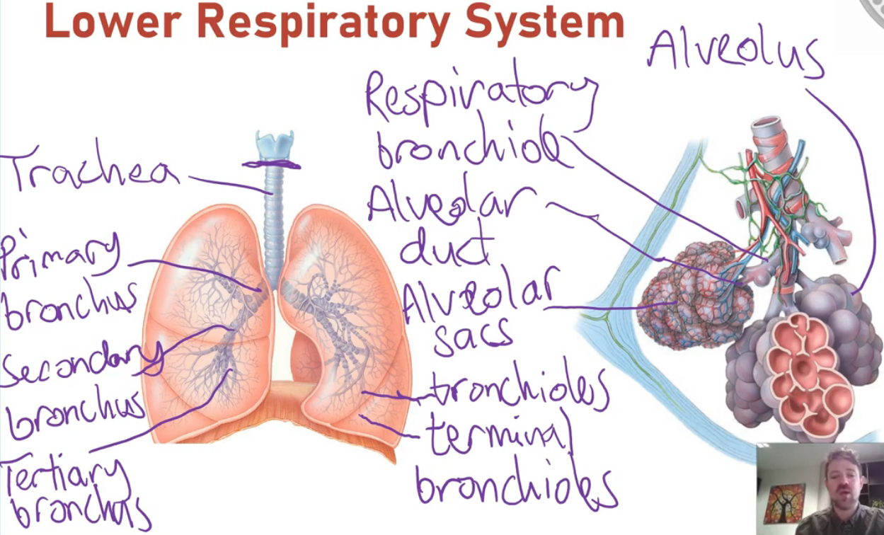
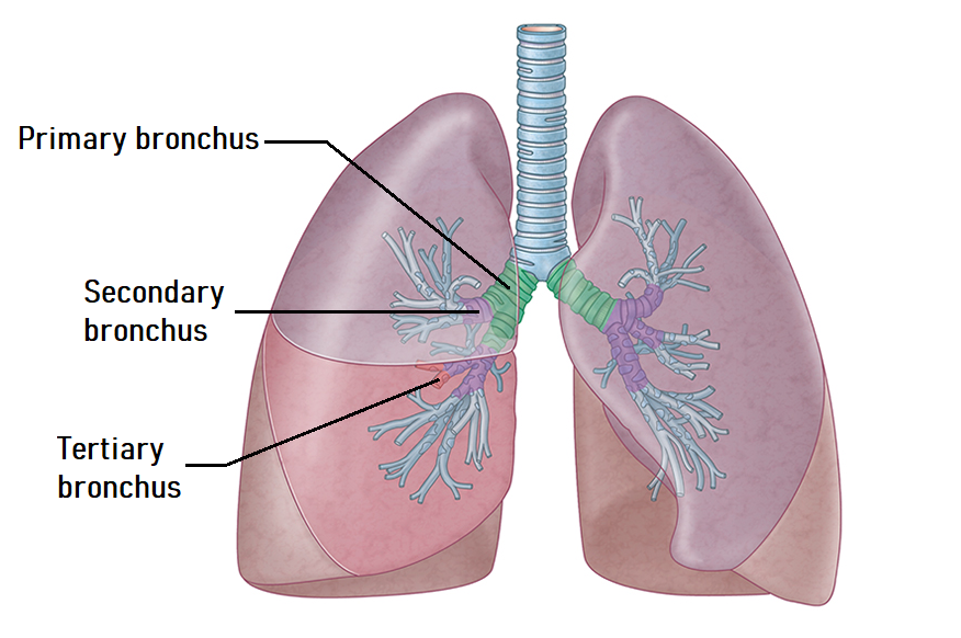
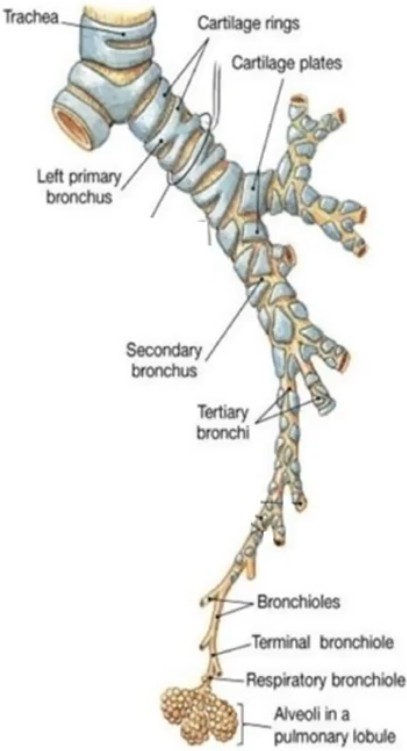
Bronchi
Inferiorly, the trachea divides into the left and right main bronchi (also called the left and right primary bronchi)
The left primary bronchus is the tube that carries air to and from the left lung, and the right primary bronchus is the tube that carries air to and from the right lung
Each primary bronchus then divides into lobar bronchi (also called secondary bronchi), which carry air to and from the respective lobes of each lung
Each secondary bronchus divides into numerous tertiary bronchi (also called segmental bronchi), which carry the air to distinct segments of the lung
These tubes continue to branch into smaller and more numerous tubes, sort of like the branches of a tree. The smaller branches penetrate the deeper areas of the lung to eventually reach the alveoli
Other airways
From the tertiary bronchi, the next branches are the bronchioles and then the terminal bronchioles
At this point, the air moves from the conducting zone to the respiratory zone and passes through the respiratory bronchioles, the alveolar ducts, the alveolar sacs and then finally into the alveoli
The structural make-up of the airway walls changes as they approach the alveoli
An important component of the airway walls is smooth muscle, which, like all muscle, can contract and relax. Similar to the smooth muscle in a blood vessel, a contraction of bronchial smooth muscle will constrict the airway, whereas a relaxation will dilate the airway
The bronchioles are innervated by the ANS. Activation of the parasympathetic NS causes smooth muscle contraction and therefore bronchial constriction (narrowing of the airway, which restricts airflow) while activation of the sympathetic NS causes relaxation of the smooth muscle and therefore bronchial dilation (widening of the airway, which allows more airflow)
alveoli
The alveolus (plural = alveoli) is the actual structure where gasses have the ability to move from the air into the bloodstream or vice versa
They can be found budding off the respiratory bronchioles or in clusters formed by the alveolar ducts and sacs. They are two types of cells that make up the walls of the alveoli; type I alveolar cells and type II alveolar cells
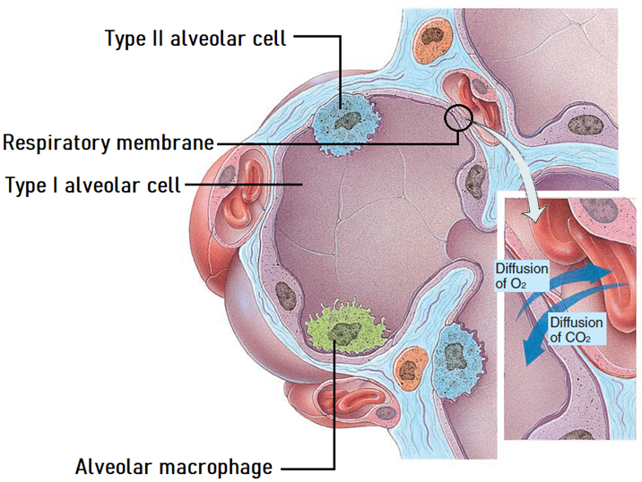
alveoli - type I cells
The more abundant squamous epithelial cells that form most of the alveolar walls. They are very thin cells that form part of what is known as the respiratory membrane (the membrane that facilitates gas exchange)
alveoli - type II cells
Less numerous and are scattered amid the type I cells. They are cuboidal epithelial cells that secrete fluid containing a substance called surfactant
Surfactant helps the lungs to inflate by reducing surface tension
alveoli - macrophages
The alveoli also contain macrophages. However, these cells are not part of the wall of the alveoli, they reside inside them
They are cells of the immune system, and they ingest and destroy foreign material that manages to make its way down to the alveolus
alveoli - respiratory membrane
There is a very close relationship between alveoli and pulmonary capillaries. This reduces the distance that gases need to diffuse to move between the air and the bloodstream
The respiratory membrane is the structure that these gases need to cross to move between the blood in the capillary and the air in the alveolus
It consists of:
The simple squamous epithelium is created by the type I alveolar cells
The simple squamous epithelium of the capillaries, called the endothelium
The fused basement membranes of these two epithelia
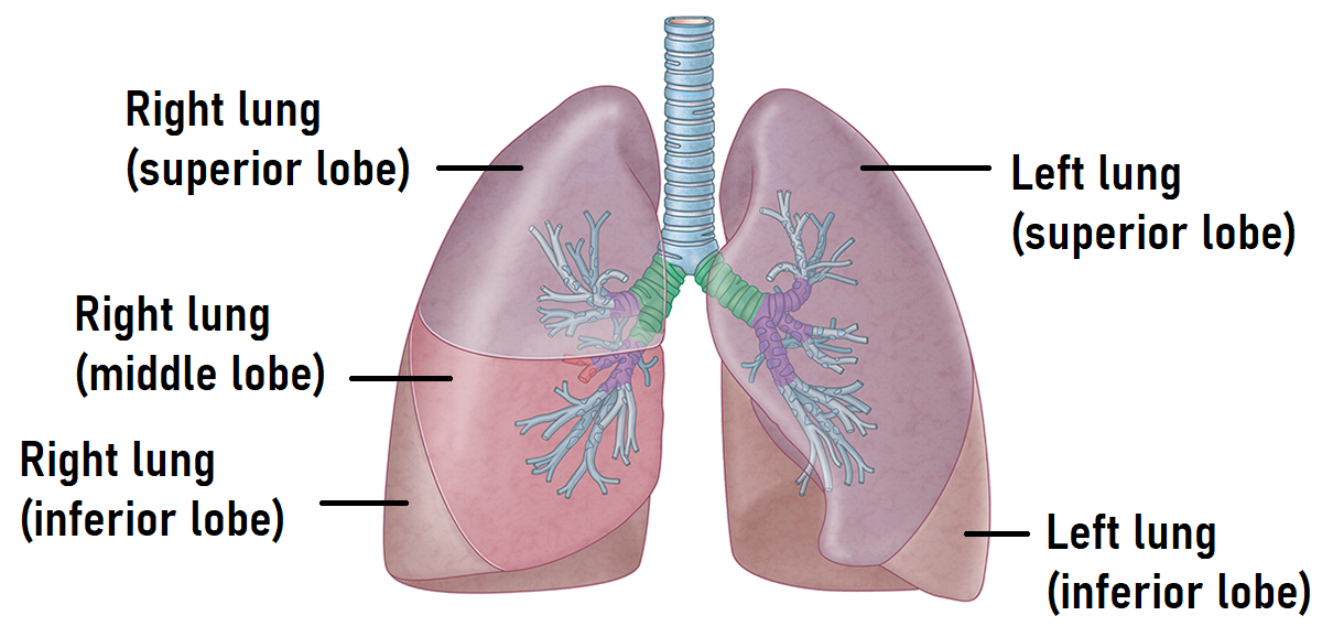
lungs
Bilateral, paired organs that reside within the thoracic cavity. They are soft, spongy, elastic organs that are divided into lobes
The right lung is divided into 3 lobes: a superior lobe, a middle lobe and an inferior lobe. The left lung is divided into 2 lobes: a superior lobe and an inferior lobe
Each lobe contains numerous air-filled tubes and alveoli, containing highly vascular connective tissue that occupies the spaces between the tubes and alveoli
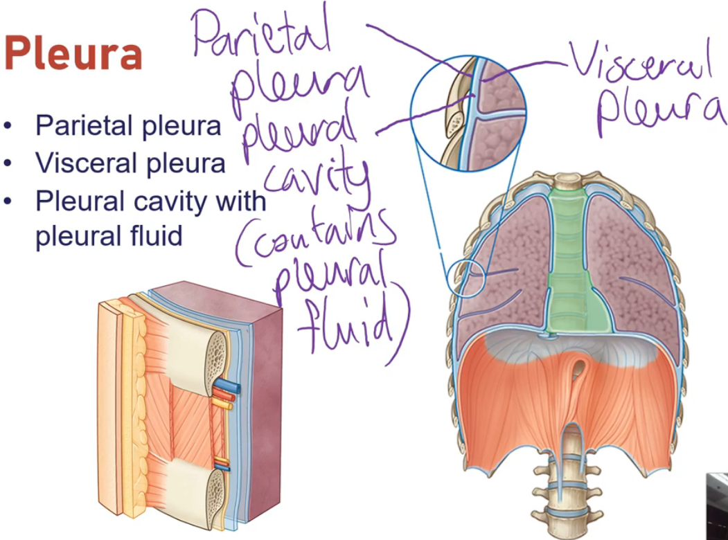
pleural membrane
The pleural membranes (also called the pleura) are thin, double-layered serous membranes that cover the lungs
The outer layer of the pleura is called the parietal pleura, and the inner layer is called the visceral pleura. The inner visceral pleura is closely adhered to the surface of the lung tissue, while the outer parietal pleura is attached to the structures that make up the wall of the thorax
There is a gap between the parietal pleura and the visceral pleura called the pleural cavity (also called the intrapleural space). A thin film of serous fluid called the pleural fluid lies within this cavity
The pleura help to reduce friction when you breathe. The pleural cavity also maintains a slightly lower pressure than the pressure inside the lung, which helps to keep the lung inflated
blood vessels of the lungs - pulmonary vessels
One of the primary roles of the pulmonary circulation is for the pulmonary arteries to carry oxygen-poor blood to the lungs for re-oxygenation (and for the removal of carbon dioxide)
The pulmonary veins then carry this newly oxygenated blood back to the heart. The heart then pumps this oxygen-rich blood into the systemic arteries, which carry blood to the tissues to supply them with oxygen (and other vital nutrients)
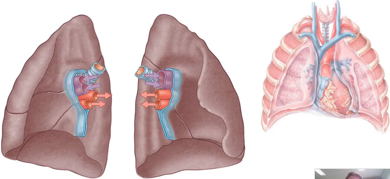
blood vessels of the lungs - bronchial vessels
All tissues of the body need to be supplied with oxygen and other nutrients to survive and function. The bronchial arteries are vessels of the systemic circulation that achieve this for the lung tissue
The bronchial veins of the systemic circulation then carry most of the resulting deoxygenated blood black to the heart (which then sends it into the pulmonary circulation for re-oxygenation)
Name a cell type within the alveoli that secretes a fluid containing surfactant
Type II alveolar cell
Name a cell type within the alveoli that is specialised for gas exchange
Type I alveolar cell
Name a cell type within the alveoli that ingests and destroys foreign material
Macrophage
Name the blood vessels that carry oxygenated blood to the left atrium of the heart
Pulmonary veins
Name the blood vessels of the system circulation that carry oxygen rich blood to the lung tissue
Bronchial arteries
Adrian Samaratunga, aged 23, goes rock climbing on the weekend and accidentally falls without a harness. He only falls 2 metres but lands on a stick which punctures his chest wall. Air begins to enter his pleural cavity. Use your understanding of the structure and function of the pleural cavity to explain what will happen to Adrian's lung when air enters the pleural cavity.
The intrapleural pressure is maintained at a slightly lower pressure than the pressure of the lungs. This helps to keep the lungs inflated. If air enters the pleural cavity then this pressure difference can be lost or reversed which can cause the lung to collapse.
John Simmons, aged 42, chokes on food that enters his airways rather than his oesophagus. During swallowing, which structure covers the air passage of the larynx?
During swallowing the epiglottis normally covers the laryngeal inlet, directing food into the oesophagus and preventing it from entering the larynx and lower respiratory tract.
Explain why the trachea has 16-20 C-shaped cartilages (rings) that prevent it from collapsing while the oesophagus is a collapsible tube?
The trachea needs to remain open at all times to allow air to enter and exit the lungs, whereas the oesophagus only needs to be open when food is passing through it on its way to the stomach.
Janice Wong's doctor tells her she has lost her voice because of a bacterial infection. Which structure of the respiratory system is likely affected?
The larynx
Aggie Chen's pharyngeal tonsils keep getting infected and she has surgery to get them removed. Where are the pharyngeal tonsils located and what is their role?
The pharyngeal tonsils are located in the nasopharynx. They are part of the immune system and play a key role in preventing and fighting infections.
Janice drinks tea that is too hot and it scalds the back of her throat. Her doctor explains she is lucky that her oropharynx contains stratified squamous epithelium. Why does the oropharynx contain stratified squamous epithelium?
It provides physical protection as well as protection against chemical and thermal trauma.
pulmonary ventilation and respiratory pressures
The mechanical process that moves air into and out of the lungs. It depends on volume changes within the thoracic cavity, which lead to pressure changes, causing gases to flow to equalise pressures
It consists of two phases:
Inspiration: Flow of air into the lungs
Expiration: Flow of gases out of lungs
Pulmonary ventilation is dependent on 3 key pressures: atmospheric pressure, intrapulmonary pressure (alveolar pressure) and intrapleural pressure
atmospheric pressure
This is the pressure exerted by the air surrounding the body
At sea level, atmospheric pressure is 760 mmHg
It provides the reference point for expressing other respiratory pressures
intrapulmonary pressure (alveolar pressure)
This is the pressure within the alveoli of the lungs
It rises and falls with the phases of breathing
Eventually, it equalises with atmospheric pressure during rest between breaths
Can be positive or negative relative to atmospheric pressure during different phases of breathing
intrapleural pressure
This is the pressure within the pleural cavity, the space between the visceral and parietal pleura
It fluctuates with the breathing phases, but is always negative relative to atmospheric and intrapulmonary pressures
It is usually 4 mmHg less than intrapulmonary pressure
This negative pressure prevents lung collapse by creating a suction effect that keeps the lungs expanded
respiratory pressures
Respiratory pressures include intrapulmonary and intrapleural pressures
They are described relative to atmospheric pressure (760 mmHg):
0 mmHg = Equal to atm pressure
-4 mmHg = 4 mmHg less than atm pressure (e.g., typical intrapleural pressure)
+10 mmHg = 10 mmHg greater than atm pressure
Respiratory types
Pulmonary ventilation
Exchange of air between the atm and alveoli
External (pulmonary) respiration
Occurs in the alveoli of the lungs
Involves the exchange of gases (O2 and CO2) between alveolar air and blood in pulmonary capillaries
Gases move across the respiratory membrane
Internal (tissue) respiration
Occurs in tissues
Involves the exchange of gases between blood and tissue cells