Ch 4 - Visual Pathways
1/26
There's no tags or description
Looks like no tags are added yet.
Name | Mastery | Learn | Test | Matching | Spaced |
|---|
No study sessions yet.
27 Terms
What is the primary role of the Geniculo-striate pathway?
It connects the retina to the lateral geniculate nucleus (LGN) and the LGN to the primary visual cortex, responsible for most visual information processing.
Where are most cones located in the retina?
the fovea, which is responsible for central vision.
Where are most rods located in the retina?
the peripheral retina, which is responsible for peripheral vision and low-light conditions.
What distinguishes the convergence factors of ganglion cells in the central vs peripheral retina?
Ganglion cells in the peripheral retina receive inputs from many rods, while ganglion cells in the central retina receive inputs from only a few cones.
What organization do On-center/Off-surround ganglion cells exhibit?
They are organized to respond actively to light in the center of their receptive field while being inhibited by light in the surrounding area.
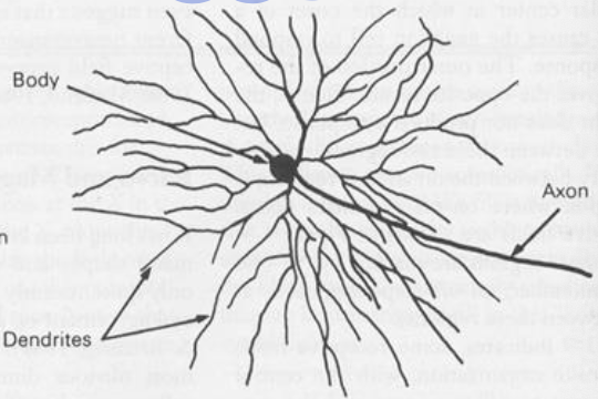
what are the two types of ganglion cells in the visual pathway?
parvo (p) cells
receive most of their inputs from cones
more common at the fovea
“see” a relatively small region
have small bodies and tight dendritic branching
sustained response; form analysis; color vision; spacial analysis
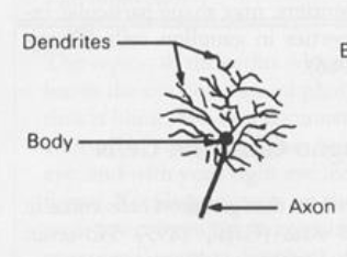
magno (m) cells
receive most of their inputs from rods
found mainly in the periphery
“see” a relatively large region of space
large bodies and diffuse branching
transient response — motion detection; depth perception
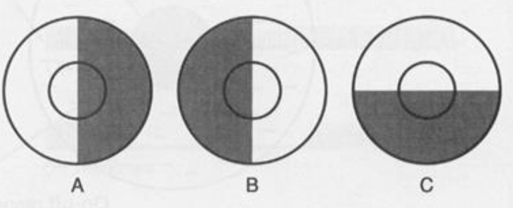
how would magno and parvo cells respond to these different stimuli?
Due to their transient nature, M cells would code changes between Stimuli A, B, and C
Due to their sustained nature, P cells would respond identically to these three stimuli
the same amount of stimulus is taking up its receptive field so it’s response is always the same
will not differentiate between patterns
How does retinotopic mapping influence spatial representation in the visual pathways?
Spatial information is preserved in the form of topographic maps, where relative distances in the visual field correspond to distances between cells in neural tissue.
how does the brain represent spacial info?
Spatial info is preserved by many visual structures in the form of topographic maps (retinotopic mapping)
retina is your first topographic map
Relative distances between points in the world roughly correspond to relative distances between cells in neural tissue
damage to the visual cortex changes depending on the location of the damage in the retinotopic map
Spatial info is distorted by the optics of the eye
Objects on our retinas are flipped horizontally and vertically relative to their spatial position in the world
Objects appearing in the left visual field are represented in the right hemisphere of the brain, and vice versa (not left and right eye! left and right hemifield!!)
what is the Geniculostriate Visual Pathway?
Connects the retina (both M & P) to the lateral geniculate nucleus (LGN) and the LGN to the primary visual cortex
Responsible for most of our visual information processing (e.g., pattern analysis)
how is the lateral geniculate nucleus organized?
retinotopically organized
consists of 6 spatially aligned layers
what is the Lateral Geniculate Nucleus (LGN)?
The LGN is a bilateral structure, you have one in your left brain hemisphere and one in your right
The responses of LGN cells do not differ from the responses of the ganglion cells projecting to the LGN (i.e., a center-surround RF organization) Lateral Geniculate Nucleus
This means that little new signal processing occurs in the LGN
80% of the inputs to the LGN are from the cortex, only 20% are from the retina
Inputs from the cortex (back-projections) far exceed outputs to the cortex
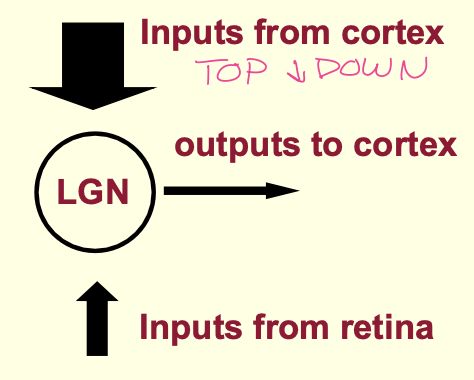
What significant difference exists in inputs to the LGN from cortex versus retina?
80% of inputs to the LGN come from the cortex while only 20% originate from the retina.
what are the “stops” of the geniculostriate pathway?
Parvo/Magno ganglion cells → optic chiasm → LGN → primary visual cortex
what is the primary visual cortex (V1-V2)?
where most low level pattern analysis occurs
last stop in the geniculostriate pathway; first place in the cortex to receive inputs from the LGN (axons of LGN synapse on PVC)
bilateral structure
very large (in comparison to the LGN)
V1 consists of layers → LGN cells project to IVc
if you follow axons of LGN they synapse on IVc
cells in this layer (IVc) have the same center-surround receptive fields found in the LGN and retina
in the surrounding layers, there are cells with more complex RFs (simple, complex, and end-stopped)
What are the three types of cells in the primary visual cortex (V1)?
simple cells
Elongated RFs rather than circular, although still center-surround antagonistic (hot dog)
Respond to stimuli having a particular spatial orientation (i.e., they are orientation “tuned”)
Function as “feature detectors” for oriented edges and bars
Simple cell RFs are constructed by selectively combining the circular RFs of layer IVc cells
complex cells
Tuned for both orientation and direction of motion
basically took the template of simple cells and added specificity for motion
Direction of motion is important, but less so than orientation
RFs are slightly larger (i.e., specific location of stimulus is less important
end-stopped cells
Tuned for orientation, direction of motion, and now the size (e.g., width or length) or shape (e.g., a corner with a particular angle) of a stimulus
RFs are larger than those found in complex cells
The farther away you look from the initial projection area (IVc), the increasingly specialized neurons become to complex patterns
What is cortical magnification in the context of V1?
A (disproportionately) large area of V1 (~10%) tissue is devoted to analyzing information from the small foveal region (.01%) of the retina
Each ganglion cell is allotted roughly the same amount of space (number of cells) in V1
What is columnar organization in the context of V1?
Cells in V1 are grouped into (at least) three types of columns: Location, Orientation, and Ocular Dominance columns
Cells within each column code for the same properties and have the same function
Different locations across the surface code for different locations in space and stimulus properties
However, all the cells at a given location (i.e., column) are coding the same thing
Hubel and Wiesel (Nobel Prize, 1981) → Described the functional organization of the primary visual cortex
Location Columns
Each column is coding for a different region of visual space
Cells within a given column are all coding for the same location in visual space
These cells all have overlapping receptive fields
Ocular Dominance Columns
Within each location column there are two ocular dominance columns
Cells within a given column respond best to stimuli at a given location as seen through a given eye (either right or left)
Orientation Columns
Within each location and OD column there are several orientation columns
Each column codes an orientation; every ~10 deg of angle is coded by a different column
Cells within a column code the same orientation
high level vs low level pathways? (name them)
High Level
“what” - ventral pathway - temporal lobe
devoted to feature analysis
“where”/”how” - dorsal pathway - parietal lobe
devoted to controlling action
Low Level
Geniculo-striate Pathway
evidence for “what” and “how” pathways?
Patient DF suffered damage to her temporal lobe (ventral pathway), leaving her with an inability to recognize objects (visual form agnosia)
damage to “what” pathway
Drawing Task
showed a model → asked to copy
could not reproduce these low level features by looking at them
could do them from memory, but when asked what they were, could not identify them
no damage to “how” / damage to “what”
Orientation Matching
“Try to match a card to the orientation of a slot” → Df could not do the task
Visuomotor “Posting”
“Put the card into the slot” → DF could do the task
where are V3 and V4?
extra-striate cortex
damage to V4 may result in what?
achromatopsia
cortical color blindness
V4 is a bilateral structure, so fdmage to only 1 hemisphere results in colorblindness in half of the visual field
DORSAL PATHWAY
what does damage to MT (middle temporal area) result in?
impaired motion perception (motion blindness) → world consists of snapshots
Newsome and Paré (1988)
Motion Detection Task: Varied the strength of a motion signal; monkeys responded when they perceived motion
Monkeys could detect motion with only 1-2% correlation
Following a lesion to MT, motion detection thresholds increased to 10-20% correlated motion (i.e., 10-20% of the dots had to be moving in the same direction in order for the monkey to detect motion).
Conclusion: Area MT is largely responsible for coding global motion perception
DORSAL PATHWAY
what does damage to right parietal result in?
spatial neglect / difficulty attending to objects appearing in the left visual field (if dam. to right)
they can still see, they just neglect what is in that particular visual field
VENTRAL PATHWAY
what is the inferior temporal (IT) cortex responsible for?
complex feature analysis
Whereas the most complex analyses in V1 involve moving edges or angles, cells in IT appear sensitive to complex objects consisting of distinct parts
Whereas V1 is organized into orientation selective columns, columns in IT contain similar shapes
IT cells are insensitive to an object’s spatial location and size (i.e., location and scale invariant)
IT receptive fields are huge (up to 1/3 of the entire visual field), and cells will respond to patterns falling anywhere within these large RFs
Some IT cells are highly selective for faces
The degree of response depends on how “good” the face is
In humans, face selective cells are found in the fusiform face area in IT cortex
VENTRAL PATHWAY
what does damage to IT cortex result in?
visual form of agnosia → difficulty recognizing objects
Many patients with this condition can copy patterns, but they cannot tell you what the patterns are
prosopagnosia → a specialized form of form agnosia resulting in a difficulty in recognizing faces
what is modularity of function?
different areas of the brain are responsible for different kinds of processing