bone quiz #3-splanchnocranium
1/194
Earn XP
Description and Tags
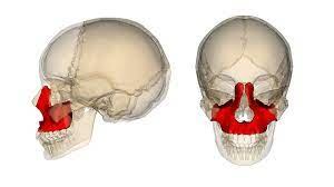
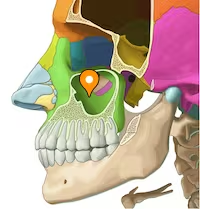
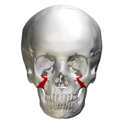
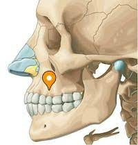
pictures and features of maxilla, palatine, vomer, inferior nasal concha, ethmoid, lacrimal, nasal, zygomatic, mandible
Name | Mastery | Learn | Test | Matching | Spaced | Call with Kai |
|---|
No analytics yet
Send a link to your students to track their progress
195 Terms
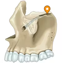
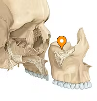
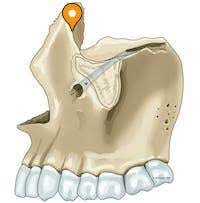
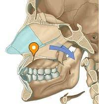
maxilla
maxillary sinus
zygomatic process
forms cheek
alveolar process
horizontal portion that holds tooth roots
alveolus
bony socket for root of tooth
gomphosis
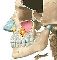
canine fossa
hollow below infraorbital foramen
canine jugum
eminence over maxillary canine root on facial surface
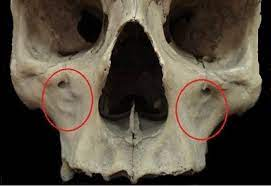
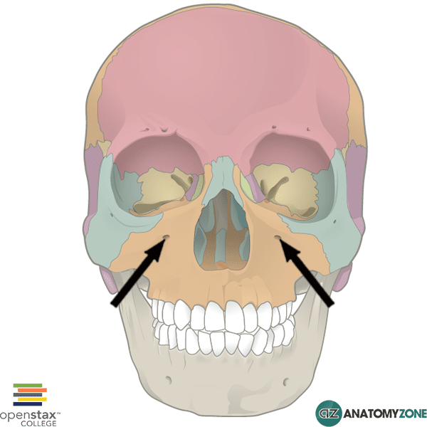
infraorbital foramen
transmits infraorbital nerve
nasal notch
anterior nasal spine
maxillary tuberosity
posterior end of alveolar process
lacrimal notch
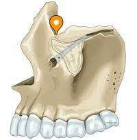
infraorbital groove
posterior half of orbital floor, connects with infraorbital foramen through the infraorbital canal
infraorbital canal
continuous with infraorbital groove and transports the infraorbital nerve and artery
anterior lacrimal crest
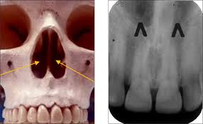
incisive foramen
anterior hard palate at midline
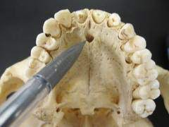
incisive canal
opening via incisive foramen
premaxilla
incisive bone for the incisor teeth

palatine torus
bony growth at the roof of the mouth
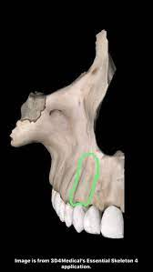
maxillary torus
outgrowths of bone on the maxilla
retained incisive (premaxilla) suture
multiple infraorbital foramina
infraorbital suture
suture above foramen
abscess
swelling and/or pus above teeth
periodontal disease
infection of the tissues that hold teeth in place
infraorbital nerve
infraorbital canal
buccinator muscle
alveolar process
horizontal plate
forms posterior third of hard palate
posterior nasal spine
attach musculus uvula
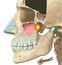
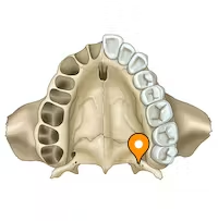
greater palatine foramen
rear corner
greater palatine vessel and nerve
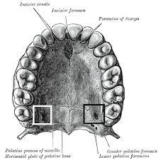
lesser palatine foramen
posterolateral corner
lesser palatine nerves
conchal crest
horizontal about halfway up perpendicular plate
for inferior nasal concha
pyramidal process
serrated groove, posterior border
orbital process
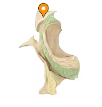
sphenoidal process
musculus uvula
retracts and elevates uvula
posterior nasal spine
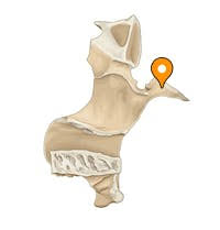
ala
wings
posterior, superior surface
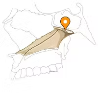
perpendicular plate
thin vertical sheet below wings

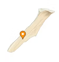
posterior border
divides posterior nasal apeture
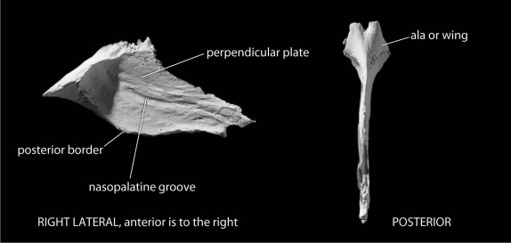
nasopalatine grooves
from nasopalatine nerves and vessels
anterior extremity
anterior point
posterior extremity
posterior point
lamina
thin vertical, extends medially
maxillary process
hook
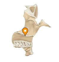
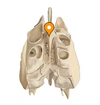
crista galli
perpendicular projection
anchors the falx cerebri
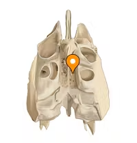
cribiform plate
roof of nasal cavities
houses olfactory nerves
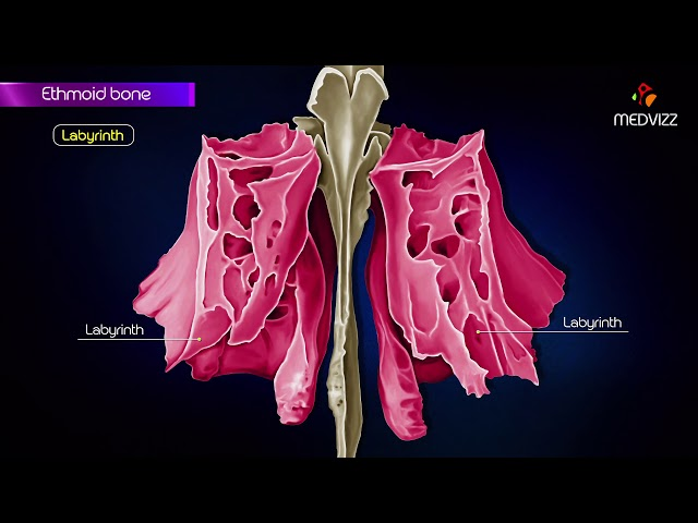
labyrinths
series of thin walled ethmoidal cells
lamina papyra
middle nasal concha
superior nasal concha
olfactory nerves
sense of smell
cribriform plate
falx cerebri
divides brain into left and right
membrane of dura mater
dura mater
membrane for brain
lateral (orbital) surface
lateral smooth surface
posterior lacrimal crest
vertical crest of the medial orbital wall
lacrimal groove
posterior, superior end of lacrimal canal
lacrimal hamulus
hammer
extension of crest
medial (nasal) surface
lateral
nasolacrimal duct
drains tears into nasal cavity
lacrimal groove
nasal foramen
facial surface
nonarticular edge
anterior
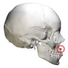
zygomaticofacial foramen
lateral surface
zygomaticofacial nerve and vessels
orbital margin
orbital surface
masseteric origin
rough inferior edge
masseter muscle
zygomaticotemporal foramen
temporal surface
zygomaticotemporal nerve
zygomatico-orbital foramina
inferolateral corner of orbital cavity
zygomaticotemporal and zygomaticofacial nerves
marginal tubercle
frontal process
vertical, separates orbit from temporal fossa
maxillary process
toward midline, inferolateral orbital margin
temporal process
posteriorly, joins zygomatic process to zygomatic arch
os japonicum
masseter muscle
elevate mandible
masseteric origin
hypophyseal fossa
deepest depression of sella
pituitary gland
middle clinoid processes
posterior clinoid processes
2 tubercles in superolateral corners of dorsum sellae
dorsum sellae
square plate-posterior boundary sella turcica
clivus
endocranial hollow-posterior of dorsum sellae
sphenoidal sinus
large paired hollows of body
sphenoidal rostrum
midline projection
articulates with alae of vomer
sphenoidal crest
continuous with sphenoidal rostrum, midline
orbital surface
lateral wall of each orbit
angular spine
inferior most projection of greater wing
foramen rotundum
maxillary nerves
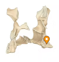
foramen ovale
mandibular nerves and accessory meningeal arteries
foramen spinosum
middle meningeal arteries and mandibular nerves
foramen vesalii
small vein
infratemporal crest
ectocranial of greater wings
form base of temporal fossae
superior orbital fissure
open spaces between inferior surface or lower wings and anterior surface of greater wings
inferior orbital fissure
space between greater wings and orbital surface of maxilla
anterior clinoid process
posterior most projections of lesser wings
tentorium cerebelli
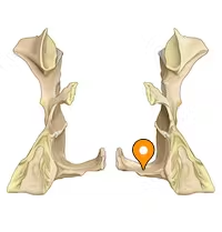
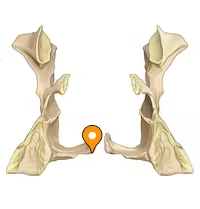
lateral pterygoid plate
thin vertical plate
medial pterygoid plate
parallel to lateral plate, more midline
medial pterygoideus muscles
pterygoid hamulus
hook-like process
pterygoid fossa
rough floored hollows between medial and lateral plates
pterygospinous spur or bridge