Oct 10th: The Occipital Lobes and Networks ✅
1/41
There's no tags or description
Looks like no tags are added yet.
Name | Mastery | Learn | Test | Matching | Spaced |
|---|
No study sessions yet.
42 Terms
Describe retina
Light-sensitive membrane in the back of eye that contains rods and cones
* The light passes through several layers of the cell before it reaches the rods and cones
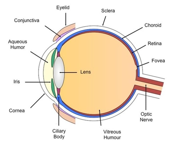
What type of neuron cells contribute to retina?
Ganglion cells, they have axons that leave the retina through the optic disc
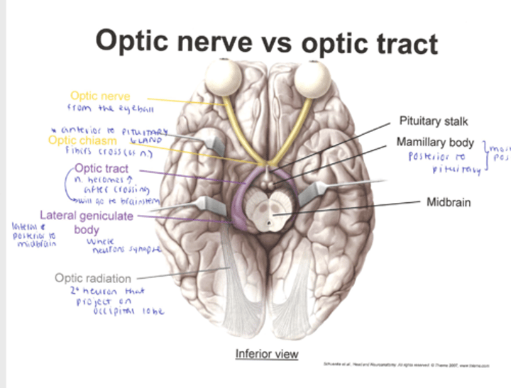
Optic nerve vs optic chiasm
Optic nerve:
* Ganglion cell axon
* Leave eye at the optic disc (which is the blind spot)
It is responsible for transmitting visual information from the retina of the eye to the brain.
Optic chiasm:
* Point of crossover for half of the visual projections
* Inferior (below, behind) to the hypothalamus
*The optic chiasm is a structure located at the base of the brain, anterior to the hypothalamus.
It is the point at which the optic nerves from each eye partially cross over or decussate (also known as crossover).
* This crossing of fibers allows for the integration of visual information from both eyes and the creation of a single, binocular field of vision.
What is a blind spot in the eye
the optic disc
(where the optic nerve exits)
Why is it a blind spot?
There are no cells to detect light on optic disc, that part of retina is blind
Describe the path in which the image processing goes from the eyeball to the brain?
Chronological
1. Eye
2. Optic nerve
3. Lateral geniculate nucleus
4. Visual cortex
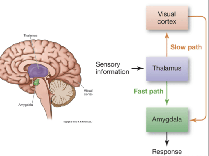
What is the difference between retino-geniculatestriate vs tectopulvinar relay pathways?
Retino-geniculatestriate Pathway
Eye - optic chiasm - lateral geniculate nucleus (LGN) of thalamus:
The primary pathway for conscious visual perception
Tectopulvinar Pathway
Eye - optic chiasm - superior colliculi - lateral posterior and pulvinar nuclei of thalamus:
For subconscious and non-conscious processing
Two lateral geniculate nuclei (LGNs) ¤ One in each hemisphere:
Relay stations that transmit visual information to the visual cortex, with each LGN serving one hemisphere of the brain. They are a PART OF retino-geniculate-striate pathway and contribute conscious perception of visual stimuli.
Each LGN receives visual input from the opposite visual field of the eye. The LGN on the right receives input from the left vice versa.

There are four visual cortex portions on the occipital lobe:
1. Primary (V1) & Secondary (V2) visual cortex
2. Association cortex (V3)
3. ¤ Association cortex (V4)
4. Middle temporal region (MT; V5)
What is the difference between primary (V1) and secondary (V2) visual cortexes?
V1: Orientation and spatial frequencies
and is the first cortical area to receive visual information.
It serves as the primary processing center for visual input. It receives raw visual data from the thalamus, the visual relay station
V2: Binocular vision
often considered the first level of extrastriate cortex, which means it receives input from V1 and processes information further.
It is involved in more complex feature processing, such as the combination of basic visual elements into more intricate patterns and shapes.
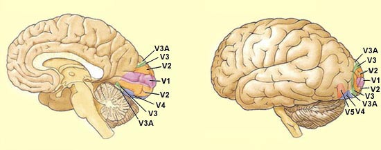
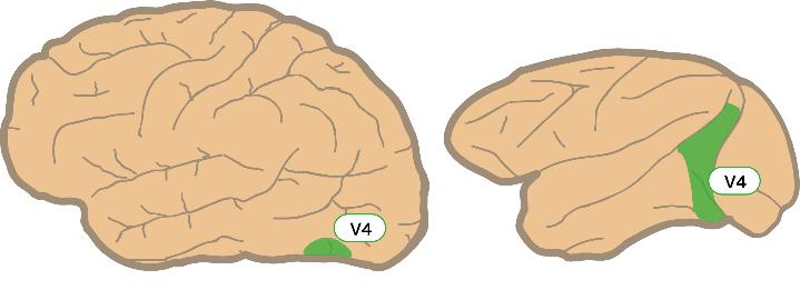
What is the difference between association cortex V4 vs middle temporal region MT; V5
V4: Associates and implements color in our vision
MT; V5: Associated and implements the motion that we see
What are three possible consequences to damage on the occipital lobe?
Hemianopia - Loss of vision in half the visual field.
Scotoma - Loss of vision in one point (see pic) .
Quadrantanopia - Loss of vision to a quarter of the visual field.

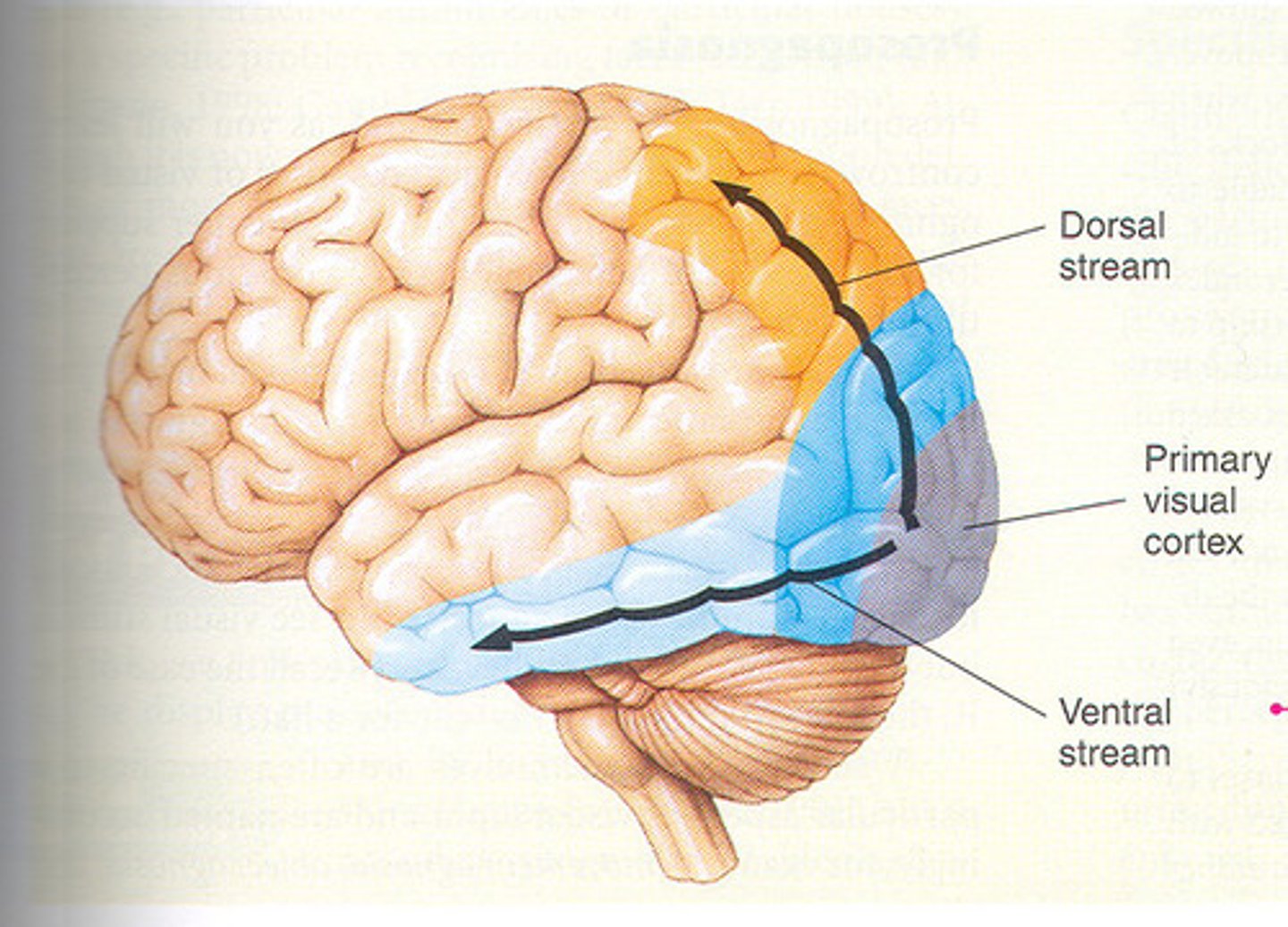
What are the two lobes that may also be involved in visual processing?
Mishkin,Ungerleider, & Macko (1983) proposed that visual processing may involve areas other than those in the occipital lobe.
¤ Temporal lobe (Ventral)
¤ Parietal lobe (Dorsal)
What is the role of the ventral stream (temporal lobe) when we receive visual processing?
The "What" pathway
¤ Recognizing objects
¤ Names and functions of objects regardless of location

What is the dorsal stream (parietal lobe) contribute to visual processing?
"Where"/"How" pathway
¤ Locations of objects, but not their names or functions
¤ How to interact with objects
What is the Inferotemporal (IT) cortex? What happens when its lesioned?
Part of cerebral cortex in lower portion of temporal lobe, important for object recognition
¤ Part of "what" pathway
If lesioned, it leads to agnosias
Agnosia: Failure to recognize objects in spite of the ability to see them
What is the complex role of IT cortex?
It responds very well to complex properties. For instance,
It responds well to complex stimuli such as:
- Face
- Hand
- Object
Does not respond to simple stimuli such as:
- Spots
- Lines
"Grandmother" cells
hypothetical neuron that represents a complex but specific concept or object. It activates when a person "sees, hears, or otherwise sensibly discriminates" a specific entity, such as his or her grandmother.
Someone specific that the person remembers, then their neuron 'activates'
Some areas of the cortex are specialized to process certain types of stimuli. What is the Parahippocampal Place Area (PPA)?
— Responds preferentially to places, such as pictures of houses
Some areas of the cortex are specialized to process certain types of stimuli. What is the Fusiform Face Area (FFA)
Responds to faces more than other objects
Some areas of the cortex are specialized to process certain types of stimuli. What is the Extrastriate Body Area (EBA)?
Specifically involved in the perception of body parts
Facial recognition is different from object recognition. For instance: double dissociation
When one perceptual function can be damaged without affecting the other. Damage to one function will not impair the other. This demonstrates that face recognition and object recognition, while connected, can be somewhat separate processes within the brain, and damage to one doesn't necessarily imply damage to the othe
Some examples include:
- Prosopagnosia
- Agnosia
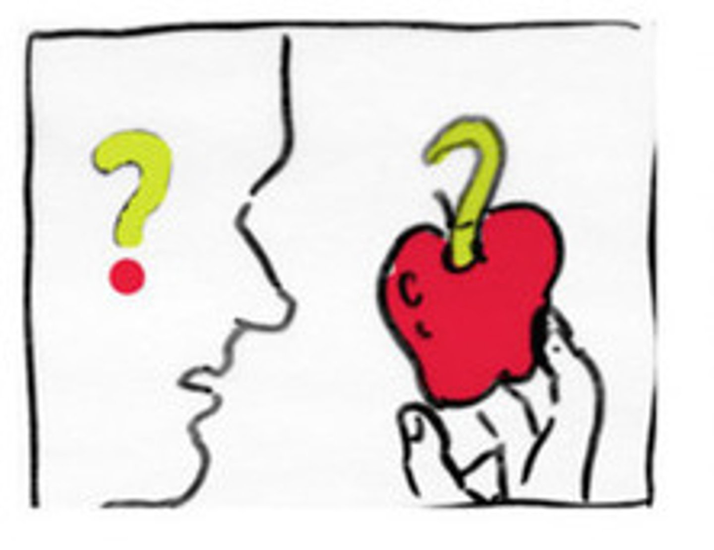
Prosopagnosia
Inability to recognize faces
Agnosia
the inability to recognize familiar objects, despite being able to see them
In facial recognition, "expertise" can lead to activation of...
FFA.
Experts in animals, cars, chess, machines ¤ When participants had significant previous expertise (e.g., birders or car enthusiasts), they were more likely to also use FFA while looking at the images.
For example, individuals who have significant expertise in particular categories, like bird watchers (birders) or car enthusiasts, are more likely to activate their FFA when they look at images related to their area of expertise. In other words, when these experts view pictures of birds or cars, their brain activity patterns may resemble those typically associated with face recognition, as observed in the FFA.
Pareidolia
Tendency to perceive meaningful images in meaningless visual stimuli. Psychological phenomenon in which people perceive familiar patterns, such as faces, animals, or objects, in random stimuli, typically in visual or auditory information. These patterns are often not actually present but are instead a result of the brain's tendency to seek and recognize familiar shapes or forms in ambiguous or random stimuli.
The most common example is seeing faces in inanimate objects, such as seeing a face in the patterns of clouds, the arrangement of objects on a shelf, or even inanimate objects like electrical sockets or cars
Pareidolia can also pick out potentially dangerous things as well

What are the left nerves of each retina show in the visual field and what hemisphere?
If need a review: https://www.youtube.com/watch?v=cG5ZuK0_qtc&ab_channel=ArmandoHasudungan
Left side of each retina (right visual field)in each Left hemisphere
Information from both eyes are combined in V1 (see flashcard). If visual disturbance affects both of the eyes, the problem will be in V1. Visual disturbances on one eye usually will be d/t level of eye, retina, or optic nerve.
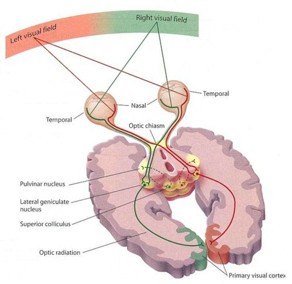
What are the RIGHT nerves of each retina show in the visual field and what hemisphere?
Right side of each retina (left visual field) in each Right hemisphere
If need a review: https://www.youtube.com/watch?v=cG5ZuK0_qtc&ab_channel=ArmandoHasudungan
Information from both eyes are combined in V1 (see flashcard). If visual disturbance affects both of the eyes, the problem will be in V1. Visual disturbances on one eye usually will be d/t level of eye, retina, or optic nerve.

What are visual perceptual disorders?
Disorder in perceiving what is in front of you.
¤ NOTdue to basic sensory deficit ¤ Problem is not an inability to see, rather it is an inability to perceive
¤ Agnosias
¤ Optic aphasia
¤ Blindsight
¤ Akinotopsia
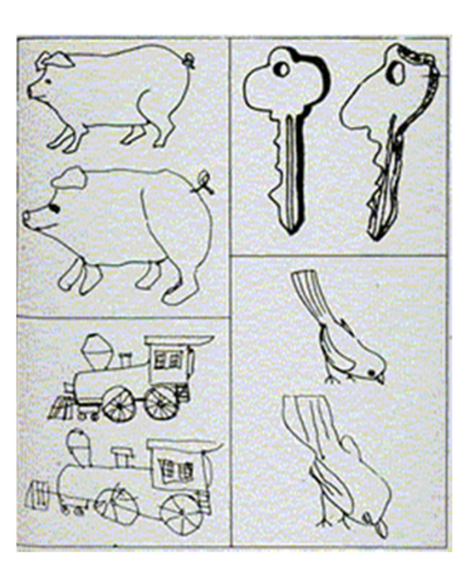
What is apperceptive visual agnosia?
¤ Inability to recognize an object due to an inability to perceive it
¤ Lack of ability to recognize visual stimuli
¤ Not due to deficit in vision, attention, language, or general intelligence
¤ RH parietal, occipital, or temporal lesions
Recognition even worse if viewpoint is unusual or if there are superfluous elements present.
The test for AVA is typically asking pt to copy a drawing. That is when they will show inability to recognize an object.
What is associative visual agnosia?
Individuals can perceive the physical characteristics of objects, such as their shape, color, and texture, but they cannot assign meaning to those objects. They have difficulty recognizing or identifying familiar objects or faces by sight alone. For example, they may see a common object like a pen but be unable to identify it or know its purpose.
This condition is often related to damage to the connections between different areas of the brain responsible for visual processing and object recognition, particularly the ventral visual stream.
Intact ability to copy drawings
Inability to ASSOCIATE objects with meanings (even if they just drew it!. They can copy it but do not know the meaning)
Deficits in drawing something from memory or when asked to name or identify function
Associated with LH lesions
They can describe what it is but cannot say what it is. They may not know what a stethoscope is but can describe it as "long cord with disc at end"
They can write but cannot read what they wrote
What is category specific visual agnosia?
There are two different types of agnosia:
1. Living vs non-living
2. Musical instruments
Specific parts of the temporal lobe have specific visual agnosia for example:
¤ People: Left anterior inferior temporal
¤ Tools: Left inferior temporal
¤ Animals: In between
Specific type of visual agnosia characterized by the selective inability to recognize objects from a specific category, even though the person's overall visual perception and cognitive functioning remain intact. This condition is related to damage or dysfunction in particular areas of the brain, often in the ventral (temporal stream) or occipitotemporal cortex.
Damage comes from: temporal lobe
Category specific visual agnosia: living things vs non-living?
¤ Living things: Few functional differences, overall high visual similarity
¤ Failure to access visual form information should impair identification
¤ Nonliving things: High functional differences, overall low visual similarity
¤ Failure to access functional information should impair identification
What is prosopagnosia
inability to recognize faces
HOWEVER, they can identify people by
- Voice
- Clothing
- Body image
- Context
¤ Cannot recognize photos of their own face
¤ Friends/family in unexpected circumstances
The 'famous faces' test where the pt is given a test to see whose famous person is this for dx of prosopagnosia. . .

What is the cause of prosopagnosia
¤ Most often after RH damage (right hemisphere)
¤ Fusiform gyrus in temporal lobe - "Fusiform Face Area (FFA)"
¤ Borders occipital lobe
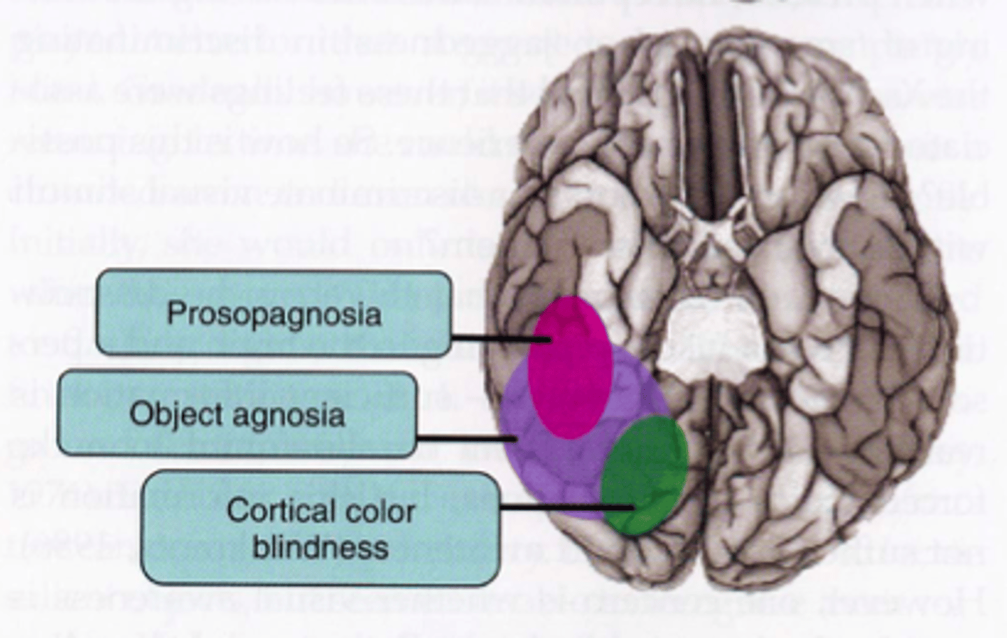
What is colour agnosia
Cannot name colours but can distinguish between colours
¤ Colour sorting test
¤ Colour in black-and-white line drawings
¤ Given coloured drawings and saying whether they are coloured correctly
What lesion is related to colour agnosia?
Lesions to the inferior occipital region
- V4 region damage
What is optic aphasia?
Naming of visual objects impaired
Recognition of objects is intact (vs. agnosia)
Specific to visually presented stimuli (vs. anomia)
Can pantomime correct use of visually presented stimuli

What lesion is related to optic aphasia?
Can pantomime correct use of visually presented stimuli. This refers to the ability of a person with this condition to demonstrate or mimic the appropriate actions associated with objects or images they see, even if they cannot verbally or in writing name or describe those objects. Optic aphasia is a specific language disorder, and individuals with this condition have difficulty processing and expressing language related to visually presented stimuli.
The lesion r/t optic aphasia:
Left-hemisphere parieto-occipital lesions
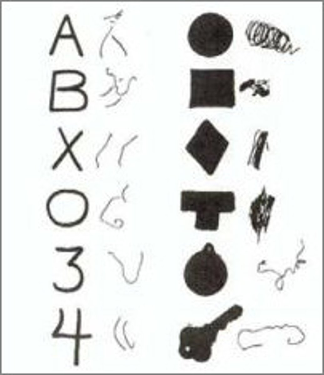
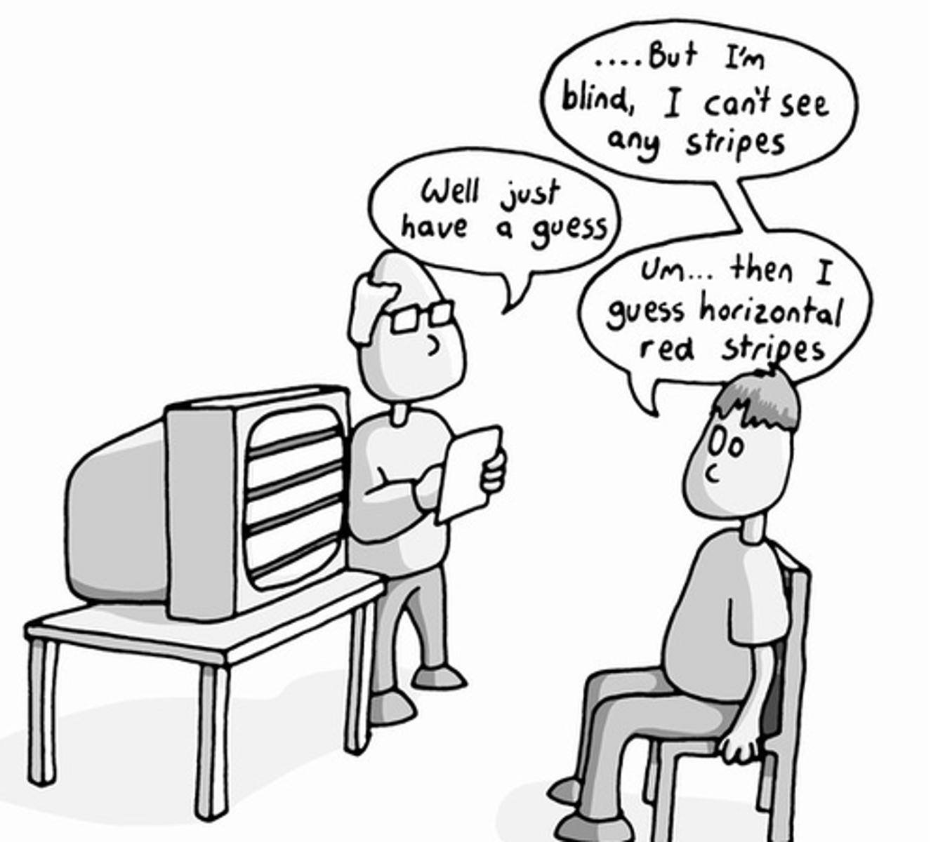
What is blindsight?
¤ Blind area of visual field - scotoma ("blind" spot occurring in opposite VF of lesion to V1)
How?
Retino-geniculate-striate visual pathway sends most information through V1 (striate cortex or primary visual cortex). If V1 is damaged, how can visual information still be processed?
¤ Unaware of perceiving information in scotoma
¤ 90% success rate in forced choice paradigm
¤ Xs & Os
¤ Priming: river bank vs. wall street
Describe the process of transmitting visual info from the eyes to the brain
Retina: The journey begins in the retina, the light-sensitive tissue at the back of the eye. The retina contains photoreceptor cells (rods and cones) that detect light and convert it into electrical signals.
Optic Nerve: The electrical signals generated by the photoreceptor cells are transmitted via the optic nerve, a bundle of nerve fibers that connect the eye to the brain.
Lateral Geniculate Nucleus (LGN): The optic nerve fibers from each eye carry visual information to a structure in the thalamus called the lateral geniculate nucleus (LGN).
The LGN is a relay station that sorts and processes visual information.
Striate Cortex (Primary Visual Cortex): From the LGN, the visual information is relayed to the striate cortex, which is also known as the primary visual cortex or V1.
The striate cortex is located in the occipital lobe at the back of the brain. This region is essential for further processing and interpreting visual information.
Higher Visual Areas: Beyond the striate cortex, the visual information is sent to higher visual areas in the brain, where more complex processing, such as object recognition and motion detection, occurs.
What is the purpose of Tecto-pulvinar pathway in blindsight?
The tecto-pulvinar pathway plays a role in blindsight by providing an alternative route for processing visual information when the primary visual cortex (striate cortex) is damaged or nonfunctional. Individuals with blindsight may be able to detect and respond to visual stimuli, such as the presence or location of objects, without consciously perceiving them.
Blindsight is a phenomenon where individuals with damage to their primary visual cortex can still respond to visual stimuli even though they are not consciously aware of what they are seeing. The tecto-pulvinar pathway represents a neural mechanism that allows some level of visual processing and response in the absence of conscious visual awareness, which is a characteristic of blindsight. It is thought to be a compensatory mechanism that helps individuals with damage to their primary visual cortex make use of residual visual information.
What is akinetopsia and what is the lesion/area that is damaged in the brain?
¤ Motion Blindness: Rare neurophysiological disorder in which affected individual has no perception of motion
¤ Patient sees the world in a series of static "frames"
What lesion of the brain is it caused by?
¤ Caused by damage to visual area V5/MT.
Visual area V5, also known as the middle temporal area (MT), is a specific region of the cerebral cortex in the brain that is involved in processing visual information related to motion and visual perception. It is located in the middle temporal gyrus, which is part of the occipital lobe of the brain
Emerging from V1 are two routes for visual processing:
Ventral - concerned with what a stimulus is
¤ Dorsal - involved in processing where a stimulus is