Veterinary Anatomy: Skull and Osteology
1/50
There's no tags or description
Looks like no tags are added yet.
Name | Mastery | Learn | Test | Matching | Spaced |
|---|
No study sessions yet.
51 Terms
Skeletal System
serves for support and protection while providing levers for muscular action. It functions as a storehouse for minerals and as a site for fat storage and blood cell formation.
Osteology
The study of the structure and function of bones.
Endoskeleton
The internal skeleton of an organism, as opposed to the exoskeleton of tortoises and turtles.
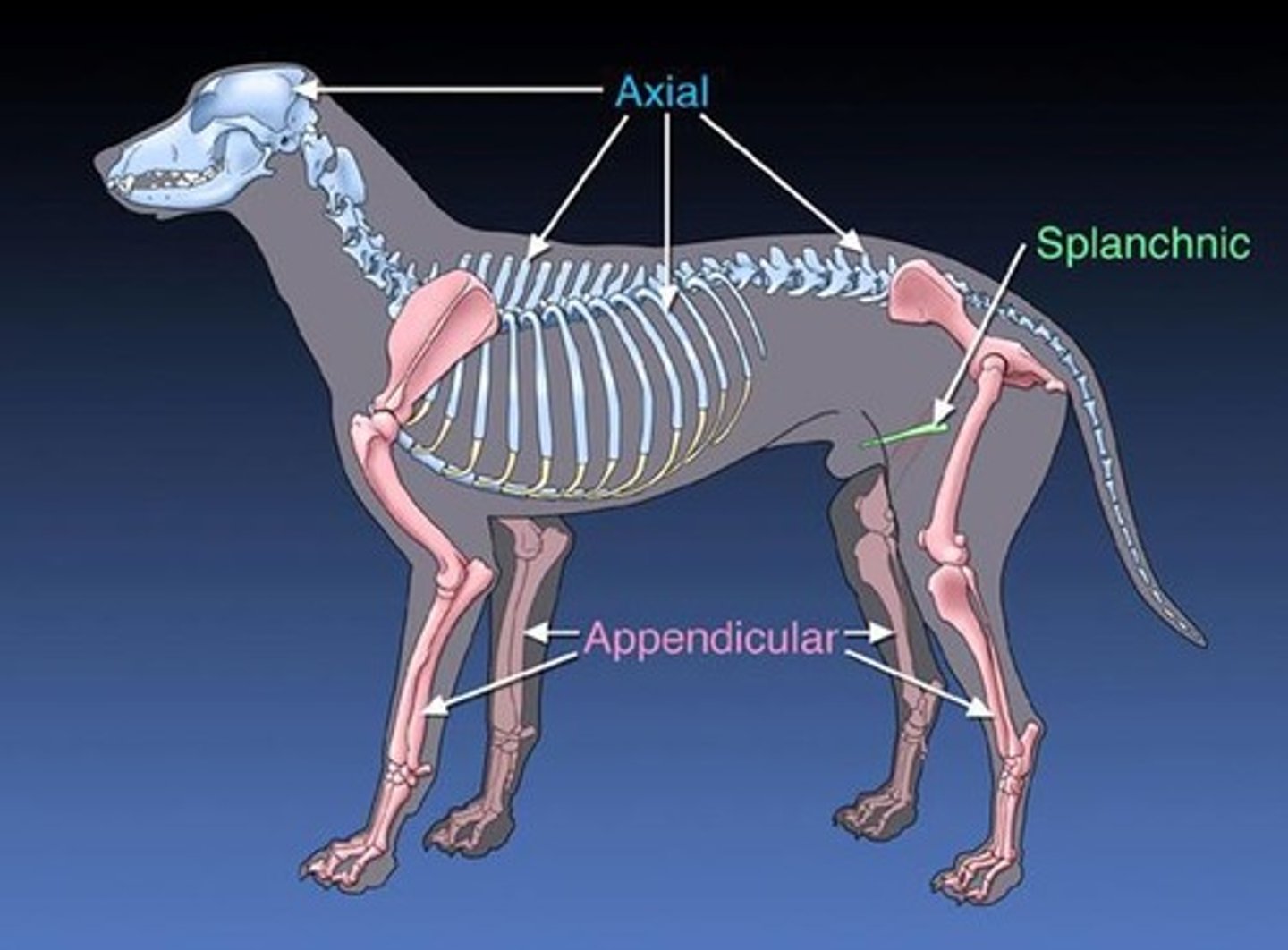
Axial Skeleton
The part of the skeleton that consists of the skull, vertebral column, ribs, and sternum.
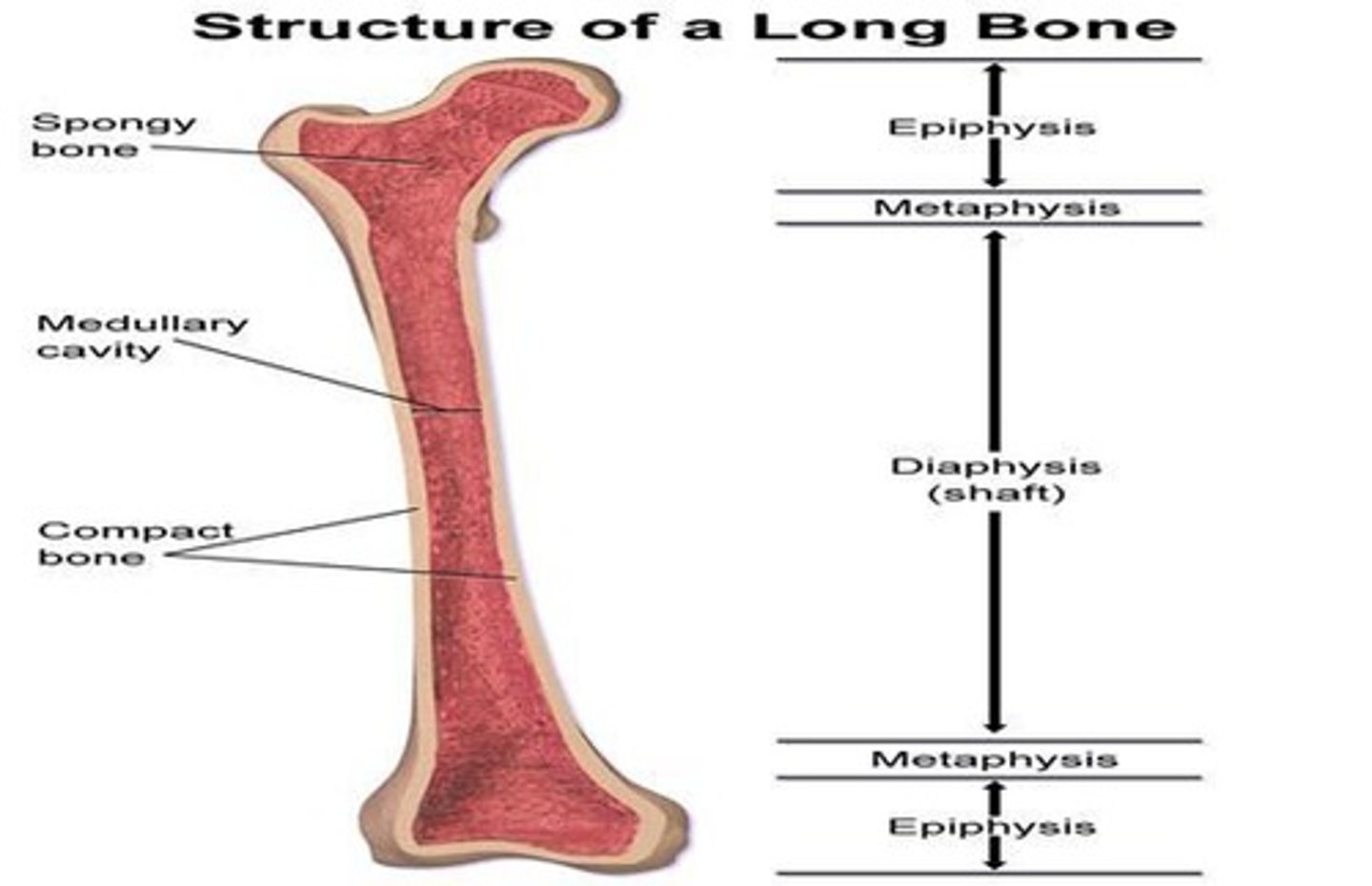
Long Bones
Bones that act as levers, each having a shaft (diaphysis) and two extremities (epiphyses).
Short Bones
Bones that diffuse concussion, such as carpals.
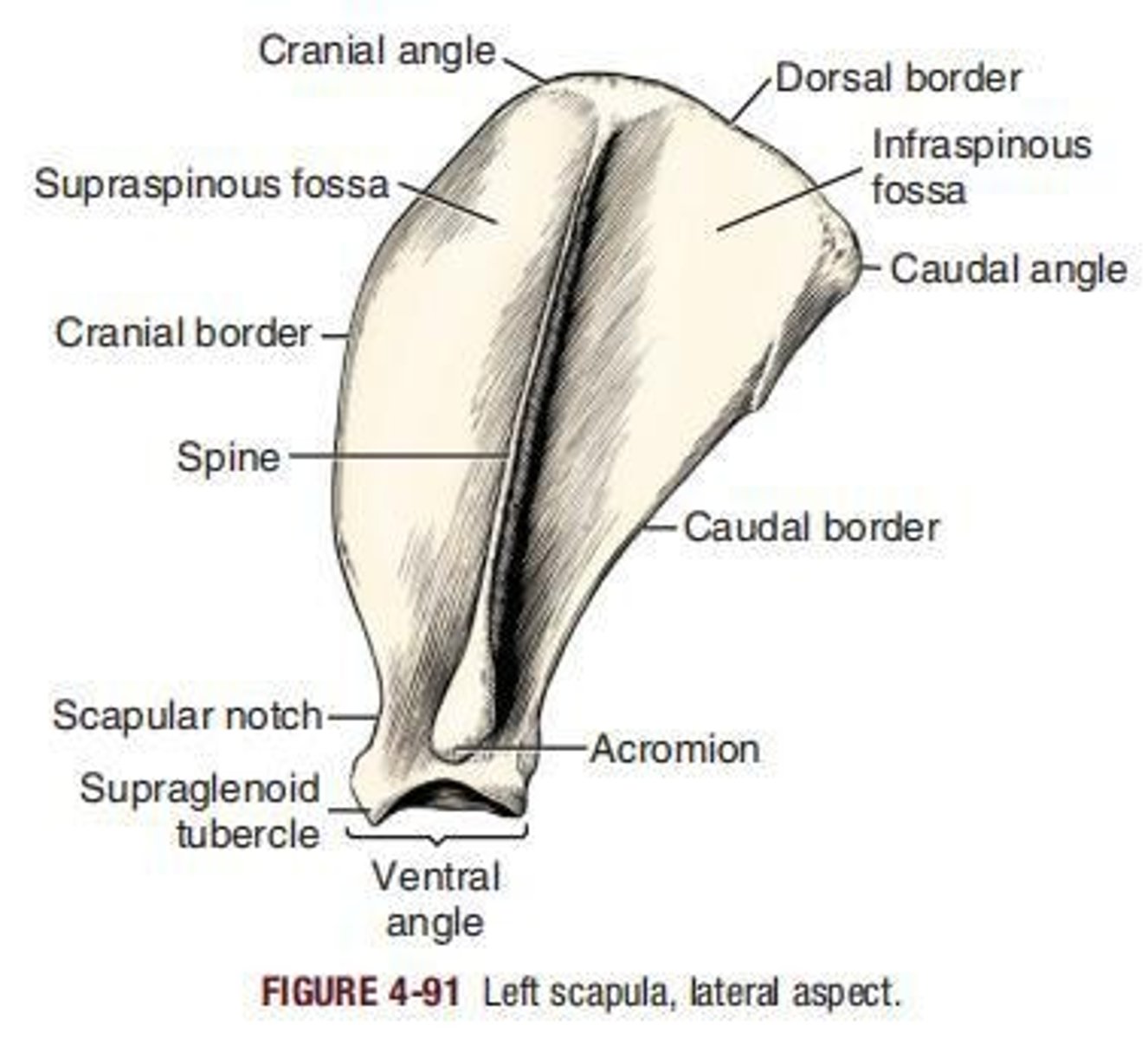
Flat Bones
Protective bones that afford a large area for muscular attachment, such as the scapula.
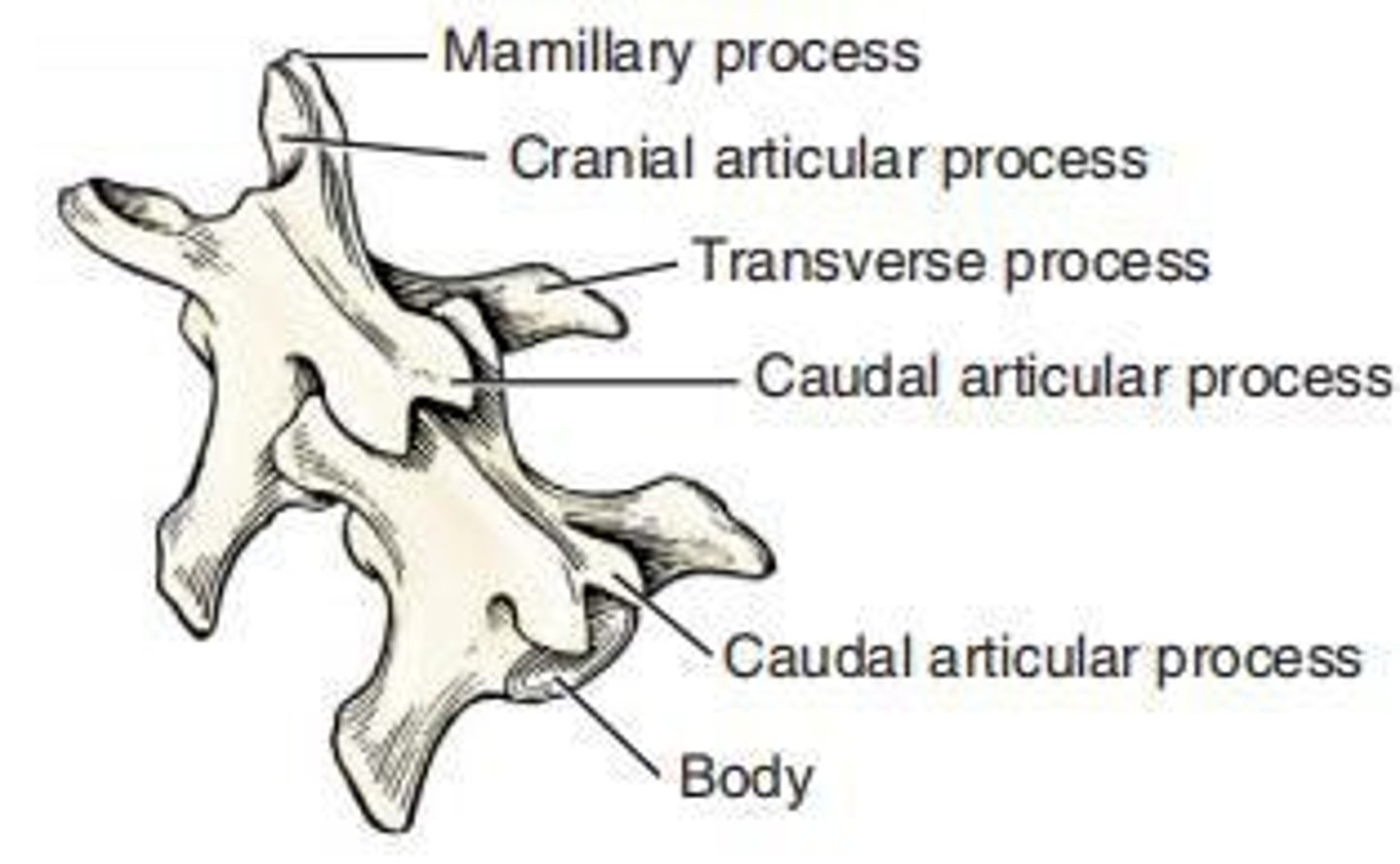
Irregular Bones
Bones with varied functions such as support and ligament attachment, exemplified by vertebrae.
Pneumatic Bones
Bones found in the skeletons of birds, lined by mucous membrane rather than marrow, and communicating with the respiratory system.
Sesamoid Bones
Bones developed in tendons to afford increased leverage, such as the patella and navicular bone.
Markings on Bones
Bumps, holes, and depressions on a bone's surface, which can be either articular or non-articular.
Canal
A tunnel through one or more bones.
Condyle
A large articular prominence.
Cotyloid cavity
A deep articular depression.
Crest
A prominent border or ridge.
Epicondyle
A prominence just proximal to a condyle.
Facet
A smooth flat surface.
Foramen
An opening through a bone.
Fossa
A small hollow.
Fovea
A shallow, non-articular depression.
Groove
A long, narrow furrow accommodating a vessel, nerve, or tendon.
Head
A rounded articular process.
Notch
A rounded articular process.
Process
Any prominent, roughened projection from a bone.
Spine
A sharp, slender process.
Trochanter
A large, blunt process found only in the femur.
Trochlea
A pulley-shaped structure.
Types of Skulls
Mesaticephalic, Dolicocephalic, and Brachycephalic.
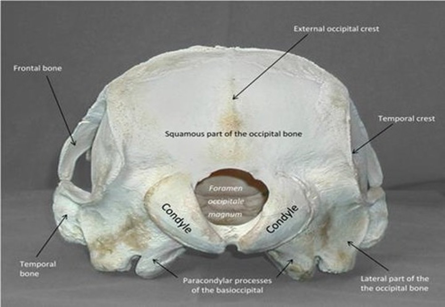
Occipital Bone
forms a ring, the foramen magnum, around the junction of the medulla oblongata and the spinal cord.
Parietal Bone
is paired and forms most of the dorsolateral part of the calvarial portion of the cranium. It articulates dorsally with its fellow and with the interparietal process of the occipital bone.
Frontal Bone
is irregular in shape, being broad caudally and somewhat narrower rostrally. Laterally, the rostral part is concave and forms the medial wall of the orbit.
Sphenoid Bones
form the rostral two thirds of the base of the cranial cavity between the basioccipital caudally and the ethmoid rostrally. Each consists of a pair of wings and a median body.
Presphenoid
The more rostral bone with orbital wings is the presphenoid (os presphenoidale); the caudal bone with the larger wings is the basisphenoid (os basisphenoidale).
Temporal Bone
forms a large part of the ventrolateral wall of the calvaria. Its structure is intricate, owing to the presence of the cochlea and the semicircular canals, and an extension of the nasal pharynx into the middle ear.
Ethmoid Bone
is located between the cranial and the facial parts of the skull, both of which it helps to form. It is completely hidden from view in the intact skull. Its complicated structure is best studied from sections and disarticulated specimens.
Incisive Bone
formerly premaxilla, has a small body (corpus ossis incisivum) rostrally with three processes: the alveolar process (processus alveolaris), the curved, tapering nasal process (processus nasalis) and the laterally compressed, pointed palatine process (processus palatinus).
Nasal Bone
is long, slender, and narrow caudally but, in large dogs, is almost 1 cm wide rostrally. The dorsal, or external, surface (facies externa) of the nasal bone varies in size and shape, depending on the breed.
Maxilla
The ___ and the incisive bone of each side form the superior jaw. The maxilla is divided grossly into a body and four processes: the frontal, zygomatic, palatine, and alveolar. It is the largest bone of the face and bears all of the superior cheek teeth.
Zygomatic Bone
formerly called jugal or malar bone, forms the rostral half of the zygomatic arch (arcus zygomaticus). It is divided into two surfaces, and two processes, the temporal process (processus temporalis) and the frontal process (processus frontalis).
Palatine Bone
is located caudomedial to the maxilla, where it forms the caudal part of the hard palate, the rostromedial wall of the pterygopalatine fossa, and the lateral wall of the nasopharynx.
Lacrimal Bone
located in the rostral margin of the orbit, is roughly triangular in outline and pyramidal in shape.
Pterygoid Bone
is a small, thin, slightly curved, nearly four-sided plate of bone that articulates with the bodies of both the presphenoid and the basisphenoid bones, but particularly with the medial surface of the pterygoid process of the basisphenoid.
Vomer
is an unpaired bone that forms the caudoventral part of the nasal septum. It contributes to the roof of the choana.
Mandible
The inferior jaw of the dog consists of right and left mandibles (mandibula) firmly united in life at the intermandibular suture (sutura intermandibularis), which is a strong, rough-surfaced, fibrous joint. Each mandible is divided into a horizontal part, or body, and a vertical part, or ramus.
Hyoid Apparatus
acts as a suspensory mechanism for the tongue and larynx. It attaches to the skull dorsally, and to the larynx and base of the tongue ventrally, suspending these structures in the caudal part of the space between the bodies of the mandible.
Basihyoid
is a transverse, unpaired bone in the musculature of the base of the tongue as a ventrally bowed, dorsoventrally compressed rod. Its extremities articulate with both the thyrohyoid and the ceratohyoid bones.
Thyrohyoid
is a laterally bowed, sagittally compressed, slender bone that extends dorsocaudally from the basihyoid to articulate with the cranial cornu of the thyroid cartilage of the larynx.
Ceratohyoid
is a small, short, tapered rod having a distal extremity that is approximately twice as large as its proximal extremity. It articulates with the basihyoid and the thyrohyoid. The proximal extremity, which points nearly rostrally in life, articulates with the epihyoid at a right angle.
Epihyoid
is approximately parallel to the thyrohyoid bone. It articulates with the ceratohyoid at nearly a right angle distally and with the stylohyoid proximally without any angulation.
Stylohyoid
is slightly longer than the epihyoid, with which it articulates. It is flattened slightly craniocaudally and is distinctly bowed toward the median plane. It gradually increases in size from its proximal to its distal end. Both ends are slightly enlarged.
Tympanohyoid Cartilage
is a small cartilaginous bar that continues the proximal end of the stylohyoid to the inconspicuous mastoid process of the skull.