Looks like no one added any tags here yet for you.
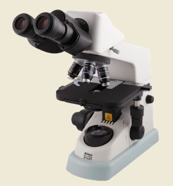
Compound Microscope
A compound microscope is defined as the type of microscope that has more than one lens. It has a combination of lenses and two optical parts known as an objective lens and an eyepiece or ocular lens.
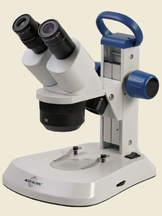
Dissecting/Stereo Microscope
Utilized to view three-dimensional objects and larger specimens, with a maximum magnification of 100x.
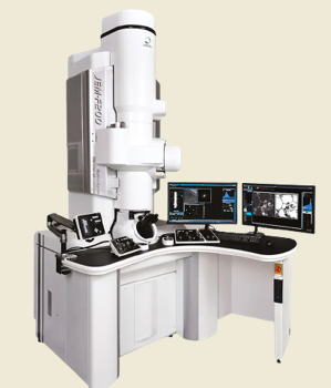
Electron Microscope
a microscope that produces an image of a specimen by using a beam of electrons rather than a beam of light. Electrons have much a shorter wavelength than visible light, and this allows electron microscopes to produce higher-resolution images than standard light microscopes.
Arm
Supports the tube/head and connects it to the base.
Eyepiece Tube/Head
Connects the eyepiece to the objective lenses.
Eyepieces/Ocular Lens
The lens at the top that you look through, usually 10x.
Nose Piece
This is the part of the microscope that holds two or more objective lenses and can be rotated to easily change power.
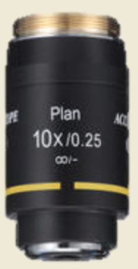
Objective Lens
Usually, you will find 3 or 4 objective lenses on a microscope. They almost always consist of 4x, 10x, 40x and 100x powers corresponding to scanner, LPO, HPO, and OIO.
Total Magnification Formula
power of the objective * power of the eyepiece
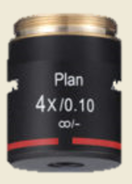
Scanning Objective Lens
low-level magnification lenses, offering only four times magnification for a general overview of the sample
Low Power Objective (LPO)
useful for examining large specimens or surveying many smaller specimens.
High Power Objective (HPO)
ideal for observing fine details within a specimen sample.
Oil Immersion Objective (OIO)
Immersion oil increases the resolving power of the microscope by replacing the air gap between the immersion objective lens and cover glass with a high refractive index medium and reducing light refraction resulting in a more detailed resolution of a sample.
Stage
The flat platform that supports the slides.
Stage Clips
hold the slides in place
Aperture
A hole in the microscope stage via which transmitted light from the source enters the stage.
Condenser
Gather light from the microscope's light source and concentrate it into a cone of light that illuminates the specimen.
Iris Diaphragm
An adjustable assembly of thin metal leaves for varying the size of openings that determine the cross section of the light ray bundle entering the condenser and the objectives.
Stage Controls
allows the movement of the stage back and forth
Course Focus
moves the stage up and down in bigger increments and brings it closer to the lens faster, bringing it into focus
Fine Focus
fine-tune the focus of the microscope to achieve optimal clarity and detail.
Illuminator
provide even, high intensity light at the place of the field aperture, through the condenser, and onto the specimen.
Base
serves as a support for microscopes