Pt 7 Animal Cardiovascular and Respiratory Systems
1/132
There's no tags or description
Looks like no tags are added yet.
Name | Mastery | Learn | Test | Matching | Spaced | Call with Kai |
|---|
No analytics yet
Send a link to your students to track their progress
133 Terms
How has the circulatory evolved through time?
Evolution
Unicellular -> multicell
All use oxygen (aerobic resp) to exchange oxygen with CO2 in env
Water: in tissue there is CO2 -> exchanged with oxygen
All devised method to exchange gases
Core = diffusion
Circulatory system
Exchange of gases supported by circulatory system -> work together for respiration (exchange gases)
Unicellular is smaller -> simple diffusion allows gas exchange
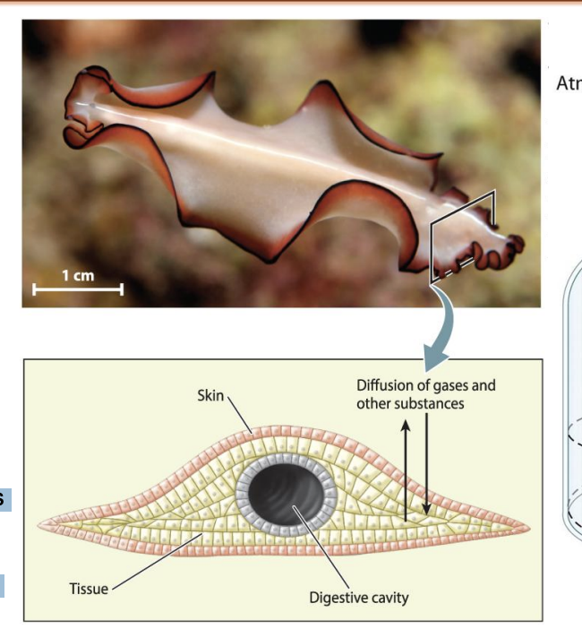
simplest invertebrate animals were composed of thin sheets of cells with a large surface area.
Multicellular have more body surface area -> needs more -> developed integrated system of gas exchange (respiratory system)
What is the respiratory system?
Multicellular have more body surface area -> needs more -> developed integrated system of gas exchange (respiratory system)
Flow of dissolved gases through medium (blood, hemolymph) to form circulatory system to transport dissolved gases to parts of body
What is diffusion?
Diffusion: movement of molecules from higher concentration to lower concen
Gas Molecule when present in high concen -> collide with each other through random motion (bronian motion) -> collide and contain kinetic energy -> move farther from the point of origin

What is the biggest issue with diffusion?
Given a large enough surface→ diffusion is an extremely effective way to exchange gases and other substances over short distances between two compartments
effective transport by diffusion is limited to short distances
Diffusion limited by distance (inversely proportional)
The farther gas molecules are apart -> less diffusion
Optimum range: up to 100 micrometer
Above: so far apart they won’t move without collisions that gave then KE to move farther -> no diffusion

What is the difference between unicellular and multicellular diffusion?
Unicellular
Only have cell membrane (single bilipid layer, 1-2 microm) -> diffusion allows unicellular to exchange gases
Multicellular
Diffusion requires interface where gas exchange takes places to be less than 100 mm

How does diffusion occur in air?
Air and water are different
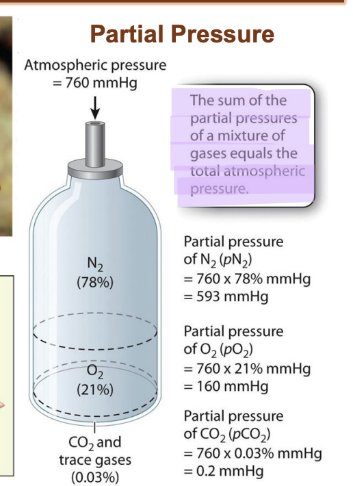
Oxygen in air: partial pressure of gas
In atmosphere -> different gases as mixture
Each gas exerts own pressure
Multiply fraction of gas present in mixture with overall atmosphere pressure = partial pressure of gas
How does diffusion occur in water?
Oxygen present in water: dissolved in water = concentration of dissolved O2
What is partial pressure?
MmHg = unit for pressure
Tells how much gas is present in a location
Important for rate of gas exchange
Can increase or decrease from partial pressure of oxygen
Valley = partial pressure is constant -> suddenly go on mountain -> change in altitude = decrease in fractional concen of O2 -> decrease in partial pressure -> body will sense change -> adjust rate of respiration -> start breathing heavily
Few days = acclimatize by increasing red blood cell production
Partial pressure of N2 changed
Dive deep in sea -> partial pressure of Nitrogen changes
Atmosphere air has N -> but does not interact in gas exchange (only O2 and CO2)
Deep in water -> N pp change -> bubble out from blood -> fatal
Divers with pressurized gas with O2 and N2 -> so Partial pressure of nitrogen does not change
What is bulk flow?
the physical movement of fluid over a given distance
circulatory system helps move dissolved oxygen into rest of body in bulk flow
Bulk flow: allows the organism to move gas into various distant parts of body
What do larger animals rely on?
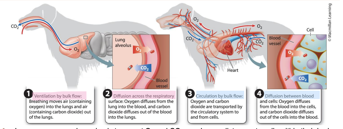
Larger, more complex animals transport O2 and CO2 over longer distances to cells within their body. → rely on bulk flow + diffusion
Larger animals have respiratory system + circulatory system
Developed because gas transport and exchange happens at designated organ system (respiratory system)

Where does diffusion occur in larger organisms?
Surfaces in the body where diffusion occurs like lungs and intestines, have high surface area to volume ratios.
Lung: interface to exchange gas form air to lung tissue
Gas exchange takes place form blood in lung and air -> dissolved oxygen in blood needs to go to the rest of the body part ->

How does ventilation occur int he resp system?
Ventilation occurs In resp system and circulation in circ system -> reaches to deeper tissues
Both steps imp
Both steps are followed by diffusion
Ventilation in lungs -> bulk lungs exchanged in lungs -> diffusion of Oxygen in lung alloviles
After circulation after dissolved oxygen reaches tissues -> dissolved O2 form blood vessel goes to tissue through diffusion
Interface in lung: air and lung blood capillary
In tissue: interface is blood and tissue interface

What occurs in the intenstine?
Diffusion also occurs In intestine
Nutrients absorbed and secretion of chemicals and transport of secreted of molecules (hormones and factors) via diffusion
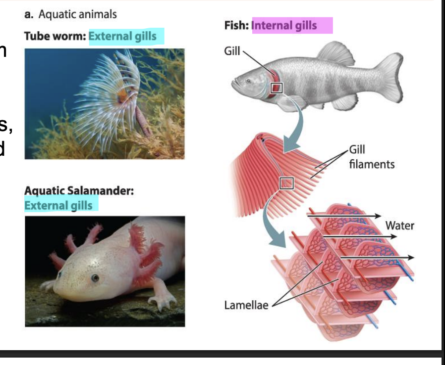
How do aquatic animals use gas exchange?
All animals obtain O2 from the surrounding air or water
take in O2 from water breathe through gills: highly folded, delicate structures that facilitate gas exchange with the surrounding water
Frogs
Respire by skin
How do poriferans exchange gas?
Poriferans
Thin epithelial layer -> respire through single layer of epithelial layer
Less than 100 mm -> gas exchange can happen in single epi layer -> efficient gas exchange
What are the structure of gills?
Highly folded membranes
Gill filaments richly supplied with capillaries
Arranged in parallel layered form
Arranged as same direction as water flow, opposite, or perpendicular from water flow
Arrangement of lamella allows rapid exchange of gases
Lamellae are very thin (1-2 microm) -> allows diffusion quickly
What are the types of gills?
Two types of gills
External gills
Tube warm + salamander
More chance of gills being dmged + very delicate -> strong water current msut dmg it
Fishes developed internal gills protected by external flap (operculum)
What is the structure of internal gills in fish?
Fish need sufficient O2 to meet the energy demands of swimming.
Fish actively pump water through their mouth and over the gills
gills located in a chamber behind the mouth cavity

What is the direction of flow for gills?
Fish greatly reduce the energy cost of moving in dense, viscous water by maintaining a continuous, unidirectional flow of water past their gills
Arrangement of gill filament is opposite of water (counter current flow) + Counter flow in lamellae of gill → help exchange gases better

What is the operculum?
In bony fishes
protective flap overlying the gills → called the operculum
expands laterally to draw water over the gills while the mouth is refilling before its next pumping cycle
Poriferans have operculum: bigger opening

What do gills consist of?
consist of a series of gill arches located on either side of the animal behind the mouth cavity and, in bony fishes, beneath the operculum.
Each gill consist of
Gill filaments that contain numerous lamellae with capillaries network moves in a direction opposite to the flow of water past the gill

What are gill filaments?
Each gill arch consists of two stacked rows of flat, leafshaped structures called gill filaments that contain numerous lamellae
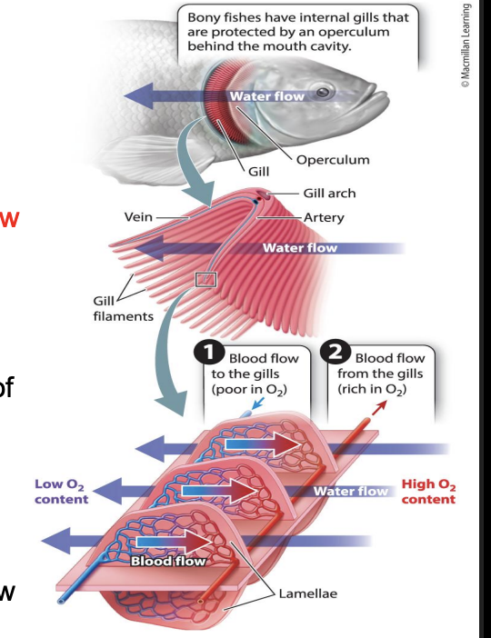
What are lamellae?
thin, sheetlike structures
Have capillaries network that moves in a direction opposite to the flow of water past the gill

What is countercurrent flow?
The type of organization in which fluids with different properties move in opposite directions
Flow in two tubes opposite
At both ends -> same temp of fluid (both tube has cold and hot at end)
Exchange is efficient -> allows end to separate
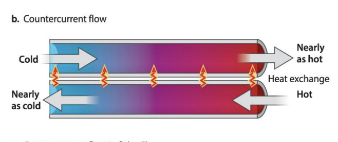
How does countercurrent exchange work in diffusion?
At the same time as countercurrent exchange: CO2 readily diffuses out of the blood vessels and into the water that leaves the gill chamber.
As a result of countercurrent exchange → fish gills can extract nearly all the O2 in the water that passes over them.
A set of blood vessels carries the O2 -rich blood away from the gills to supply the fish’s body
Fish gills
Blood flows in lamellae -> moves low concentration of Oxygen to higher concen of O2
Water flows over in opposite direction (high oxygen concen -> passes through gills -> becomes low O2
exchange of O2 -> allows to maintain region of low and high O2 concen
Separates oxygenated blood from deoxygenated blood
A set of blood vessels carries the O2 -rich blood away from the gills to supply the fish’s body
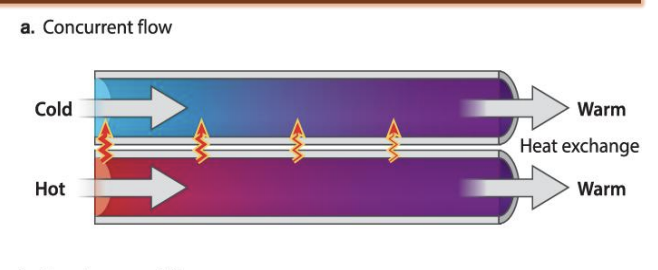
What is concurrent exchange?
less efficient than countercurrent exchange
a steep concentration gradient is not maintained along the length of both tubes.
Instead, both tubes will reach an average temperature.
Concurrent
Keep close together -> transfer of heat energy due to flow of hot and cold water in tubes
As flow continuous -> some time -> heat exchange at both of end of tube -> water is the avg temp btwn two
Ends the energy merges -> avg of two physical factors
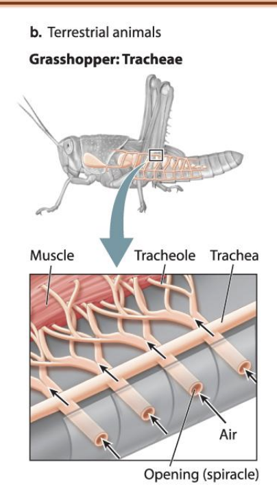
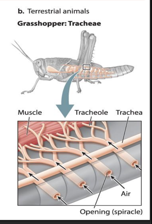
What is the system that terrestrial insects evolved for gas exchange?
Lower animals in invertebrates
Use tubes (trachea)
Trachea: a system of air tubes that branch from openings along their abdominal surface into smaller airways
What do many terrestrial animals use for gas exchange?
Many terrestrial animals, such as reptiles, birds, and mammals, have internal lungs for gas exchange.
The respiratory surfaces of more complex and active animals are highly folded → creating a large surface area within a small space
These surfaces are only one or two cell layers thick —> providing a diffusion distance of as little as 1 to 2 μm.
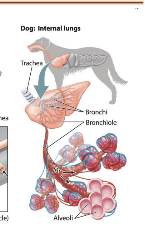
What is the structure of the lung system?
Vertebrates: lung system
Starts with trachea -> divides into bronchi -> bronchiole -> alveoli sac
Separate into alveoli (single layer structure that allows interface for gas exchange)
Contain air breathed in
Single layer epithelial layer is where gas exchange takes place
What are the steps to insect breathing?
Because of their small size → insects do not have a respiratory surface → instead employ a direct pathway of air transport that gets air directly to their tissues.
breath through trachea
insects rely on a two-step process of ventilation and diffusion to supply their cells with O2 and eliminate CO2 .

How does insect breathing work?
Air enters an insect through openings (spiracles) along either side of its abdomen
spiracles can be opened or closed to limit water loss and regulate O2 delivery
Diffusion occurs at the cell:
O2 supplied by the fine airways diffuses into the cells
CO2 diffuses out and is eliminated through the insect’s tracheae
mitochondria of the flight muscles and other metabolically active tissues are located within a few micrometers of tracheole airways.
Because O2 and CO2 diffuse rapidly in air, the tracheal system delivers O2 at high rates.
Ventilation:
Once airs enter trachea (enters in opening of skins (spiracles))
Spiracles allow entry of air into trachea
Trachea branch out to multiple tracheole -> reach other tissues and muscles
At the muscle and junction of tracheole -> help of diffusion -> gas exchange ->
After CO2 comes out of trachea
What kind of ventilation do vertebrates use?
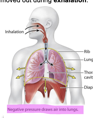
most land vertebrates inflate and deflate to move fresh air with O2 into the lungs and expire stale air with CO2 out of the lungs
Use Tidal ventilation
What is tidal ventilation?
The low density and viscosity of air enables these animals to breathe by tidal ventilation
Tidal ventilation allows movement of low density to enter body -> gas exchange takes place
Requires less muscular energy
Air is drawn into the lungs during inhalation and moved out during exhalation
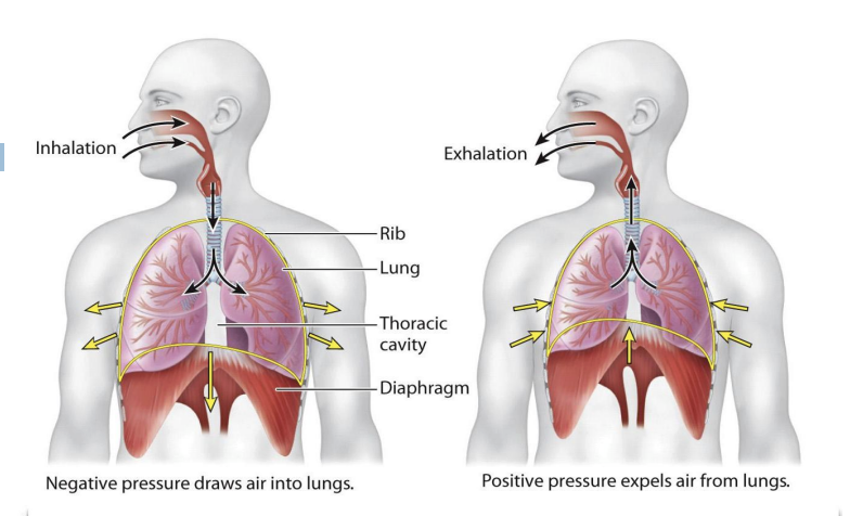
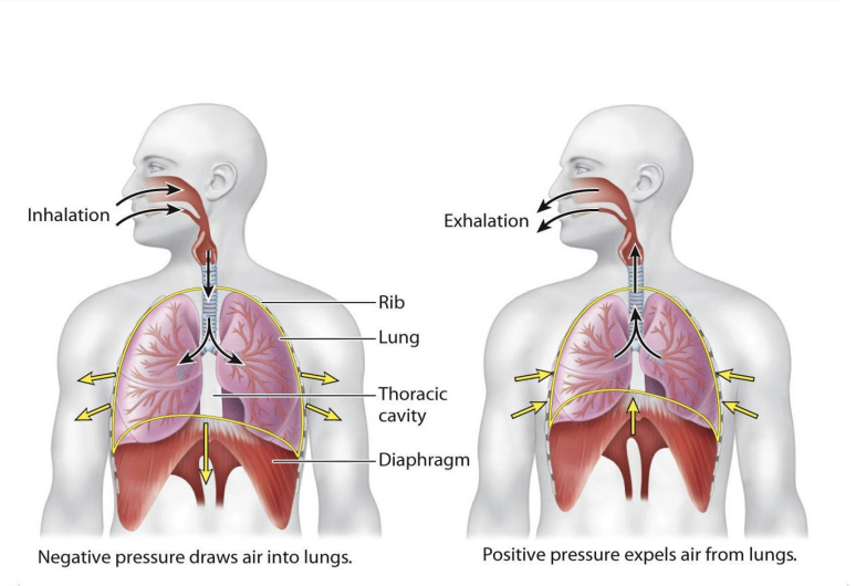
What is inhalation?
Mammals and reptiles expand their thoracic cavity to draw air into their lungs.
The expansion of the lungs causes the air pressure inside the lungs to become lower than the air pressure outside the lungs.
The resulting negative pressure draws air into the lungs.
Inhalation is active that requires contraction of diaphragm
How is inhilation active?
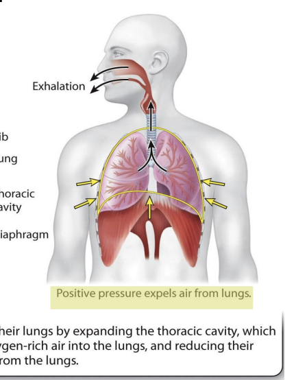
What is exhilation?
exhalation is passively driven by the elastic recoil of tissues that were previously stretched during inhalation.
The contraction of the lungs causes the air pressure inside the lungs to become higher than the air pressure outside the lungs.
The resulting positive pressure forces air out of the lungs.
Exhalation is passive because lungs is elastic property from elastic tissue -> inhalation in complete lungs recoil back and exhale air with CO2
How is inhalation and exhalation related?
Lungs expand –> initiation of inhalation is triggered by contraction of diaphragm
Diaphragm = largest muscle in human body
When it contracts -> pulls the thoracic cavity and expand it -> creates negative pressure in the lungs
Pressure from high pressure moves to low concentration
Air from inhalation moves into the lungs -> alveoli where gas exchange take place
When lungs filled with air -> pressure changes -> exhale the air needed (contains CO2 -> exhalation)
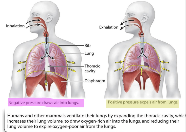
What is the diaphragm?
a domed sheet of muscle located at the base of the lungs that separates the thoracic and abdominal cavities.
What happens during relaxed breathing?
In mammals, inhalation during normal, relaxed breathing is driven by contraction of the diaphragm
At rest
Diaphragm allows breathing (mostly using diaphragm)
Exhalation occurs passively by the elastic recoil of the lungs and chest wall
What are intercostal muscles?
attached to adjacent pairs of ribs
assist the diaphragm by elevating the ribs on inhalation and depressing the ribs during exhalation
used for active excersize
What happens to breathing during excersize?
During exercise, other muscles assist with inhalation and exhalation.
Intercostal muscles assist the diaphragm by elevating the ribs on inhalation and depressing the ribs during exhalation
Lungs require support of intercostal muscles present btwn ribs
Intercostal muscles contract -> lift rib cage up -> expand thoracic cavity farther
Contraction of diaphragm allows expansion of thoracic cavity
When speaking or excersing needs better exchange -> intercostal muscle expands thoracic cavity
action of the intercostal muscles helps to produce larger changes in the volume of the thoracic cavity →
increasing the negative pressure that draws air into the lungs during inhalation
+ assisting elastic recoil of the lungs and chest wall to pump air out of the lungs during exhalation
Intercostal muscle and diaphragm work together
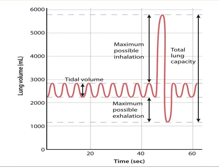
What is the functional capacity of lungs?
When inhale or exhale air -> certain volume of air that can exhale or inhale that can be recycled
Functional capacity of lungs (how much it can inhale and exhale, how much of air is always present in the lungs)
Some air stays in alveoli -> lungs never collapse = residual air
Portion of residual air is recycled with each breath (mixture of previous residual air and new one)

What is tidal volume?
Tidal volume
At rest: amnt of air you inhale and exhale = .5 liter every cycle
Each breath inhale .5 liter of air and exhale .5 liter of air = tidal volume

What is the ventilation rate?
With a breathing frequency of 12 breaths per minute
(breathing frequency × tidal volume) is 6 liters per minute.
Ventilation rate can be changed by either increasing breathing frequency, tidal volume, or both.

What is the total lung capacity?
Maximum volume of air = around 5600 mL
Maximum air exhale by forceful exhalation by 800 mL
Both maximum inhil and exhal = total lung capacity
During diseases condition -> total lung capacity can change
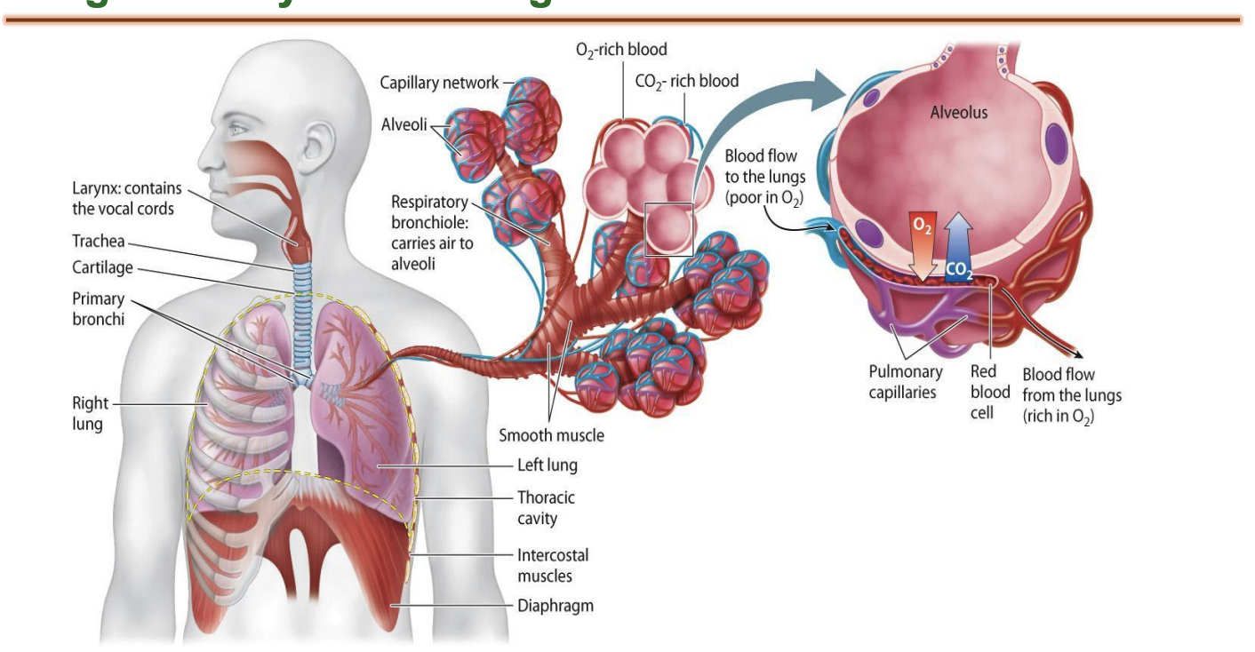
What is the whole system of respiration?
Hair in nose prevents the entry of any particulate matter in air to enter resp system
If large particular matter enters nose -> hair traps it -> sneeze to expel
Air moves back into nasal cavity
Air inhaled moves in nasal cavity to humidify (tirbulates?)
Air inhaled is brought to body temp for efficient gas exchange in lungs
Air humidified and temp brought closer to body temp in nasal cavity -> towards pharynx
Air moves deeper into the trachea: composed of elastic cartilage -> flexibility and prevents from collapsing
Trachea lies interior to the food pipe or esophagus
Trachea do not collapse because of presence of elastic cartilage rings
Air goes down from trachea -> trachea bifurcates into two primary bronchi
Primary bronchi divide into terminal bronchi
Terminal is 1 millimeter
Separates more into alveoli (single epithelial layer air sacs surrounded by capillaries)
Pulmonary capillary surrounds single alveoli (alveolus)
Single epithelial layer (1-2micro) -> gas exchange
Oxygen form air diffuse form alveolus into pulmonary capillaries and Co2 diffuse out of pulmonary cavity into alveolus -> CO2 is exhaled

What is the Larynx?
Pharynx allows for speech and control flow of air in the
lower resp system= larynx (adams apple)
Vocal chords: noises

What is the trachea?
Point of bifurcation is sensitive (sensory receptors that can initiate cough reflex)
Lined by pseudo stratified columnar ciliated epithelial cells
Cilia pushes back any matter or pathogen trapped in air -> push towards esophagus to be digested and cleared out
Smoking makes cilia dysfunctional
What kind of respiration do birds have?
Tidal ventilation has expansion and deflation of lungs
Birds not tidal ventilation because lungs are not flexible -> rigid and do not inflate or deflate

What is the structure of bird lungs?
Lung + air sacs
anterior air sac
Posterior air sac
Both surrounded by flight muscles
Contraction of flight muscles contract

How does bird respiration work?
Birds inhale air -> air first goes to posterior air sac -> Contraction of flight muscles contract -> compresses air sac -> pushes fresh air into lung
In lungs gas exchange takes place -> deoxygenated air goes into anterior air sac -> contraction of flight muscles -> compression of anterior pushes deoxygenated air out of body through trachea and mouth
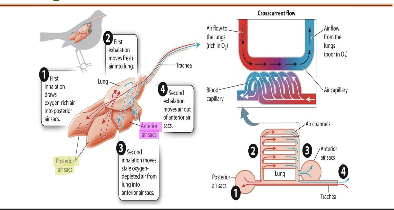
How is blood circulated in birds?
Birds: blood capillaries are perpendicular to the alveoli sac -> crosscurrent flow
Allows gas exchange and efficient
Lungs always received fully oxygenated blood
Perpendicular exchange allows efficient exchange of gases
Bird lung always have oxygenated air
Residual volume in human lungs, not always fully oxygenated air

What controls respiration?
Animals adjust their respiratory rate to meet their cells’ changing demand for O2 .
unique: by both the voluntary and involuntary components of the nervous system
What does homeostasis depend on for breathing?
often depends on sensors that monitor the levels of the chemical being regulated
sensors = chemoreceptors
What are chemoreceptors?
located within the brainstem
in sensory structures called the carotid and aortic bodies.
sense CO2 and H+ concentrations
Bifurcation definition?
the division of something into two branches or parts.
What are carotid bodies?
sense O2 and proton (H+ ) concentrations of the blood going to the brain.
At bifurcation of common carotid are carotid bodies
Carotid and aortic monitor the oxygen and acid level of the blood going to the brain
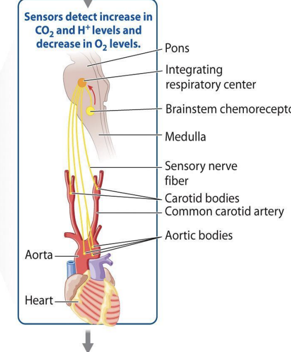
What are aortic bodies?
monitor their levels in blood moving to the body.
in the aorta
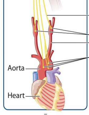
What is the aorta?
Aorta: largest blood vessels that comes out from heart
Supplies oxygenated blood to whole body
When blood goes out of level -> Aorta body and branch of Aorta goes towards neck (common carotid artery)
What happens when concentration of CO2 in blood is too high?
chemoreceptors in the brainstem stimulate motor neurons → activate the respiratory muscles to contract more strongly or more frequently.
Stronger or faster breathing rids the blood of excess CO2 and increases the supply of O2 to the body.

What parts of the brain are used for respiration?
Brain: pons and medulla

What is the medulla?
regulates essential, involuntary life-sustaining functions, including breathing, heart rate, blood pressure, and digestion
What are pons?
Pons: resp center that controls the transition of inhalation controls the transition of inhalation to exhalation
Controls the rate of respiration and in the brain stem

What is the brain stem?
Brain stem chemo receptors
Chemo receptors sense the concentration of CO2 and acidity in the brain
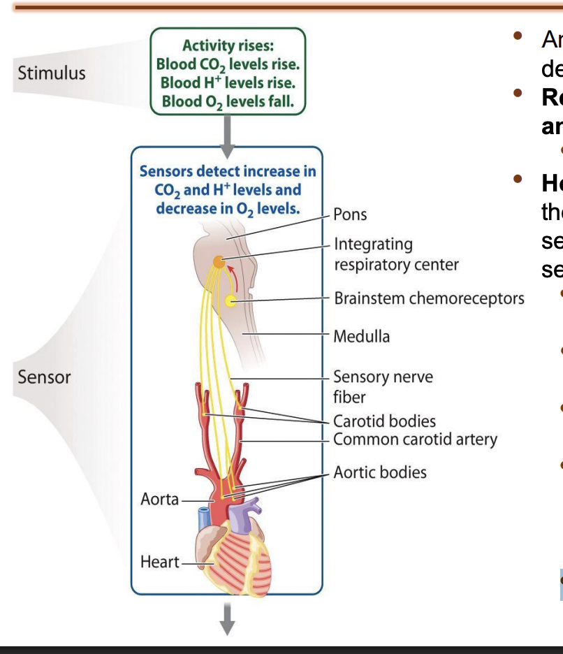
How does chemoreceptors, carotid, and aortic work together?
Chemoreceptors carotid and aortic check level of O2, CO2, and acidity
During aerobic resp use lots of oxygen -> end product can create accumulation of acid (lactic acid)
Exercise lactic acid produced -> change of pH of blood detected by chemoreceptors and ceratoid bodies
If concen of CO2 is too high -> resp center in pons to increase rate of resp and to clear out the excess CO2 by exhalation
Hyperventilation to increase removal of CO2 and inhalation of O2
Negative feedback that increase rate of resp

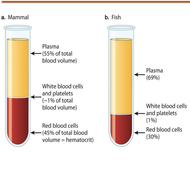
What is mammalian blood made of?
composed of plasma, white blood cells, platelets, and red blood cells.
What is hematocrit?
fraction of total blood volume that is red blood cells
Fish vs human hematocrit levels?
A human has a hematocrit of 45%
whereas a fish has a hematocrit of 30%.
Less hematocrit
Higher plasma fraction
Mammal and fish blood
Plasma
1% white blood cells and platelets
Red blood cells
Fraction of redblood cells = hematocrit

What is blood plasma?
Blood plasma is the fluid portion of blood without the cells.
Plasma not used for transfusion because it has low oxygen carrying capacity

What happens with low hematocrit levels?
Blood with a lower hematocrit flows with less resistance but carries less oxygen.
It can hold only as much O2 or CO2 as can be dissolved in solution.

What determines how much O2 can dissolve in solution?
a measure of its solubility.
CO2 is 30% highly soluble in solution compared to O2
In blood, less soluble oxygen compared to CO2
CO2 is 30x times more soluble than oxygen → only about 0.2 ml of O2 can be carried in 100 ml of blood.

Why do mammals have less plasma?
Dissolved concen of oxygen is low compared to CO2 -> compensated by the presence of higher hemocrit in mammals
Red blood cells have hemoglobin
Hemoglobin binds to oxygen molecule
High carrying capacity -> more mole of oxygen are carried and stored by red blood cells than plasma
Why do fish have more plasma?
Fishes have more plasma because they use dissolved O2 in water (less O2 in water than air)
Fish have lower metabolic rates, meaning they require less oxygen transport via red blood cells, allowing them to function with a higher proportion of low-viscosity plasma.
When dissolved in water -> pp of O2 is low
Rely more on oxygen dissolved in plasma = higher plasma concen -> Higher plasma volume + dissolved O₂ helps compensate for the lower oxygen availability in water (and lower hemocrit) → still provide efficient gas exchange capacity

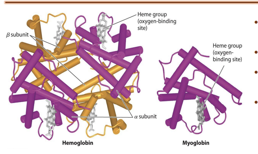
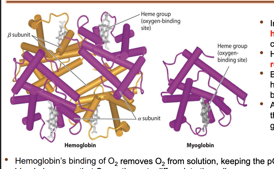
What is hemoglobin?
hemoglobin evolved as a specialized ironcontaining, or heme, molecule for O2 transport
the cells and the blood their red appearance
Has two subunits
A subunit: made of proteins
B subunit: main oxygen-carrying protein in adult human red blood cells
+ Heme group

What is a heme group?
contains an iron atom,
reversibly binds one O 2 molecule
Given time point 1 mol of heme bind to 4 O2
What does hemoglobin do in blood?
By binding O2 and removing it from solution, hemoglobin increases the amount of O2 in the blood a hundredfold.
After O2 diffuses into the blood, it diffuses into the red blood cells and binds to the heme groups in hemoglobin.
Hemoglobin’s binding of O2 removes O2 from solution → keeping the pO2 of the red blood cells below that of the blood plasma so that O2 continues to diffuse into the cell.
removal of O2 from the plasma → keeps the pO2 of the plasma below that of the lung alveolus → O2 continues to diffuse from the lungs into the blood.
Because of its greater solubility → CO2 is transported in solution within the blood
What happens to most CO2 in the body?
Most of the CO2 (about 95%) in the blood is converted to carbonic acid → dissociates to form bicarbonate ions (HCO3– ) and protons.
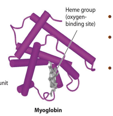
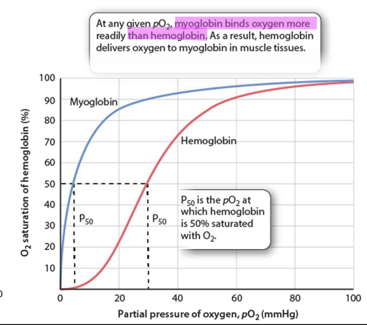
What is myoglobin?
a monomer that contains only a single heme group
a specialized O2 carrier within the cells of vertebrate muscles.
What happens in myoglobin?
Myoglobin protein that is present in skeletal muscles
Slow twitch fibers have more myoglobin
Myoglobin
Only one heme -> only one oxygen can bind
Affinity to bind with Oxygen is higher
What happens in hemoglobin?
4 Heme groups → higher carrying capacity for oxygen
What happens during excerize?
Hemoglobin that dissociates from oxygen and provides oxygen to tissue
Once oxygen is replenished -> usage of oxygen in tissue level creates low partial pressure -> allows myoglobin to release at low pp
When exercising, using oxygen bound to hemoglobin followed by myoglobin
Why slow twitch fibers can go for longer
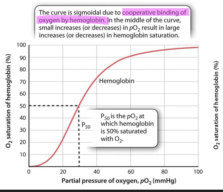
What does the dissociation curve of hemoglobin look like?
Sigmoidal due to the cooperative binding of oxygen by hemoglobin
Begins with slowly -> follows exponential phase -> platues out at 100% near saturation
Graph
Begins with slowly -> follows exponential phase -> platues out at 100% near saturation
Hemoglobin has four heme that combines to 4 oxygen atom
When first atom binds to first heme -> brings conformational changes in heme -> changes its affinity and increases the affinity of the next heme to bind more rapidly to the next oxygen
Next oxygen binds quickly -> cycle repeats
Confirmational changes allows for oxygen to bind with higher affinity = incorporated binding
Region during early phase is the undergoing of corporates binding
As soon as first O2 bind with first hemo -> picks up pace -> all four heme occupied by O2 -> saturates
P50 = out of the 4 heme, 2 are occupied by O2
What does the curve of myoglobin look like?
Only one heme -> no incorporative binding -> does not have a sigmoid curve -> able to bind more strongly to oxygen
Add very low oxygen pressure allows for release
At low pp of O2 -> myoglobin releases oxygen

How does myoglobin and hemoglobin work together?
at any pO2, myoglobin binds oxygen more readily than hemoglobin → hemoglobin delivers oxygen to myoglobin muscle tissues
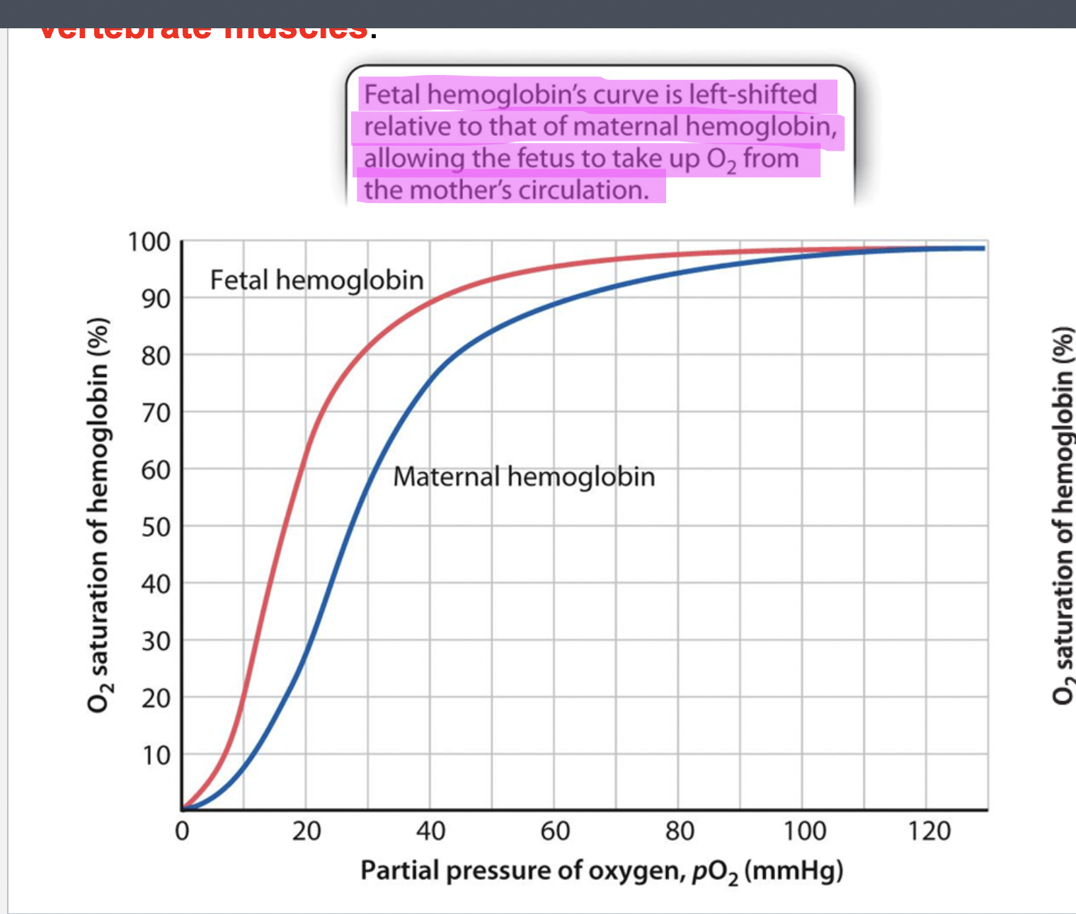
What does fetal hemoglobin levels look like?
Fetal hemo curve is shifted left compared to maternal hemo
allows fetus to take up O2 from mother’s circulation
Fetal hemoglobin
During digestion period -> placenta provides nutrients and oxygen to growing fetus
Even through fetus and maternal circulation is close (placenta connects circulation -> closed network with mother)
Pp is not favorable for rapid exchange
To overcome during digestional period
Fetal hemoglobin have higher affinity to oxygen (less than myoglobin but more than maternal hemoglobin)
Fetal shifts towards left-> allows high affinity in response to unfavorable pp of oxygen
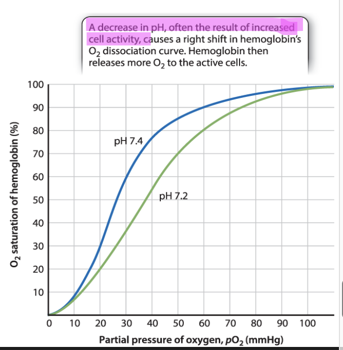
How does decrease in pH effect hemoglobin?
Decreased pH often result of increased cell activity
Decrease in pH caused by metabolism -> affects disassociation of O2
When pH = 7.4 (physiological pH of blood)
PH more acidic
Right shift of dissociate curve
Allows hemoglobin to release more O2 to cells
Right shift in hemoglobins O2 disassociation curve → hemo releases more O2 to active cells
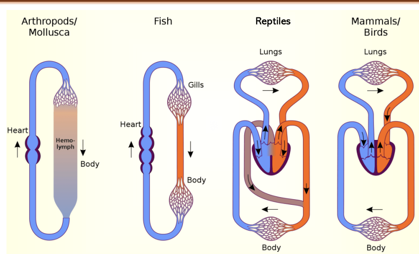
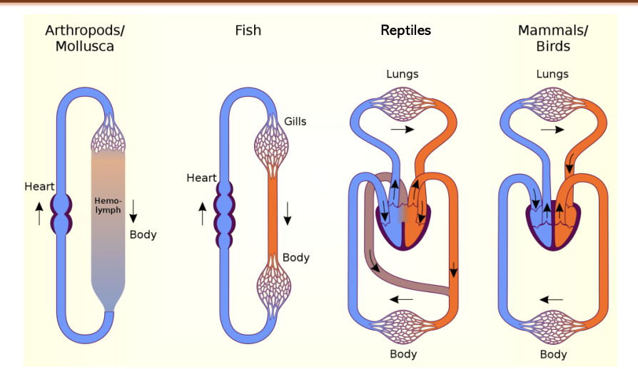
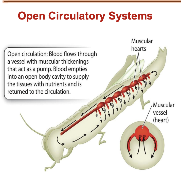
What is an open circulatory system?
Invertebrates
Still have blood visceral organ and heart to pump blood
Visceral organs bathed in blood
Contraction of muscles around periphery help in pushing the oxygenated blood towards the heart -> pump the oxygenated blood though the aorta
Open circulatory system supported by tracheal resp system
Trachiel tubes bathing the visceral organs and muscles -> tracheal systems allow the efficient gas exchange in the open circulatory system

How does circulation work in the circulatory system?
Blood flows through vessel with muscular thickenings (act as pump) → blood empties into open cavity to supply tissues with nutrients → returned tot he circulation
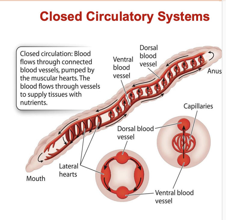
What is a closed circulatory system?
Vertebrates
Segmentation requires better circulatory system for distribution of oxygen throughout segments
Ventral blood vessel and dorsal blood vessel
have lateral hearts
Ventral blood vessel and btwn dorsal are blood capillaries that allows blood exchange to occur form tissue to pulmonary circulation to tissue

How does circulation occur in closed circulatory systems?
Blood flows through connected blood vessels
pumped by muscular hearts
Blood flows through vessels to supply tissues with nutrients
What is laminar flow?
Blood vessels
Blood that flows into blood vessels called laminar flow
Laminar flow
Cylinder of pipe and water flowing through it
Water flow through -> flows in concentric layer
Inner most layer of pipe is the one that faces the least amnt of resistance to flow
Inner most layer flows the fastest
Ex
Fish has streamline shape -> provides middle part to be the farther end
Fluid flow in the concintric layer = laminar flow
What is needed for blood to flow through pipes?
pressure (P) is required to overcome the resistance (R) to flow.
What is the rate of blood flow?
rate of blood flow is governed by P/R,
rate of blood flow increases with an increase in pressure
decreases with an increase in resistance.
What is the resistance to flow determined by?
determined in part by the fluid’s stickiness (viscosity)
and vessel’s length.
Longer vessels of a given size impose greater resistance.
main factor: vessel’s radius (r).
What is resistance?
proportional to 1/r^4
if a vessel’s radius is reduced by half, its resistance to flow increases 16 times
What are the kinds of vessels?
Arteries
veins
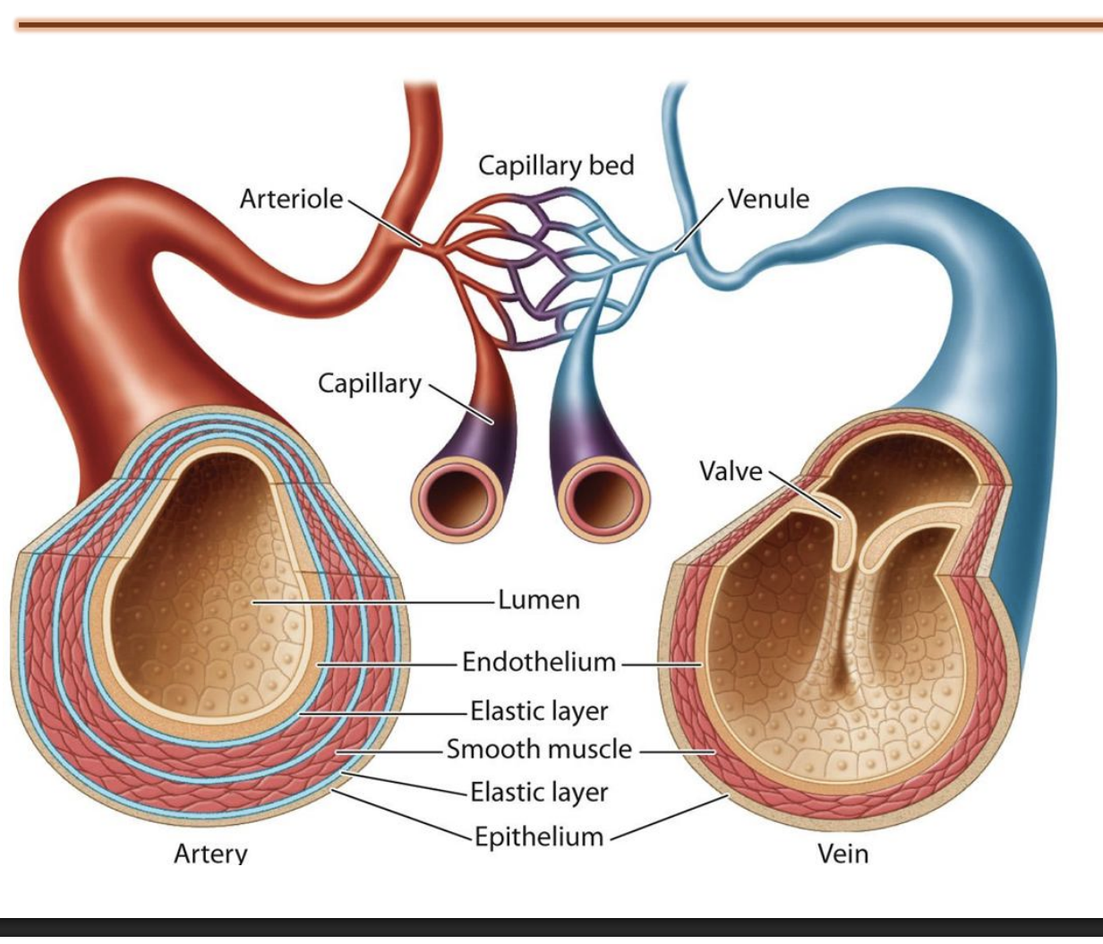
What are arteries?
Artery carry oxygenated blood
Two layers of elastic tissue -> withstand high blood pressure and be able to maintain structure
Recoiling ability: high blood pressure expand -> return to normal and recoil to normal size

What are veins?
Veins carry deoxygenated blood
Walls: collect deoxygenated blood from extremities of body and push it towards the heart
Since veins are working against gravity -> require mechanism to prevent backflow of blood -> walls prevent back flow

What is the structure of vessels?
Both artery and veins divide to form smaller blood vessels
Artery -> arteriole -> capillary bed
Veins -> no elastic layer but have walls -> prevent the back flow blood -> divide into venule -> capillary bed
Capillary bed have arteriole and venule end

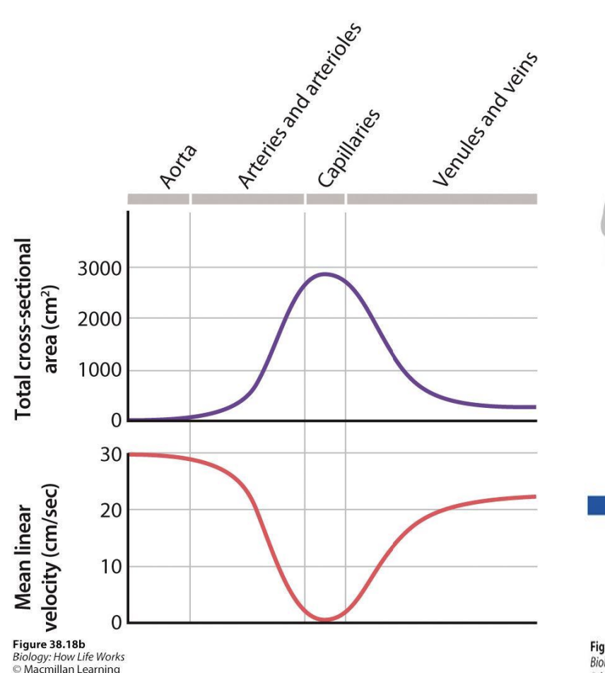
Which parts have the largest surface area?
Since capillaries are last branching part of arities and veins -> huge surface area
Max in capillary and least in aorta and veins