Physics to know for Exposures Class
1/37
Earn XP
Description and Tags
Fall '25
Name | Mastery | Learn | Test | Matching | Spaced |
|---|
No study sessions yet.
38 Terms
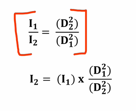
The inverse square law:
as distance increases, radiation intensity decreases
The SI unit of exposure for ionizing radiation is the:
coulomb per kilogram (C/kg)
this unit measures the amount of electric charge produced by ionizing radiation in a kilogram of air and has largely replaced the older unit, the roentgen (R)
The SI unit of absorbed dose for ionizing radiation is the:
gray (Gy)
which is defined as the absorption of one joule of radiation energy per kilogram of matter
in other words: “One gray means one joule of energy has been absorbed by each kilogram of the material”
The SI unit of dose equivalent for ionizing radiation is the:
sievert (Sv)
it measures the biological effect of radiation, taking into account not just the amount of energy absorbed but also the type of radiation and its impact on human tissue
The SI unit of radioactivity for ionizing radiation is the:
becquerel (Bq)
one becquerel is defined as one nuclear decay (or disintegration) per second.
What is Frequency (f)?
the number of waves passing by a given point per second
is measured in hertz (Hz) = 1 cycle per sec
What is a Wavelength?
the distance between any two successive points on a wave
represented by the Greek letter lambda λ
is measured from peak to peak (crests) or trough to trough (valley
If the wavelength of a beam of electromagnetic radiation increases, then its frequency must:
decrease
High frequency =
short wavelength
Low frequency =
long wavelength
X-rays originate in the:
electron shells
Gamma rays originate in the:
nucleus
Higher energy photons (e.g., x-rays and gamma rays) act more like:
Lower energy radiation (e.g., radio waves, microwaves, visible) act more like:
waves
Types of Ionizing Radiation:
— Alpha Particles (does the most damage if gets in)
— Beta Particles (less damage)
Electromagnetic Radiation is:
X-rays / Gamma Rays
Radioactivity is the process by which:
— an atom with excess energy in its nucelus emits particles and energy in order to regain stability
— this is known as: Radioactive Decay
Half Life:
the time it takes for ½ of the remaining atoms in an amount of the element to decay
What is the purpose of the protective housing?
— it provides mechanical support, electrical insulation, a thermal cushion, and absorbs leakage radiation
— reduces leakage to less than 100 mR per hour, as required by regulation.
The oil bath (draws heat away) and cooling fans are inside the:
House Tube
Heat from the x-ray production is removed through conduction by the heat traveling from:
the tube to heat tolerant materials
The anode is the positive end of the tube consisting of:
a target and an induction motor
The cathode is the negative end of the tube and consists of:
the focusing cup and filaments with its supporting wires
Glass envelope of tube has a high heat resistance, generally made of:
pyrex
X ray tube gives us three things needed for x-ray production:
— a high potential difference (kVp)
— a vehicle for electrons (mAs)
— and a target (anode)
What material is commonly used for the anode target and why?
Tungsten—due to its high atomic number, thermal conductivity, and high melting point
What are the two types of anodes?
rotating (used for high intensity) and stationary (used in dental/portable units)
What is the purpose of a rotating anode?
to spread heat over a larger surface area and allow higher intensity x-rays
STATOR:
made up of electromagnets arranged in pairs around the rotor (stationary)
ROTOR:
made of an iron core (ferromagnetic) surrounded by coils
What is the Line Focus Principle?
angling the anode allows for a large actual focal spot and a small effective focal spot
Large Focal Spot:
- Better heat dissipation
- Worse recorded detail
- Best for chest, abdomen x-rays
- (larger more spread out body parts, less actual bones that you are concerned with and viewing)
Small Focal Spot:
- Better recorded detail
- Worse dissipation of heat
- Better for extremities
The size of the actual focal spot depends on the size of the:
cathode filament being used
Actual focal spot size is the physical area on the target that is:
exposed to electrons from the tube current
The effective focal spot is the area projected onto the:
— patient and film
— it’s the x-ray beam as seen from below the tube
When there are body parts that do not have uniform thickness, position the thicker part of anatomy on which side?
the CATHODE side will result in an image with uniform density
Heat units are calculated by multiplying:
kVp x mA x s x c x number of exposures
*C= correction factor