Cell adaptations, ageing and accumulations
1/19
There's no tags or description
Looks like no tags are added yet.
Name | Mastery | Learn | Test | Matching | Spaced | Call with Kai |
|---|
No analytics yet
Send a link to your students to track their progress
20 Terms
What are the 5 mechanisms cells can use to adapt to sub-lethal injury
Hypertrophy = increase in cell size (post-mitotic cells)
Hyperplasia = increase in cell number
Atrophy = decrease in cell size
Metaplasia = differentiating into a different type of cell
Dysplasia = abnormal differentiation
What is atrophy a physiological response to?
Lack of use
Lack of innervation
Lack of stimulation
Describe the metaplasia that occurs in asthma
Ciliated respiratory epithelium in bronchi changes to goblet cells to produce more mucous. Too much metaplasia = maladaptive metaplasia = too much mucous produced causing breathing difficulties
Describe the metaplasia that occurs in the prostate secondary to testicular cancer
Unclear pathogenesis
Cancer produces oestrogen
Columnar glandular epithelium changes to stratified squamous
Prostate enlargement = urination problem and accumulation of debris cause prostatitis
Explain the metaplasia caused by vitamin A deficiency in parrots
Squamous metaplasia of respiratory and upper GIT epithelium
Mechanism poorly understood
Normal ciliated pseudostratified and glandular epithelium change to stratified squamous epithelium = produces keratin = hyperkeratosis (oral cavity, nasal lacrimal duct, upper alimentary tract, respiratory tract)
Keratin can block glands causing large keratin granulomas and rhinolith formation
Features of a dysplastic epithelium
Increased number of poorly differentiated or immature cells
5 microscopic features of dysplasia
Anisocytosis
Poikilocytosis (variation in cell shape)
Hyperchromatic nuclei
Karyomegaly
Increased size/number of nucleoli
Mitotic figures
= looks like neoplasia, so can be difficult to differentiate
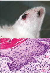
What is this?
Actinic keratosis (frequently develops into carcinoma)
What are
Labile cells
Quiescent cells
Senescent cells
Labile = constantly dividing cells (bone marrow, enterocytes, keratinocytes)
Quiescent = not currently dividing but can be easily stimulated to divide (liver)
Senescent = cells that are told to not divide but remain metabolically active (tumour suppressor genes or damaged DNA)
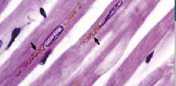
What is this pigment accumulating in aged post-mitotic cells and describe what it is and 2 pathological causes
Lipofuscin
Accumulation of undigestible lipoproteins and pigments
Pathology:
lipofuscinosis (genetic)
Phalaris poisoning (plant, ruminants)
Can cause impaired function of neurones in excessive amounts
Example causes of hypopigmentation (2)
Vitiligo
Melanin intolerance - some skin diseases of dermo-epidermal junctions e.g. pemphigus
Name the 4 haematogenous blood and bile pigments and what are they
Haemoglobin = red staining due to contact with blood
Haemosiderin = brown in macrophages due to recently phagocytosed blood at areas of bleeding - seen histologically
Haematoidin = green in chronic bruising
Bilirubin = yellow in jaundice
What is porphyria and what changes does it cause
Heme synthesis disorder
Deposition of porphyrin pigments in tissues
Teeth and bones turn pink, and fluoresces under UV light
What porphyrin-related condition is seen in Limousin cattle?
Congenital erythropoietic poryphria
Caused by a different enzyme
No tooth discolouration
Causes photosensitivity
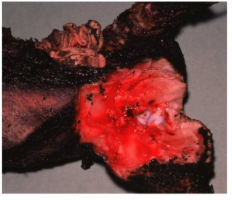
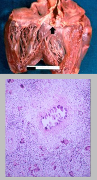
Cause of this muscle pigmentation
Carbon monoxide poisoning - carboxyhaemoglobin
Describe causes of methaemoglobinaemia
Methaemoglobin is constantly being formed then reduced to haemoglobin via the methaemoglobin reductase pathway
Increased methaemoglobin formation due to marked oxidant exposure *red maple in horse. can progress to haemolytic crisis)
Associated with Heinz bodies
Blood and mucous membranes appear brown
Describe cause of haemoglobin imbibition
Freezing and thawing of cadavers causes erythrocyte lysis, resulting in haemoglobin leaking out and staining nearby tissues
Static clotted blood in large blood vessels and atria can leak haemoglobin due to RBC lysis
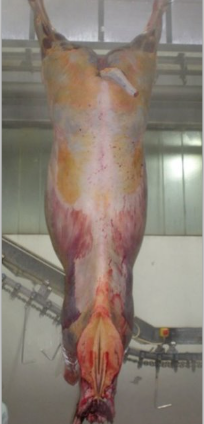
Name and explain the cause of this pigmentation on a horse.
Carotenoids
Present in leafy vegetables
Yellow colour in adipose tissue of the animals that eat the vegerables.
Usually only seen in horses and Channel Island cattle, as other breeds seem to metabolise them in a different way
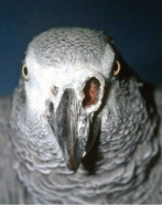
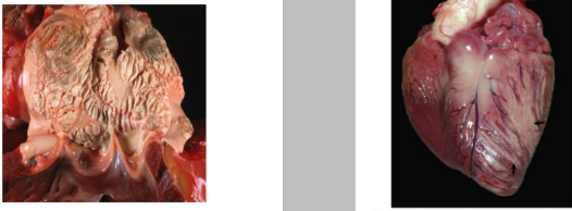
What is pathological calcification and name and explain 2 types
Deposition of calcium salts in soft tissue - usually phosphates and carbonates
Metastatic calcification (left) = due to increased calcium levels
Dystrophic calcification (right) = secondary to necrosis
Describe the gross and histological appearance of calcification
Gross:
palpable calcium granules
feel knife grating upon sectioning
chalk-white colour usually. Can be yellowish/green due to eosinophils if parasites are involved
Histo:
dark blue colour
microtome mark due to shattering of tissue
Von Kossa stain turns it black