Nervous system I - Nervous Tissue
1/17
There's no tags or description
Looks like no tags are added yet.
Name | Mastery | Learn | Test | Matching | Spaced | Call with Kai |
|---|
No analytics yet
Send a link to your students to track their progress
18 Terms
Axon of multipolar neuron
Location:
Central nervous system
Peripheral nervous system
Description:
Single, cylindrical process extending from soma (cell body) of neuron
Arises from axon hillock
Usually without branches near cell of origin
Terminates as axon terminal on other neurons or effectors
Function:
Conveys efferent nerve impulses (i.e., away from soma)
Conveys information to other neurons or effectors
Also known as:
Nerve fiber
Comment:
Axons with diameter greater than 2 µm usually myelinated
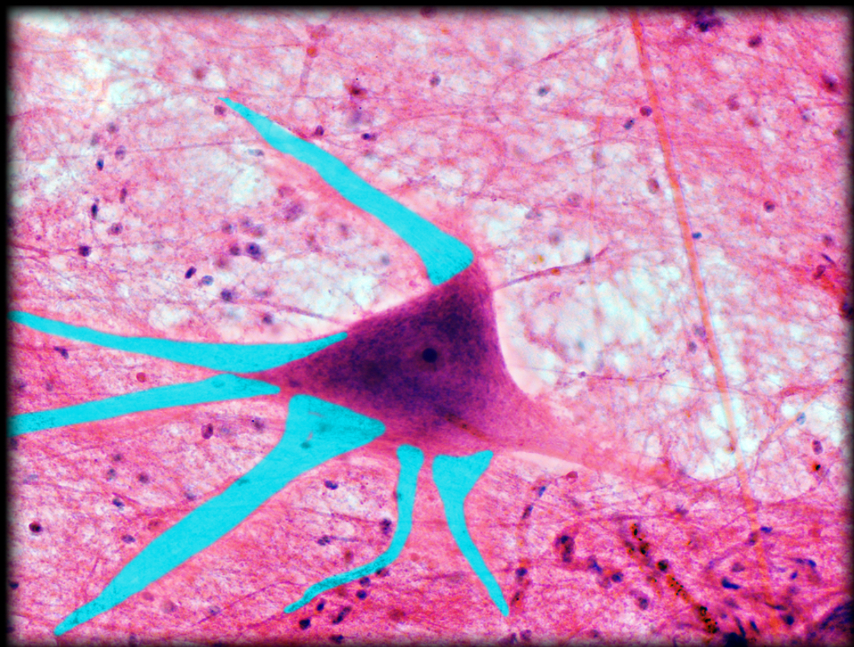
Dendrites of multipolar neuron
Location:
Central nervous system
Ganglia of autonomic nervous system
Description:
Tapered, highly branched processes extending from soma (cell body) of neuron
May have specialized sensory receptor (e.g., pressure receptor in hypodermis)
Function:
Convey afferent nerve impulses (i.e., towards soma)
Receive information from other neurons
Comment:
Not myelinated
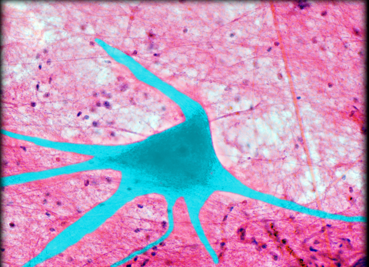
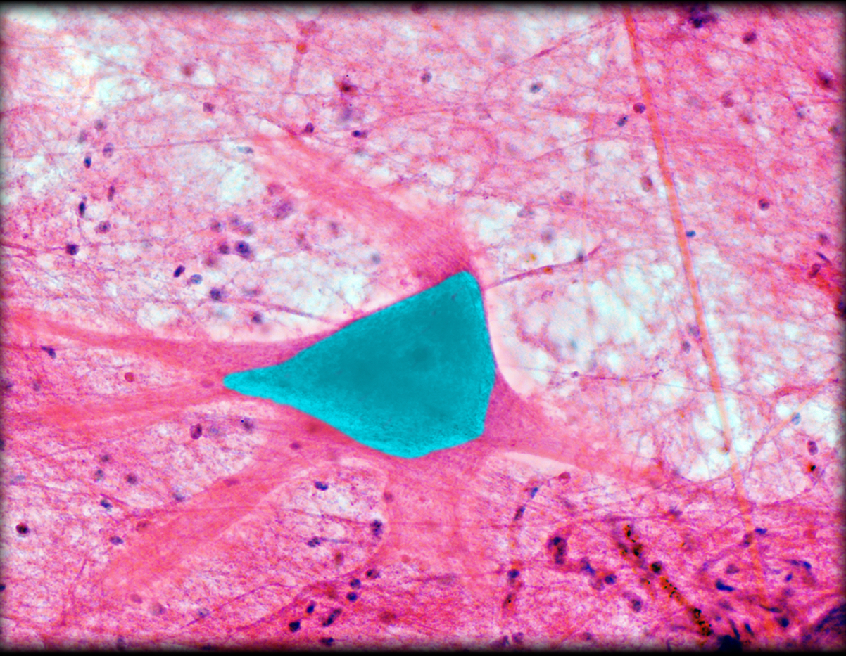
Multipolar neuron
Location:
Central nervous system
Ganglia of autonomic nervous system
Description:
Composed of soma (cell body), multiple dendrites, and a single axon
Function:
Soma contains nucleus and other cytoplasmic organelles that support neuron structure and function
Dendrites convey afferent nerve impulses (i.e., towards soma)
Axon conveys efferent nerve impulses (i.e., away from soma)
Comment:
Neurons are the functional unit of nervous system
Three structural categories of neurons; unipolar, bipolar, and multipolar
Soma of multipolar neuron
Location:
Central nervous system
Ganglia of autonomic nervous system
Description:
Cell body of neuron
Two types of processes: dendrites and axons
Contains Nissl bodies
Function:
Nucleus and other cytoplasmic organelles support neuron structure and function
Comment:
Axons convey efferent nerve impulses (i.e., away from soma)
Dendrites convey afferent nerve impulses (i.e., towards soma)
Nissl bodies (dark-staining region) represent aggregates of rough endoplasmic reticulum
Cytoplasm of Schwann cell
Location:
Schwann cell (soma and outermost layer of myelin sheath)
Description:
Amorphous substance between plasma membrane and nuclear membrane
Contains nucleus and other organelles
Contains cytoskeleton, enzymes, nutrients, and other proteins
Also known as:
Cytoplasm also known as cytosol
Comment:
Inner layers of myelin sheath lack cytoplasm
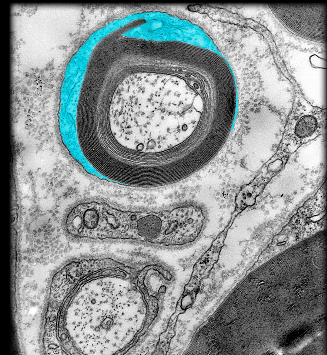
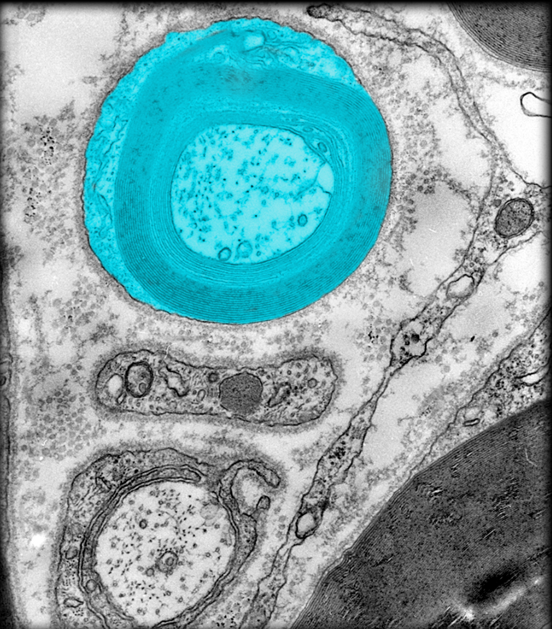
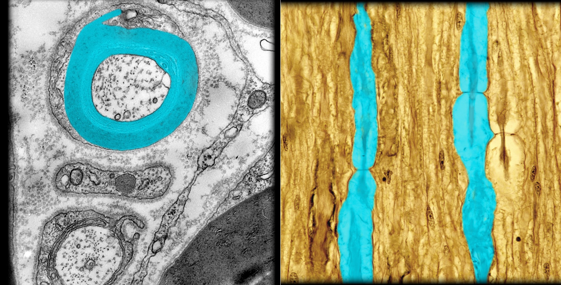
Myelin sheath
Location:
Surrounds myelinated axon
Description:
Formed by oligodendrocyte in central nervous system or Schwann cell in peripheral nervous system
Cell membrane wrapped spirally (up to 100 times) around axon
Lacks cytoplasm between myelin layers
Composed of a series of short segments (0.2-1.0 mm) of myelin wrappings on axon
Myelin sheath segments are called internodes
Function:
Maintains action potential conduction along axon
Myelin serves as electrical insulator
Increases speed of nerve impulse conduction
Also known as:
Neurilemma (neurolemma)
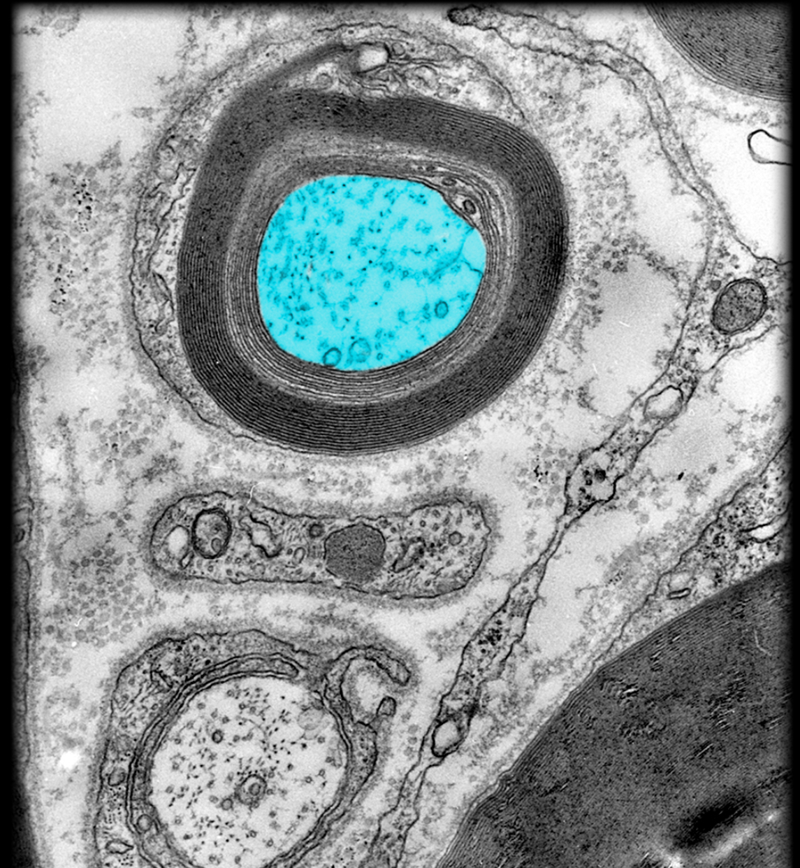
Myelinated axon
Location:
Central nervous system (CNS)
Peripheral nervous system (PNS)
Description:
Axon (usually 2 µm) wrapped by myelin sheath (layered spirals of myelin)
Function:
Conveys efferent nerve impulses (i.e., away from soma)
Conveys information to other neurons or effectors
Myelin sheath increases speed of nerve impulse conduction
Comment:
Nerve impulses travel faster (3-15 m/sec) in small myelinated axons than in unmyelinated axons of similar size (0.5-2.0 m/sec)
Myelinating cell is oligodendrocyte (CNS) or Schwann cell (PNS)
Aggregates of myelinated axons in CNS known as white matter
Schwann cell
Location:
Peripheral nervous system
Description:
Neuroglial cell
Function:
Forms myelin sheath around a single axon
Myelin sheath increases speed of nerve impulse conduction
Structural and metabolic support for axons
Also known as:
Neurolemmocyte
Comment:
Myelin sheath is cell membrane wrapped spirally (up to 100 times) around axon
Oligodendrocyte in CNS can form myelin sheath around more than one axon
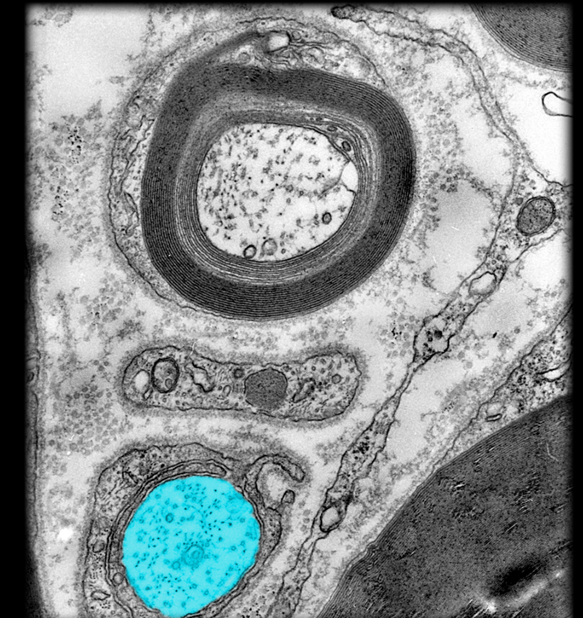
Unmyelinated axon
Location:
Central nervous system (CNS)
Peripheral nervous system (PNS)
Description:
Axons lacking a myelin sheath
Unmyelinated axons rest in invaginations of Schwann cell or oligodendrocyte
Function:
Conducts nerve impulses
Comment:
Nerve impulses travel slower in unmyelinated axons (0.5-2.0 m/sec) than in small, myelinated axons of similar size (3-15 m/sec)
Myelinating cell is oligodendrocyte (CNS) or Schwann cell (PNS)
Mitochondrion in presynaptic terminal
Location:
Presynaptic terminal
Description:
Membrane-bound organelle
Function:
Synthesize adenosine triphosphate (ATP)
Comment:
Origin of mitochondria as an organelle: thought to be prokaryotic organisms (like bacteria) that formed symbiotic relationship with anaerobic eukaryotic cells: "mitochondria" received protection and nutrients, the cell received a chemical energy source
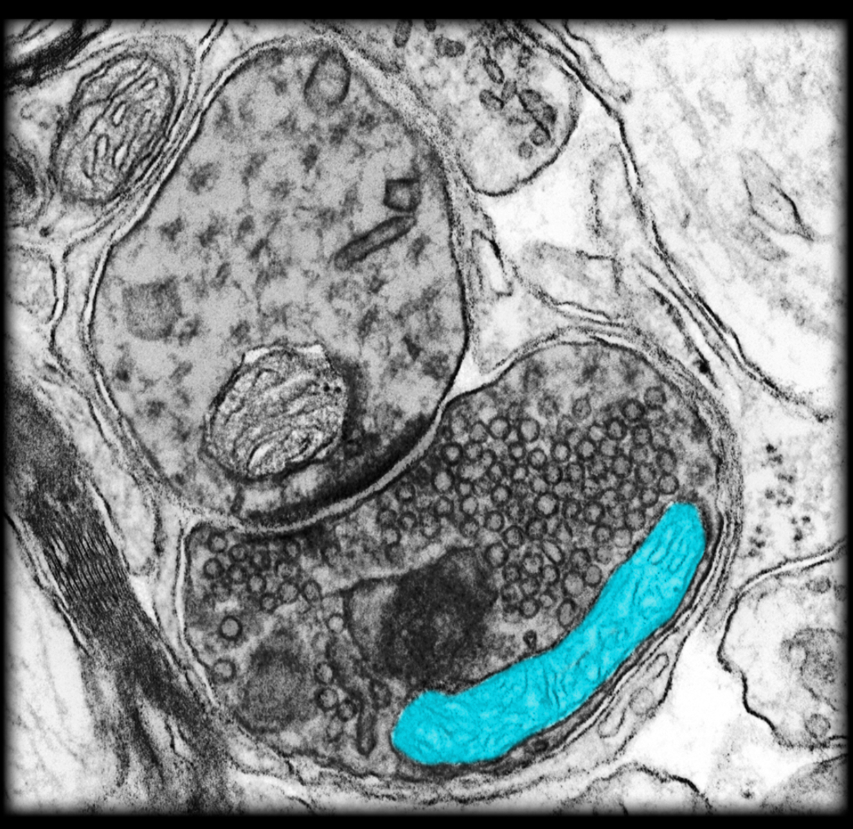
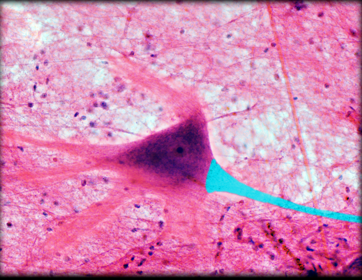
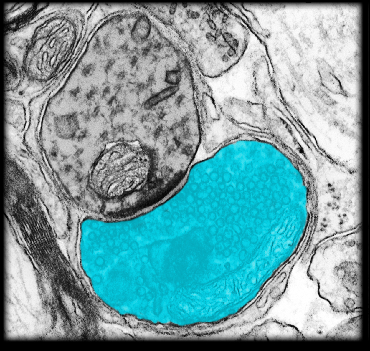
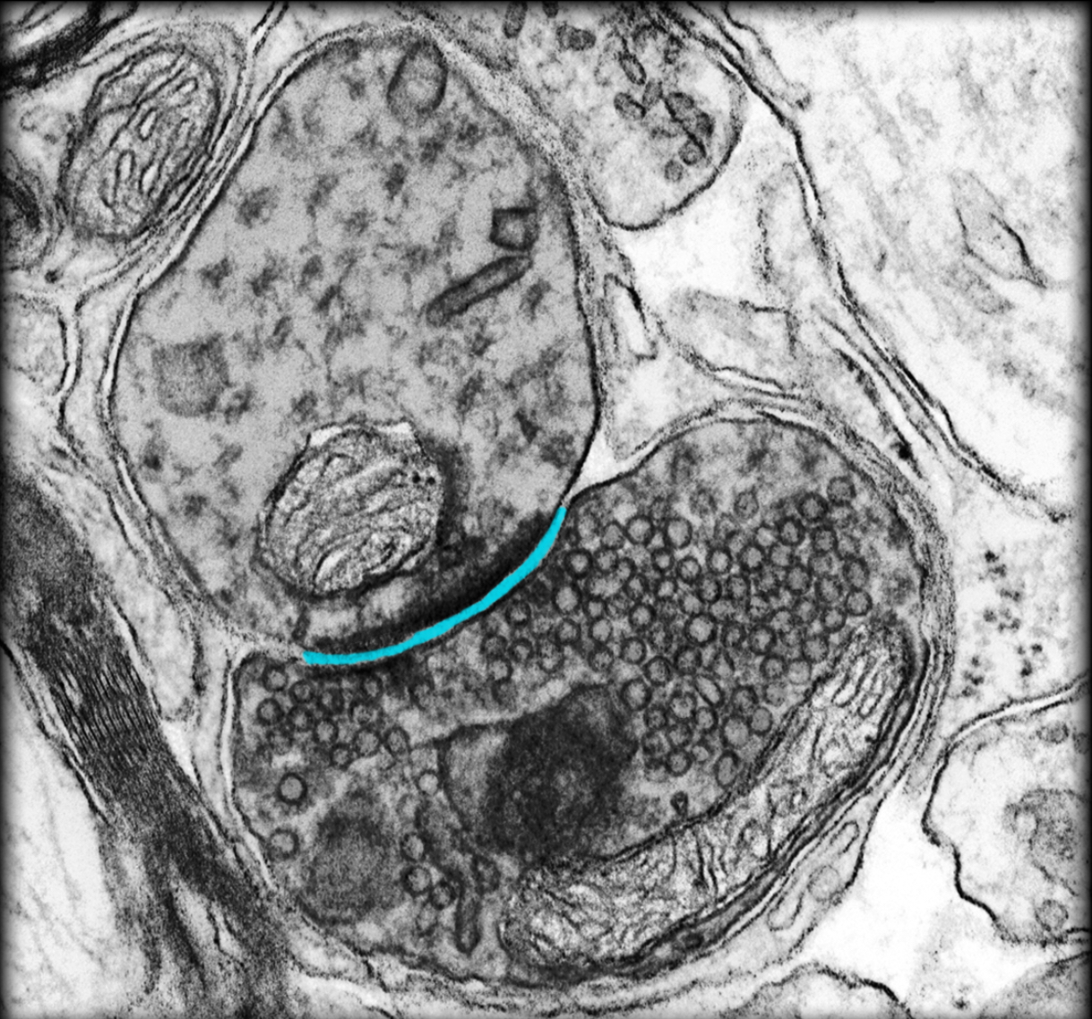
Presynaptic terminal
Location:
Distal end of axon
Description:
Bulbous swelling containing synaptic vesicles
Contains presynaptic membrane
Contains mitochondria and synatic vesicles
Function:
Delivers neuronal impulses to (chemical) synapse
Accumulates and recycles synaptic vesicles (containing neurotransmitter)
Comment:
Presynaptic terminal is unmyelinated
Synapse
Location:
Junction between axon or dendrite and an effector
Description:
Contact between two neurons or a neuron and an effector
Includes presynaptic membrane, synaptic cleft, and postsynaptic membrane
Function:
Transmit neuronal impulse by use of neurotransmitter (chemical synapse)
Comment:
Synaptic effectors include neuronal dendrites, axons, or somata, and non-neuronal cell types (e.g., muscle)
Neurotransmitters are chemical agents (e.g., acetylcholine, norepinephrine) released by presynaptic cell at chemical synapse
Electrical synapse involves current (ion) flow through gap junctions between adjacent cells
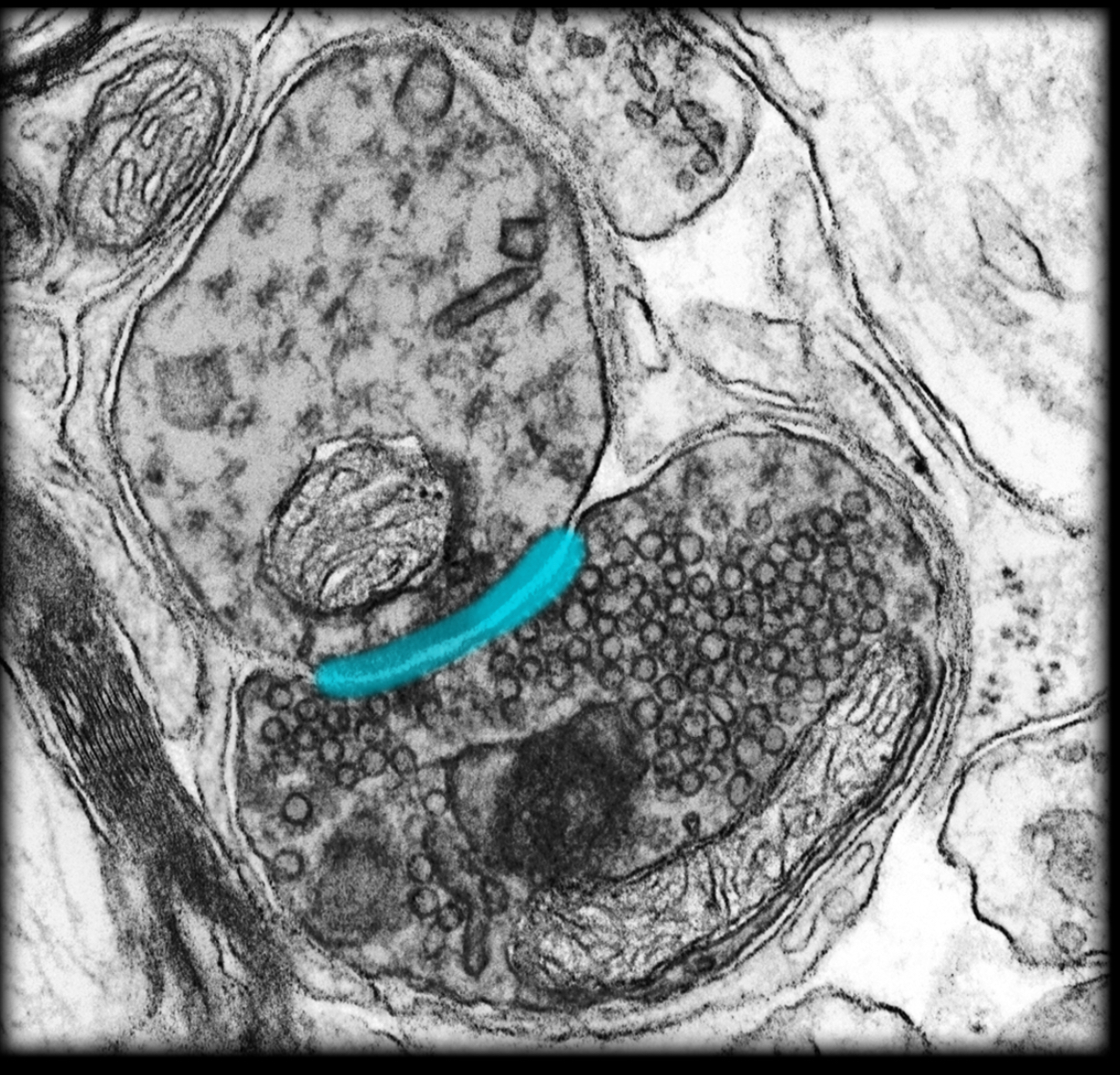
Synaptic cleft
Location:
Chemical synapse
Between neurons, or between neuron and effector
Description:
Narrow intercellular gap
Defined by presynaptic and postsynaptic membranes
Function:
Neurotransmitter released from presynaptic membrane diffuses across cleft to postsynaptic membrane
Comment:
Neurotransmitters are chemical agents (e.g., acetylcholine, norepinephrine) released by presynaptic cell at chemical synapse
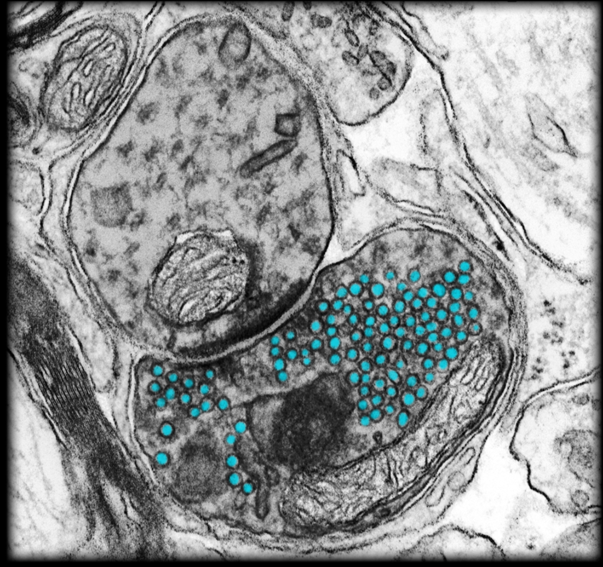
Synaptic vehicles in presynaptic terminal
Location:
Presynaptic terminal
Description:
Membrane-bound organelle
Small sac containing neurotransmitters
Function:
Storage and release of neurotransmitters
Comment:
Neurotransmitters are chemical agents (e.g., acetylcholine, norepinephrine) released by presynaptic cell at chemical synapse
Myelin sheath
Location:
Surrounds myelinated axon
Description:
Formed by oligodendrocyte in central nervous system or Schwann cell in peripheral nervous system
Cell membrane wrapped spirally (up to 100 times) around axon
Lacks cytoplasm between myelin layers
Composed of a series of short segments (0.2-1.0 mm) of myelin wrappings on axon
Myelin sheath segments are called internodes
Function:
Maintains action potential conduction along axon
Myelin serves as electrical insulator
Increases speed of nerve impulse conduction
Myelinated axon
Location:
Central nervous system (CNS)
Peripheral nervous system (PNS)
Description:
Axon (usually 2 µm) wrapped by myelin sheath (layered spirals of myelin)
Function:
Conveys efferent nerve impulses (i.e., away from soma)
Conveys information to other neurons or effectors
Myelin sheath increases speed of nerve impulse conduction
Comment:
Nerve impulses travel faster (3-15 m/sec) in small myelinated axons than in unmyelinated axons of similar size (0.5-2.0 m/sec)
Myelinating cell is oligodendrocyte (CNS) or Schwann cell (PNS)
Aggregates of myelinated axons in CNS known as white matter
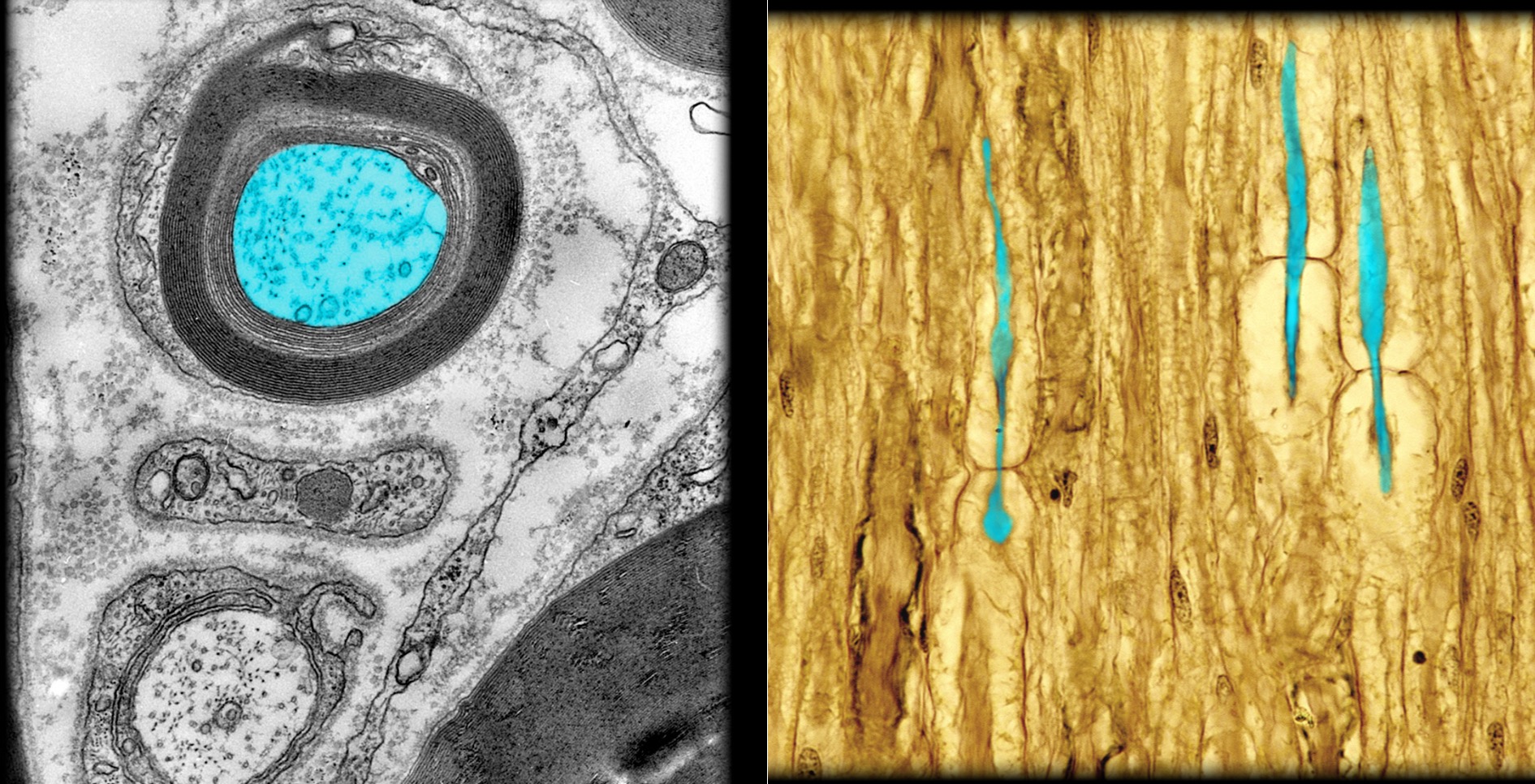
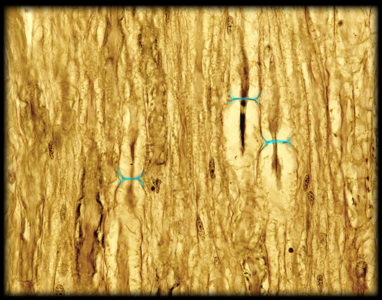
Node of fancier
Location:
Myelinated axon
Description:
Cleft between internodes of myelin sheath
Function:
Rapid nerve impulse conduction
Impulses "jump" from node to node (a process called saltatory conduction)
Comment:
Myelin sheath segmented (segments called internodes)
Latin: saltare = to jump
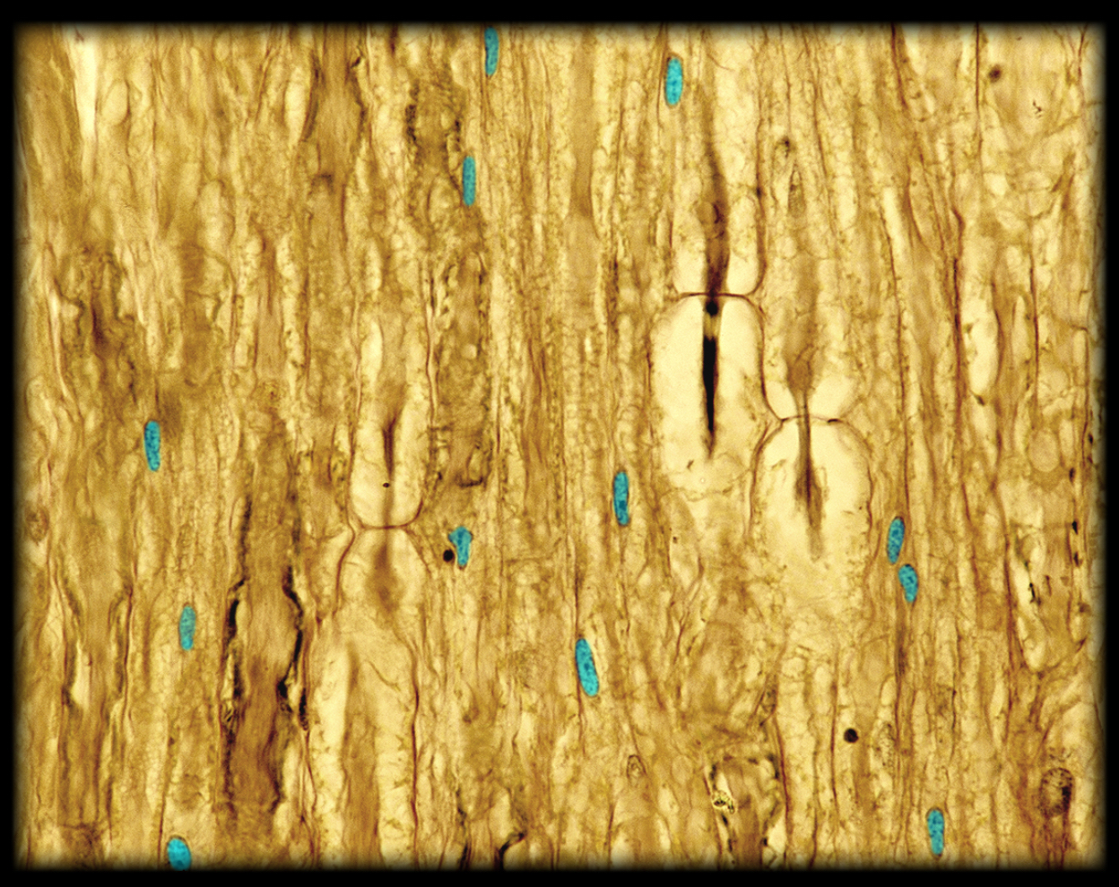
Nucleus of Schwann cell
Location:
Soma (cell body) of Schwann cell
Description:
Largest organelle (5-10 µm in diameter)
Spherical or ovoid structure
Nuclear envelope comprised of double membrane (i.e., two lipid bilayers)
Contains cellular DNA and nucleolus
Function:
DNA replication
DNA transcription into mRNA
Ribosomal RNA synthesis and ribosome subunit assembly (in nucleolus)
Comment:
Staining of DNA with basic histological dyes like hematoxylin depends on transcriptional activity: DNA undergoing active transcription, called euchromatin, stains lightly because it is less folded and, therefore, less dense; transcriptionally inactive DNA, or heterochromatin, is tightly packed and condensed and, therefore, stains more darkly
Micrometer (µm), also known as a micron, is 1/1,000 of a millimeter