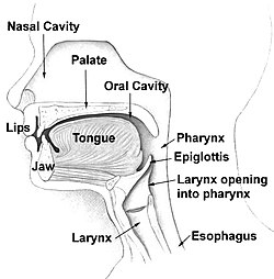
Pharynx
Throat
Muscular passageway for food and air
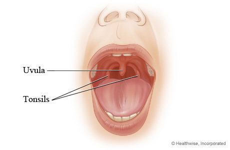
Uvula is found here
Tonsils are found here
Ulvula
Responsible for sealing off nasal cavity during swallowing
Tonsils
Responsible for trapping and removing bacteria/pathogens entering throat
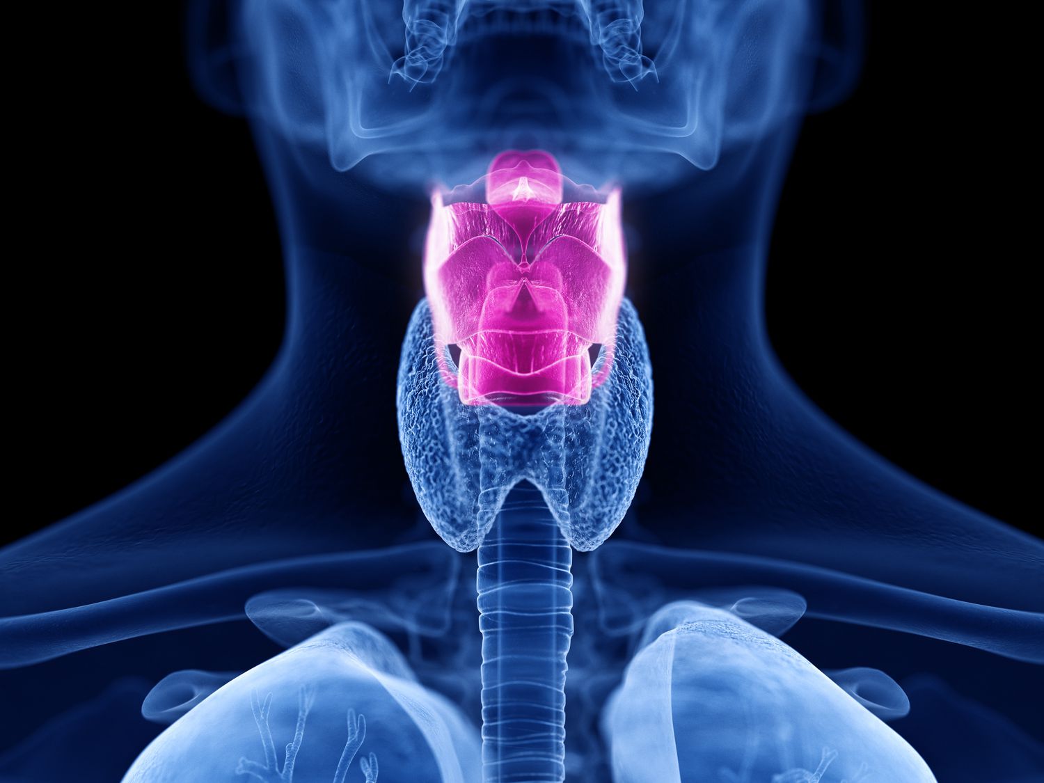
Larynx
Contains vocal cords
Epiglottis found here
Epiglottis
Responsible for sealing off trachea during swallowing
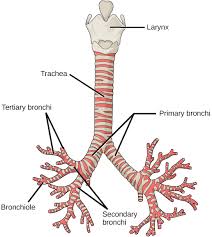
Trachea
Windpipe
Made up of hyaline cartilage
Lined with ciliated cells continuously beating opposite of the flow of air to filter and sweep material out of the lungs
Bronchi
Two large branches off of the trachea
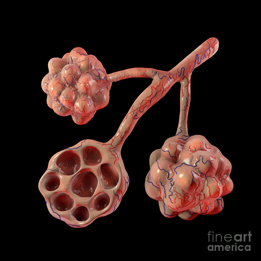
Bronchioles
Smaller branches off of bronchi- found in lungs
Lead to alveoli (gas exchange)
Alveoli
Very tiny air sacs that are the site of gas exchange
Gas exchange
Supplies oxygen for cellular respiration and disposes the waste product carbon dioxide
Where does gas exchange happen?
Between the capillaries of the circulatory system and the alveoli of the respiratory system
Mouth
Used for eating and speaking
Nasal Cavity
Filters and warms air to make moist before entering the lungs
Diaphragm
Muscle that contracts and flattens that created a vacuum for air to enter the lungs
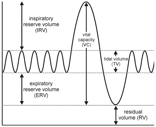
Tidal Volume (TV)
The volume of air breathed in and out without conscious effort
Inspiratory Reserve Volume (IRV)
The additional volume of air that can be inhaled with maximum effort after a normal inspiration
Expiratory Reserve Volume (ERV)
The additional volume of air that can be forcibly exhaled after normal exhalation
Vital Capacity (VC)
The total volume of air that can be exhaled after a maximum inhalation
VC= TV+IRV+ERV
Residual Volume (RV)
The volume of air remaining in the lungs after maximum exhalation (the lungs cannot be completely emptied)
Total Lung Capacity (TLC)
VC+RV
Minute Ventilation
The volume of air breathed in 1 minute (TV)(breaths/minute)
What is required for respirtation?
Larger surface area, thin membrane, moist respiratory surfaces
Pressure Gradient
O2 and CO2 diffuse where there pressures are higher to go to where they are lower
ALWAYS HIGH TO LOW GRADIENT
Steps of Gas Exchange
Inhalation
Oxygen diffuses from alveoli into deoxygenated RBCs
always flows with gradient
RBCs delivers oxygen to body, transports CO2 back to lungs
What is made of simple squamous epithelium?
Alveoli and capillaries (very thin membrane)
Oxyhemoglobin
Oxygen that attaches to hemoglobin in RBCs
Gas Transportation of Oxygen
Oxygen attaches to protein hemoglobin in RBCs
Gas Transportation of Carbon Dioxide
Transports as bicarbonate ions (70%)
Bound to hemoglobin (23%)
Dissolved in plasma (7%)
Inspiration
Air flowing into lungs
Expiration
Air leaving lungs
Intercostalis
Pulls ribs to elevate rib cage
Serratus Dosrsalis
Depresses rib cage
What does RBCs convert carbon dioxide ions to?
Carbonic Acid
Is Carbonic Acid stable?
No, must convert to bicarbonate ions and hydrogen ions (BUT NOT ALL)
What happens to the remaining hydrogen in RBCs?
Turns acidic, can cause shift in pH, but hemoglobin will bind to the ions to maintain pH levels.
Why is carbon dioxide converted for transportation in RBCs?
The conversion into bicarbonate allows for the continued uptake of carbon dioxide in the blood, due to the concentration gradient
What happens to the bicarbonate buffer system once it reaches the lungs?
Process reverses as it reaches the lungs
Benefits of Bicarbonate buffer system
Maintains pH levels
Maintains carbon dioxide concentration gradient
Respiratory Disorders
Chronic Obstructive Pulmonary Disease (COPD)
Emphysema
Chronic Bronchitits
Blood
Found in arteries and veins throughout the body
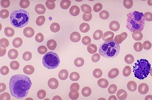
Red blood cell
Biconcave disks, anucleate (dark pink)
White blood cell
Nucleated, larger but less numerous than RBCs (purple)
Platelet
Tiny, fragments of cells responsible for clotting blood after an injury (tiny dots)
Plasma
Fluid matrix, contains various of substances
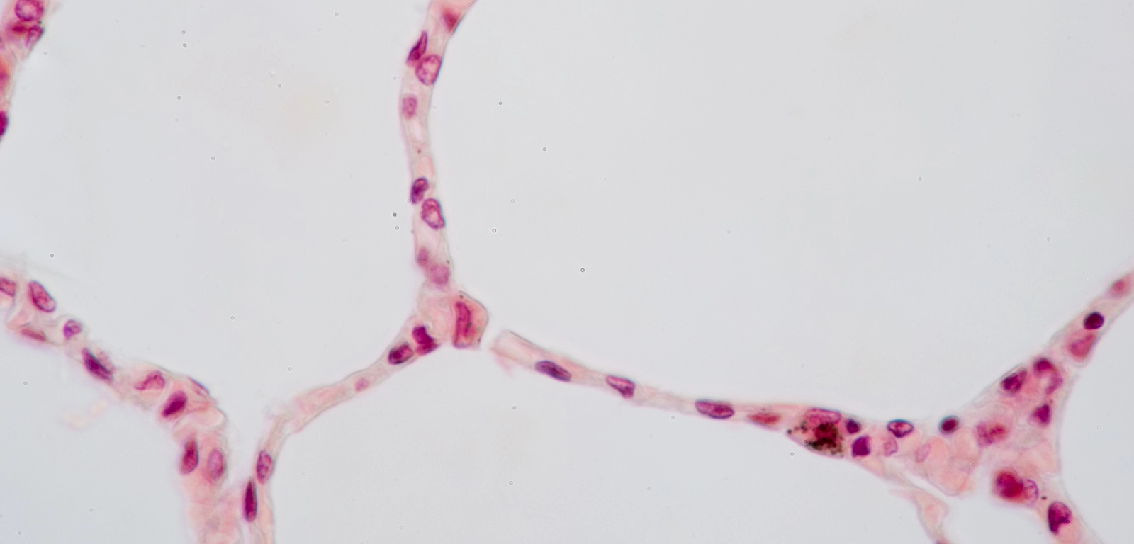
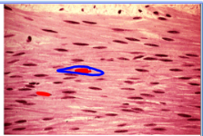
Simple Squamous
Found in alveoli
Empty space
Air
Nuclei in Simple Squamous
dark purple
Individual cell in Simple Squamous
Light pink, “squashed” shape (like a fried egg)?
Red blood cell in Simple Squamous
Dark pink, space between squamous layers, away from air space
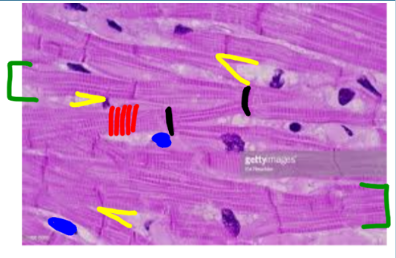
Cardiac Muscle
Lining atria and ventricles in the heart, myocardium
Nucleus in Cardiac Muscle
Blue
Muscle Fiber in Cardiac Muscle
Green, singular strand
Striation
Thin, small, vertical grooves (Red)
Intercalated disk in Cardiac muscle
Darker and less frequent than striations (Black)
Branch
A fork in the fibers (Yellow)
Smooth muscle
Lining hallow organs and lining arteries and veins