Looks like no one added any tags here yet for you.
State the purpose of the microscopic examination of urine.
The identification of formed elements
State an advantage and a disadvantage of performing urine microscopics based on macroscopic screening.
Advantage: Low cost
Disadvantage: Lack of standardization
List the seven chemical parameters commonly used as markers in macroscopic screening.
Blood
Protein
Nitrite
Leukocyte Esterase
Glucose
List the three CLSI recommendations for urine macroscopic screening.
Requested by Physician
Lab-specified patient population
Any abnormal physical or chemical results
What is an important step in specimen preparation to ensure an adequate urine sediment?
thoroughly mix patient specimen
Why is 10-12 ml indicated for a urine volume to perform an accurate urine microscopic?
to obtain an appropriate amount of sediment (1ml)
Why is the centrifugation speed or centrifugal force (RCF) important in standardization?
optimal amount of sediment without damaging elements
What is the recommended RCF and time of centrifugation?
400 RCF for 5 mins
Why is RCF a more accurate way to determine optimum speed of centrifuge?
It accounts for the differences in diameter of centrifuge tubes
What is the desired ratio of urine volume to sediment?
1 part sediment per 12 parts urine
Why should the amount of supernatant poured off be controlled?
to prevent accidental loss of sediment
Why would it be important to control the amount of sediment examined?
to obtain an accurate normal range
Discuss the advantages of using commercial system slides for sediment examination.
to control the amount of sediment
What magnifications should be used for sediment examination? What is the purpose of each magnification?
10x: Casts, general ID
40x: ID
Which cell can be used to focus on the correct plane in a urine specimen?
Epithelial Cell
Which urine elements are reported by lower power field? And high power field?
10x: casts
40x: RBCs, WBCs
If the sample is red, turbid, and tests positive for Blood and Protein, what microscopic elements can you expect?
RBCs
If the sample is turbid and tests positive for Protein, Nitrite, and Leukocyte Esterase, what microscopic elements can you expect?
WBCs
If the sample is turbid and tests negative for everything else, what microscopic elements can you expect to see?
Epithelial cells
If the sample is cloudy and tests positive for Protein, what microscopic elements can you expect to see?
Casts
If the sample is turbid, has a higher pH, and tests positive for Nitrite, and Leukocyte Esterase, what microscopic elements can you expect to see?
Bacteria
If the sample turbid, has color, an acidic pH and tests positive for Bilirubin, what microscopic elements can you expect to see?
Crystals
What are some advantages to using stain on urine sediment?
Increases visibility
Easier ID of nucleus, cytoplasm, and inclusions
List 6 urine sediment stains and their use
Crystal Violet: provides clearer delineation of structure and contrasting colors of the nucleus and cytoplasm
Acetic Acid: enhances nuclear detail of WBCs & epithelial cells
Lipid Stains: stains Triglycerides and neutral fats
Gram Stains: differentiation of gram-positive and gram-negative bacteria. ID of bacterial casts
Hansel Stain: urinary eosinophil detection
Prussian Blue Stain: Stains hemosiderin granules in a blue color
How does cytodiagnostic urinalysis differ from routine microscopic examination?
Cytodiagnostic urinalysis uses permanent slides
List 6 types of microscopy discussed for urine examination and list one function for each.
Bright-field: Routine urinalysis
Phase-contrast: enhances visualization of elements with low refractive index
Polarizing: aids in ID of cholesterol in oval fat bodies, fatty casts, and crystals
Dark-field: aids in ID of Treponema Pallidum
Fluorescent: allows visualization of naturally fluorescent or microorganisms stained by a fluorescent dye
Interference-contrast: produces a 3D microscopy image and layer-by-layer imaging of a specimen
What should the light intensity of the microscope be for examining urine sediment?
Low light
How is the light intensity regulated on the microscope?
Light condenser knob
What does birefringent property refer to in an object?
the ability to refract light in 2 directions
List two urine elements that have birefingent properties.
artifacts and crystals
List 6 formed elements that may be observed in urine sediment.
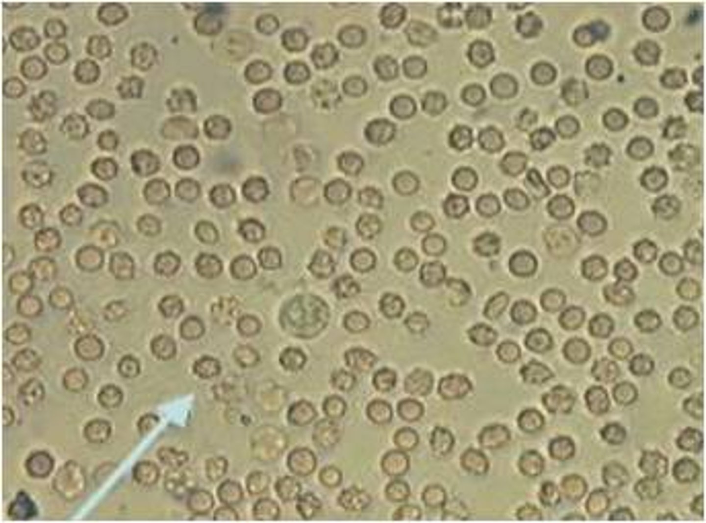
RBCs
WBCs
Crystals
Epithelial cells
Casts
Yeasts
What is the significance of red cells in urine?
Damage to glomerular membrane or vascular injury to genitourinary tract
Which elements may be confused with rbcs microscopically?
Yeasts
Oil droplets
Air bubbles
Starch
Which chemical test on dipstick should correlate with microscopic rbcs in urine?
Blood
Specific Gravity
Color
Describe the appearance of rbc in hypersthenuric urine and hypothenuric urine.
Hypersthenuric: crenated
Hypothenuric: Ghost cells
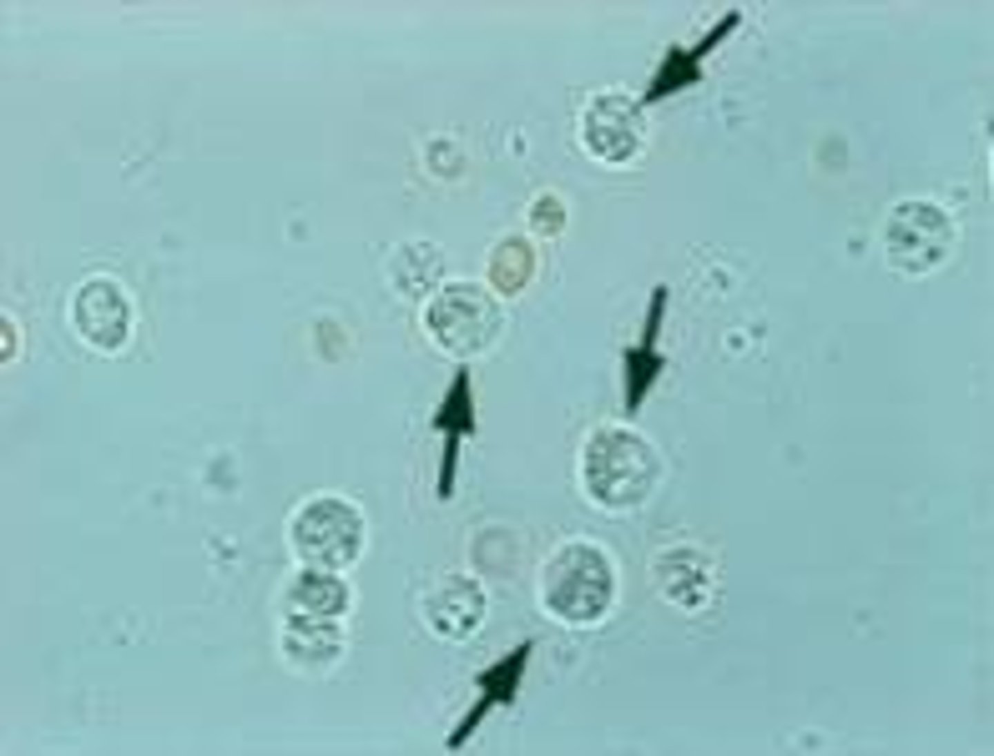
What is the significance of white cells in urine?
Infections
Immune system response
What is a glitter cell?
WBC that expands in hypotonic urine, exhibits Brownian movement
Which WBC is the predominant white cell found in urine?
Neutrophils
Which chemical test on dipstick should correlate with microscopic wbcs in urine?
Clarity
Leukocyte Esterase
Nitrite
List and briefly describe the three types of epithelial cells seen in urine, and state their sitesof origin.
Squamos: Vagina, male and female urethra
Transitional: bladder, renal pelvis, calyces, ureters, upper male urethra
RTE: renal tubules
Which of the three epithelial cells is significant and why?
RTE
Indicates tubular necrosis
Which type of urine collections may limit the number of contaminated squamous epithelial cells in urine sample?
midstream clean-catch
Name and describe a clinically significant form of squamous epithelial cells. Describe the appearance of cell.
Clue cell
Granular cell appearance
What is the significance of the different shapes of renal tubular cells?
The different shapes indicate where they originated from
How is the WBC and RTE cells differentiated?
RTE cells are larger
Name three substance RTE may absorb and show up in RTE appearance.
Heavy metals
Drug induced toxicity
Hemoglobin and Myoglobin toxicity
What are oval fat bodies and state the clinical significance. Describe one identificationusing polarized light.
Fat laden Rt cells
Nephrotic syndrome, glomerular syndrome, RT cellular death
Maltese cross formation
What would be a possible indication if bacteria and wbc are seen in urine?
UTI
What is the most common significance of bacteriuria in the absence of WBCs?
Specimen left at room temperature too long
Which chemical test on dipstick should correlate with microscopic bacteria in urine?
Alkaline pH
Leukocyte Esterase
Nitrite
Why are yeast cells misidentified as rbcs? Which dipstick test should be correlated for rbc?
Neither contain a nucleus
Blood
Which dipstick test may be positive if yeast is present in fresh urine?
Glucose
Leukocyte Esterase
List 3 parasites that may be found in urine and indicate significance.
Trichomonads: Sexually tramsitted
Schistosoma Haematobium: bladder cancer
T. Vaginalis: vaginal inflammation
Why do casts vary in size and composition?
Vary depending on what they pick up
What is the primary constituent that all casts have in common?
Uromodulin
State the primary condition required for the formation of casts.
Stress or exercise
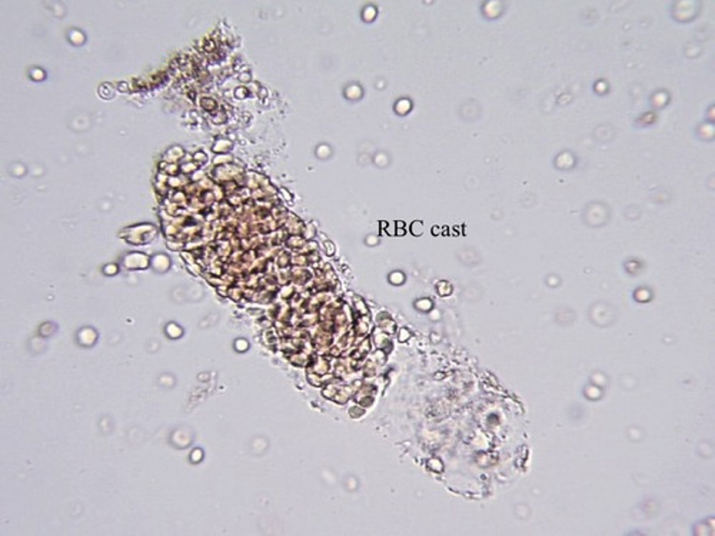
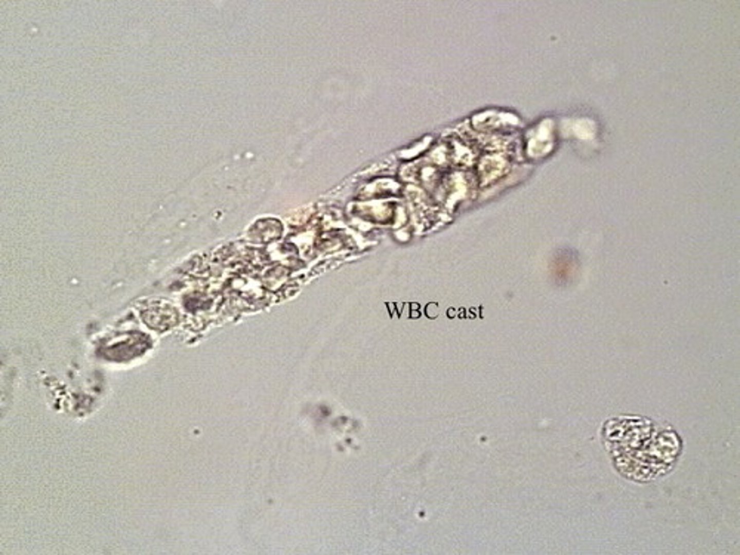
Explain the difference in the clinical significance of free RBCs or WBCs versus RBC castsand WBC casts in the urine sediment.
Casts indicate issues with the nephron
RBC casts indicate bleeding in the nephron
WBC casts indicate infection in the nephron
Describe two methods by which granular casts are formed.
Lysomes excreted by RTE cells
Disease states
Disintegration of cellular casts and tubular cells
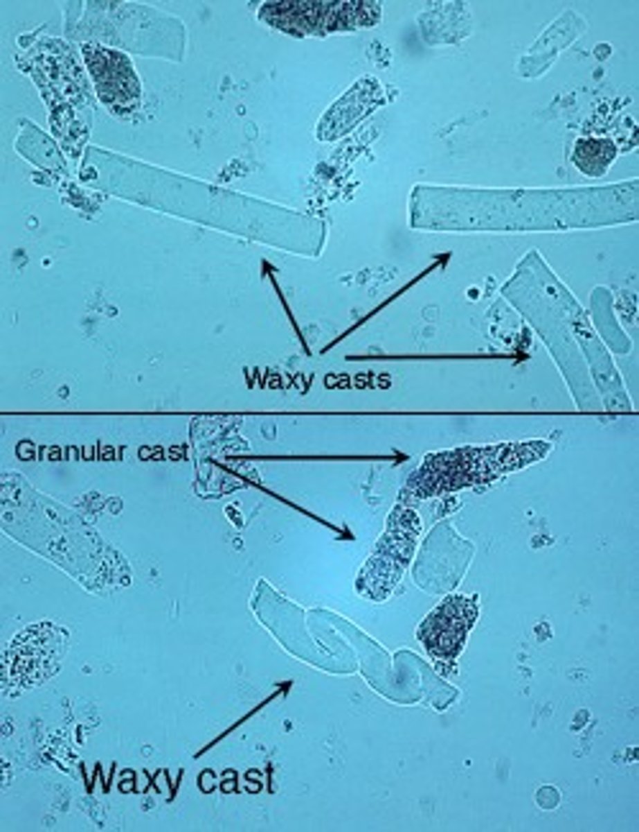
What do waxy and broad casts have in common?
Both are caused by urine stasis
Since most urinary crystals are not clinically significant, why is it necessary to identify them?
A few abnormal types can signify liver disease, inborn errors of metabolism, and damage to tubules
Why is pH important in identifying crystals?
Different crystals form under different pHs
At which pH do abnormal crystals precipitate?
Acidic urine
Which microscopy characteristic is valuable in crystal identification?
Shape and color
How can amorphous urates and amorphous phosphates be dissolved to aid obscured field?
Amorphous Urates: warming in a water bath
Amorphous Phosphates: Acetic acid
Which pH is amorphous urates found? Amorphous phosphates?
Amorphous Urates are in acidic urine
Amorphous Phosphates are in alkaline urine
What are the most common normal crystals observed in acidic urine?
Amorphous Urates
Uric acid
Acid urates
Sodium urates
Calcium Oxalate
What are the most common normal crystals observed in alkaline urine?
Amorphous Phosphates
Triple Phosphate
Calcium Phosphate
Calcium Carbonate
Ammonium Biurate
How is the identity of abnormal urines confirmed?
Patient disorders and medications
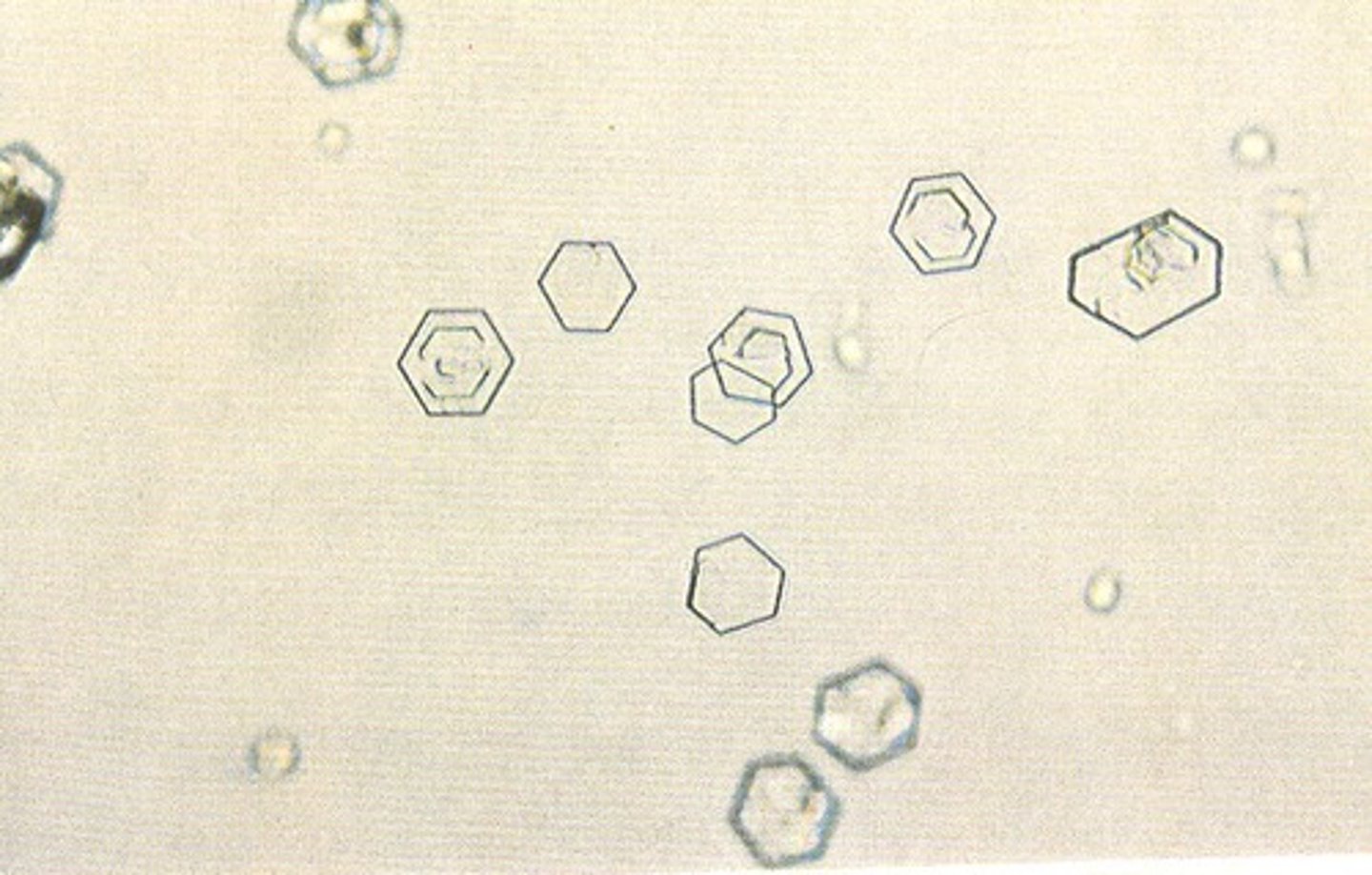
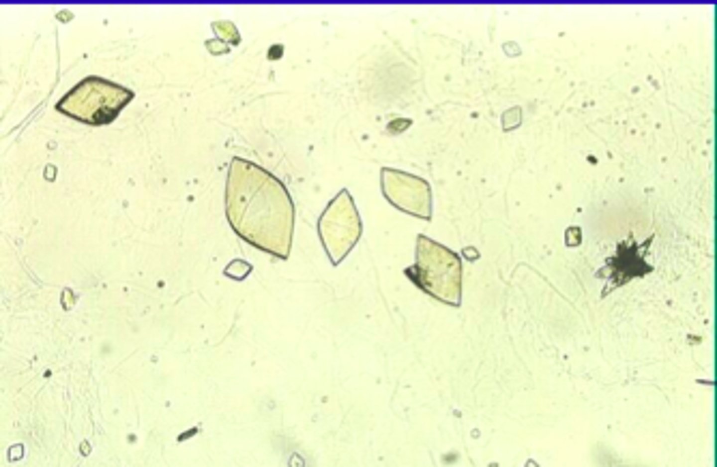
List 8 abnormal crystals that may be found in urine.
Cystine
Cholesterol
Radiographic Dye
Tyrosine
Leucine
Bilirubin
Sulfonomide
Ampicillin
What 3 urinary crystals may be seen with liver disease?
Tyrosine
Leucine
Bilirubin
Why would iatrogenic compounds precipitated in urine considered significant?
They can be caused by a variety of compounds
Name 4 possible artifacts seen in urine. What are two distinguishing microscopiccharacteristics of artifacts?
Fibers: longer, more refractile
Starch granules: highly refractile spheres, dimpled center
Air bubbles: highly refractile, resemble RBCs
Pollen grains: spheres with cell walls, occasional concentric circles
Name the identifying characteristics of: Amorphous Urates
Thin needles and seen in synovial fluid
Name the identifying characteristics of: Uric acid
Rhombode, Rosettes
Name the identifying characteristics of: Calcium Monohydrate
Ovoid calcium oxalate
Name the identifying characteristics of: Calcium Oxalate
Envelopes calcium oxalate
Name the identifying characteristics of: Amorphous Phosphates
White precipitate
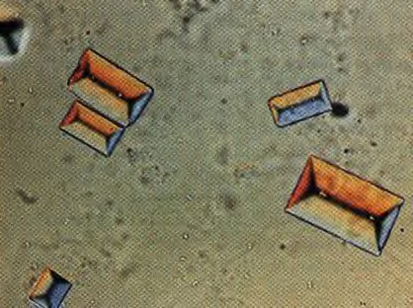
Name the identifying characteristics of: Triple Phosphate
"Coffin lids"
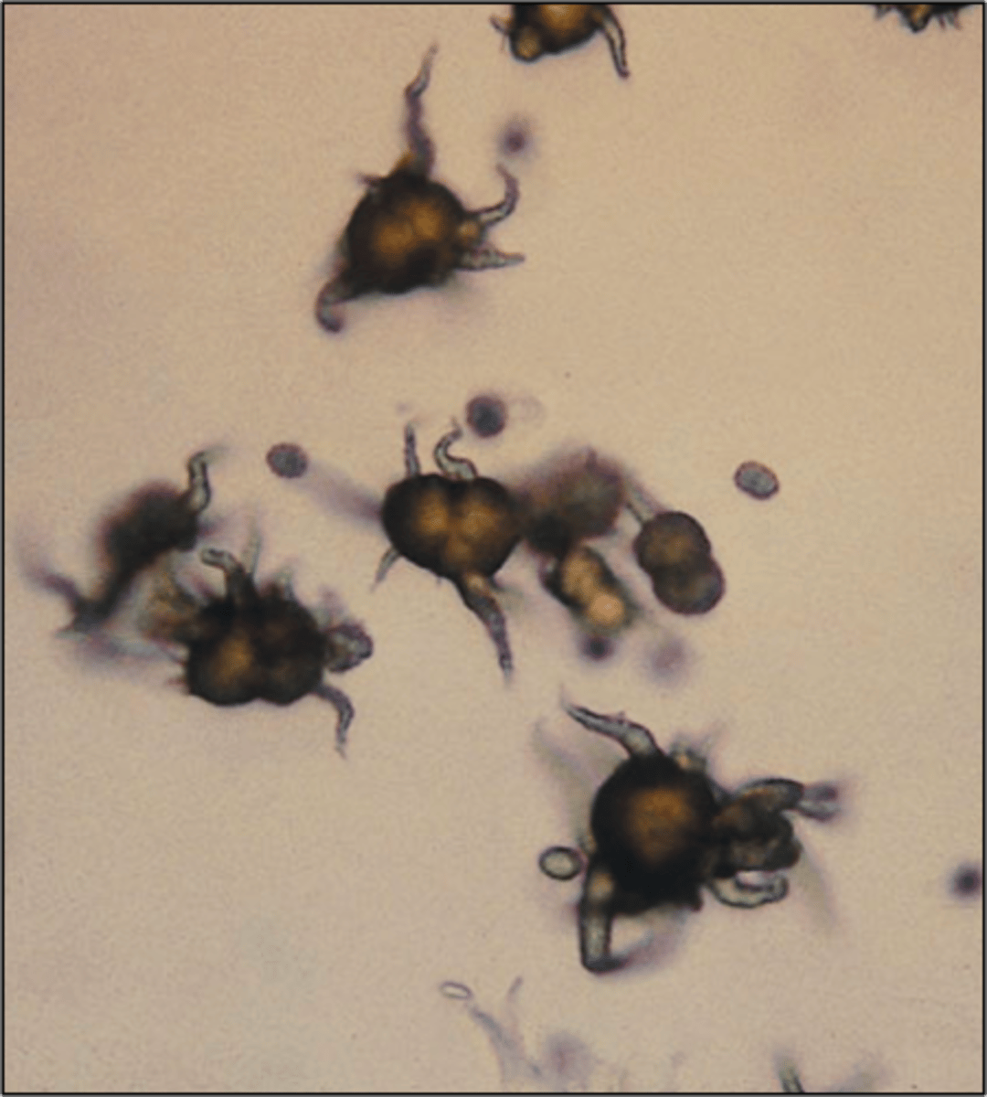
Name the identifying characteristics of: Ammonium Biurate
Thorny apple phosphate
Name the identifying characteristics of: Calcium Phosphate
Thin prisms or flat rectangular plates
Name the identifying characteristics of: Calcium Carbonate
Dumbbell or spherical shape
Name the identifying characteristics of: Cystine
Hexagonal plates dye
Name the identifying characteristics of: Cholesterol
Notched corners
Name the identifying characteristics of: Tyrosine
Fine needles with clumps or rosettes seen in liver disease
Name the identifying characteristics of: Leucine
Concentric circles and radial striations
Name the identifying characteristics of: Bilirubin
Bright yellow clumped needles/granules
Name the identifying characteristics of: Ampicillin
Colorless needles with bundles following refrigeration
Name the identifying characteristics of: Radiographic Dye
Flat plates
High specific gravity/refractometer
Name the identifying characteristics of: Sulfonomides
Needles, rhombics, whetstones, sheaves of wheat, and rosettes with colors ranging from colorless to yellow-brown
RBCs
WBCs
Cystine
Uric Acid
Hyaline Casts
Triple Phosphate
Ammonium Biurate
What are reagent strips?
Strips that consist of chemical-impregnated absorbent pads