Phys Lab 5
1/76
There's no tags or description
Looks like no tags are added yet.
Name | Mastery | Learn | Test | Matching | Spaced | Call with Kai |
|---|
No analytics yet
Send a link to your students to track their progress
77 Terms
when you are cold your blood vessles _____
dialate
what is resting heart rate determined by
rate of SA node
effects of innervation by parasympathetic nervous system
What does the SA node want to set the resting heart rate to
100 bpm
What is the true avg resting heart rate due to parasympathetic NS
75 bpm
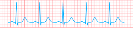
what is the range of normal values for resting heart rate
60-100 bpm
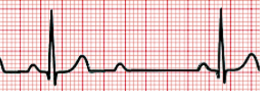
bradycardia
resting heart rate below 60 bpm
tachycardia
resting heart rate above 100 bpm
what act on the SA node to increase hr / rate of action potentials
sympathetic ns
epiniephrine
what is the formula to estimate HR max
220 - age =
what makes the lub sound
closure of atrioventricular valves
when does LUB sound happen (timepoint)
when ventricular systole begins
what makes the DUB sound
closure of semilunar valves
when does DUB sound happen (timepoint)
ventricular diastole begins
where should you place stethescope to hear the LUB sound
apex of heart
where should you place stethescope to hear DUB sound
second intercostal space
how does inspiration and expiration effect HR
inhale - increases HR
exhale - decreases HR
what is conduction system of the heart made of
non contractile cardiac muscles that spread electrical impulses
what control the contraction of cardiac muscles
conduction system
where is the SA node located
right atrium
what is known as the pacemaker
Sinoatrial node
where is AV (atrioventricular) node located
base of atrium
lable
.
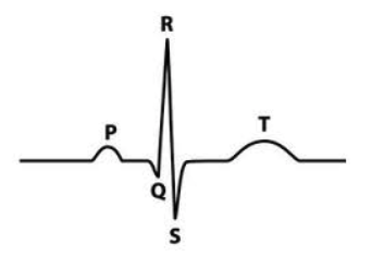
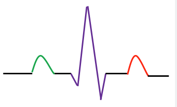
P wave is due to
atrial depolarization
(SA node fires and depol wave spreads through atria)
QRS wave is due to
ventricular depolarization
(depol wave spreads from AV node through the ventricles)
Is atrial repolarization seen on ECG
no, it is hidden by QRS
T wave due to
ventricular repolarization
P-Q interval located where
from start of P to start of Q

what happens during P-Q interval
atria contracts and AP spreading through the AV node
S-T interval located
end of S to start of T wave
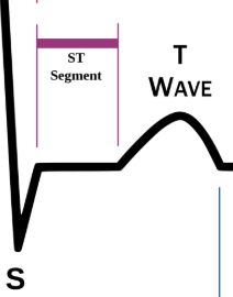
what happens during S-T interval
ventricles are depol and contracted, blood ejected from ventricles into arteries
T-P interval
period of time between cardiac cycles when heart at rest (atria and ventricles)
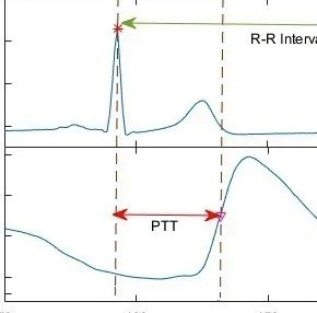
R-R interval located
interval between R waves
what does R-R interval represent
time it takes for one cardiac cycle
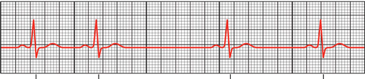
what is ECG
graphic record of electrical activity ( all AP generated by nodal and contractile cells)
arrhythmias
irregular rythems caused by abnormal impulse formation or conduction
conduction delays
slower then normal electrical transmission through hearts conduction pathway
what does Bradycardia look like on ECG
longer T-P interval
what does Tachycardia ECG look like
shorter T-P interval
Heart block
atrial impulses fail to conduct through (AV, bundles, Purkinje)
what does heart block look like on ECG
dropped beats, P waves not followed by QRS
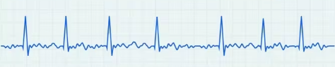
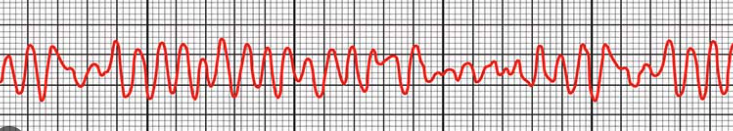
Fibrillation
uncoordinated signaling and contraction of atria or ventricles
What does atrial fibrillation look like
multiple irregular waveforms where P value would normally be (no P waves visible)
what does ventricular fibrillation look like
rapid irregular waveforms with no P, QRS, T waves
does atypical pattern always mean unhealthy
not always
R-pulse interval
time it takes from ventricular depolarization to contraction causing pulse pressure
how to calculate HR from ECG
chart speed / QRS distance (mm/beat) = beats per second
60 seconds x beats per second = HR/min
perfusion
the passage of blood to deliver O2 and nutrients
systole
force that blood exerts on arterial walls during contraction
diasole
force that blood exerts on arterial wall during relaxation
blood pressure
force that blood exerts on vessel walls (normally artieries)
systolic pressure
pressure in large arteries at peak ventricular ejection
diastolic pressure
pressure in large arteries during ventricular relaxation
BP is meassured in what?
mm Hg
120/80
what is systolic and diastolic pressure
120 - systolic
80 - diastolic
what is average normal BP
120/80
Hypertension is classified as
> 140/90 with stethoscope
> 135/85 with automatic BP device
what can cause short term hypertension
caffne, nicotine, drugs
chronic hypertension is caused by
aging, lack of exercise, stress
Sphygmomanometer
instruemnt used to obtain BP using auscultatory method.
auscultatory method
listening to sounds of body with stethescope
what arterie is used to meassure BP
bracial artery
what sounds are used to meassure BP with spygmomanometer
sounds of Korotkoff
when is systolic and diastolic pressure meassured with BP cuff
sounds detedcted - systolic
sounds disapear - diastolic
what causes sounds of Korotkoff
turbulent blood flow
pulse pressure
actual working BP (systolic - diastolic = PP)
Mean artieral pressure (MAP) represents
pressure with which blood is being delivered to organs
what is normal MAP
70 - 110 mmHG
what MAP would need medical intervention
under 60 mmHG
what happens if MAP to low
may not reach organ or pass to slowly for adequate delivery
what happens if MAP to high
blood passes through organs to fast for exchange
MAP can be calculated by
PP/3 + Diastolic pressure = MAP
turbulent flow
chaotic / irregular flow
laminar flow
steady flow
what happens to systolic and diatolic pressure during exercise
systolic - increases
diastolic - stable
where is first heart sound heard on ECG
QRS
where is 2nd heart sound heard
T wave