Neuro exam 3: Speech
1/45
There's no tags or description
Looks like no tags are added yet.
Name | Mastery | Learn | Test | Matching | Spaced |
|---|
No study sessions yet.
46 Terms
What are the two main functions of language?
Comprehension (sensory/perceptual)
speaking (motor function).
How does the brain express language?
using syntactic rules (grammar)
words to convert internal thoughts into external speech or gestures.
What does the internal conceptual–intentional interface represent?
Concepts, intentions, and reasoning — the mental side of language (meaning and ideas).
What does the external sensory–motor interface control?
Perception and production of speech sounds and gestures — the physical side of language.
What links the internal and external language systems?
The syntactic and lexical system, which organizes grammar and words for externalization (turning thought into speech).
Which area connects internal meaning to external speech?
Broca’s area
Which area processes word meaning and comprehension?
Wernicke’s area, part of the conceptual-intentional interface.
What connects Broca’s and Wernicke’s areas, and what happens if it’s damaged?
The arcuate fasciculus; damage causes conduction aphasia (inability to repeat words accurately).
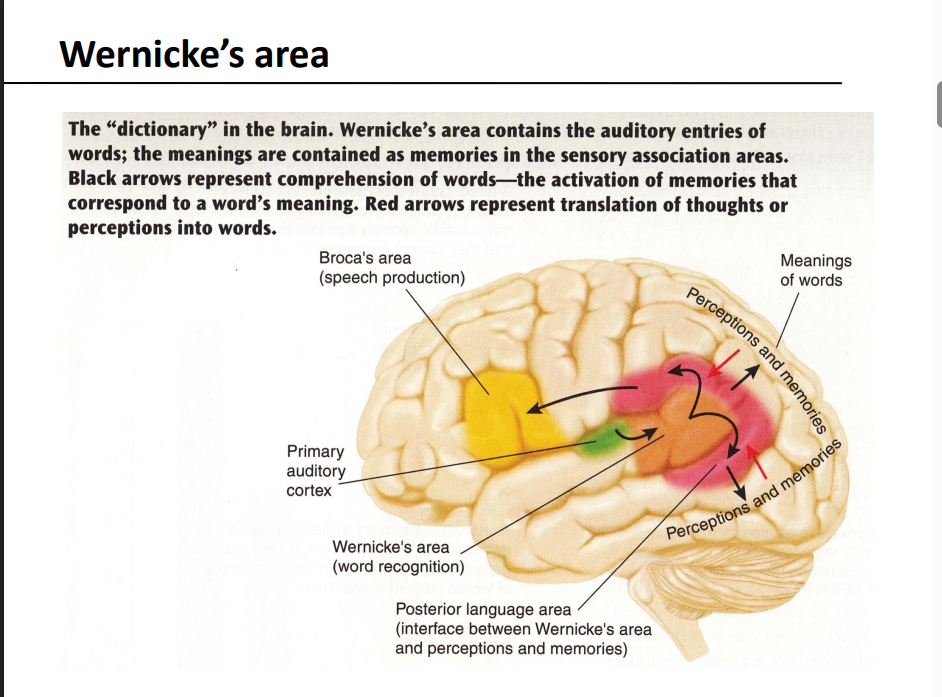
What kind of information does Wernicke’s area contain?
The auditory entries of words — it acts like a “dictionary,” linking sounds of words to their meanings stored in sensory association areas.
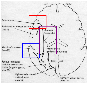
What is the arcuate fasciculus?
white matter fiber tract that connects Wernicke’s area to Broca’s area, allowing communication between understanding and speaking.
What does the arcuate fasciculus enable?
allowing us to repeat, paraphrase, and produce meaningful language based on what we hear or read.
What happens if the arcuate fasciculus is damaged?
Conduction aphasia — individuals can understand and speak, but cannot accurately repeat heard words or sentences.
What other brain areas support language connections besides Broca’s and Wernicke’s?
Angular gyrus – (reading/writing).
Supramarginal gyrus – links sound and meaning.
Inferior parietal lobe – integrates multiple sensory language inputs.
How does visual information (like reading) reach Wernicke’s area?
Via the visual cortex → angular gyrus → Wernicke’s area pathway, allowing written words to be converted into understood language.
What is the pathway for hearing and speaking a word?
Auditory cortex → Wernicke’s area → Arcuate fasciculus → Broca’s area → Motor cortex
What is the pathway for reading and then speaking a word?
Visual cortex → Angular gyrus → Wernicke’s area → Arcuate fasciculus → Broca’s area → Motor cortex
What happens when the FOXP2 gene is mutated?
causes impairment of orofacial movements, preventing proper word sounds and resulting in severe language difficulties
What does the FOXP2 gene normally do?
encodes a transcription factor that regulates other genes needed for brain and lung development, vocal motor learning, and language.
How does human FOXP2 differ from that of other species?
human version differs by 2 amino acids from chimpanzees, gorillas, and rhesus monkeys, and by 3 amino acids from mice.
What are the shared and human-specific functions of FOXP2?
Shared: Sensorimotor integration, vocal motor learning guided by auditory feedback (seen in birds too).
Human-specific: Adds language and advanced auditory-guided vocal learning.
what is Broca’s aphasia
Expressive aphasia — speech is slow, labored, and non-fluent, but comprehension remains intact.
Where is the lesion located in Broca’s aphasia, and what does it affect?
Lesion in left posterior frontal lobe (Broca’s area, BA 44–45) → motor planning deficits that impair speech production.
How is Broca’s aphasia different from dysarthria?
Broca’s = language production problem (planning words),
Dysarthria = motor control problem (moving mouth/tongue muscles).
what is Wernicke’s aphasia
Receptive (sensory) aphasia — Impairment of language comprehension, both written and spoken.
What are common features of Broca’s aphasia?
Halting, effortful speech
Tendency to repeat phrases/words (perseveration)
Disordered syntax, grammar, and word structure
How does speech sound in Wernicke’s aphasia?
Speech is fluent, with normal grammar and syntax, but it’s meaningless or nonsensical — words are often inappropriate or made up.
Where is the lesion located in Wernicke’s aphasia?
Lesion in Wernicke’s area (Brodmann area 22) in the left superior temporal gyrus
What are notable behavioral signs of Wernicke’s aphasia?
Comprehension not intact
Fluent but meaningless speech
Denial of deficit (anosognosia) — patients often unaware they’re not making sense
What is conduction aphasia?
language disorder where speech is fluent and comprehension is good, but repetition is very poor due to disrupted communication between Broca’s and Wernicke’s areas.
What brain structure is typically damaged in conduction aphasia?
The arcuate fasciculus
What are the key speech features of conduction aphasia?
Fluent, meaningful speech
Good comprehension
Very poor repetition (especially for unfamiliar or nonsense word
What functions are impaired in global aphasia?
Speech production, repetition, comprehension, and naming — all impaired due to widespread left hemisphere damage.
What is the hallmark feature of anomic aphasia?
Difficulty naming objects (word-finding problems) despite fluent, grammatically correct speech and normal comprehension/repetition.
How does transcortical aphasia differ from Broca’s and Wernicke’s aphasias?
Repetition remains intact because the arcuate fasciculus is spared.
Motor type: Like Broca’s (non-fluent speech).
Sensory type: Like Wernicke’s (poor comprehension).
What abilities are affected in sensory aphasia?
Fluent speech and normal repetition but poor comprehension and naming
lesions near temporal-parietal sensory association areas.
The_______ is dominant in about 90% of all people for speech
left hemisphere
How can early brain lesions affect speech lateralization?
Lesions early in life can cause language functions to shift to the right hemisphere, showing brain plasticity.
split-brain patients?
People who have undergone transection of the corpus callosum and anterior commissure
Purpose of Split-Brain Surgery
treatment for severe epilepsy, preventing seizures from spreading between hemispheres.
What remains intact in split-brain patients despite the surgery?
Bilateral thalamic projections, allowing some interhemispheric coordination through subcortical pathways.
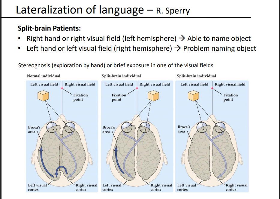
In split-brain patients, when can they name an object correctly?
When the object is in the right hand or right visual field, which projects to the left hemisphere (where language centers are located).
Why can’t split-brain patients name an object seen with the left visual field or felt with the left hand?
the right hemisphere perceives it, but language centers (Broca’s/Wernicke’s) in the left hemisphere can’t access that information without the corpus callosum connection.
What does the split-brain experiment demonstrate about visual fields and hemispheric processing?
Each visual field projects to the opposite hemisphere, showing that language is lateralized to the left hemisphere in most people.
Where is the planum temporale located?
It’s the cortical area just posterior to the auditory cortex within the Sylvian fissure (temporal lobe).
how does the planum temporale differ between the left and right hemispheres?
The left planum temporale is usually larger than the right, reflecting the left hemisphere’s specialization for language.
What is the planum temporale associated with?
involved in language processing and auditory perception, especially related to speech and sound localization.