Lecture 6 - Medulla
1/66
There's no tags or description
Looks like no tags are added yet.
Name | Mastery | Learn | Test | Matching | Spaced |
|---|
No study sessions yet.
67 Terms
name and view
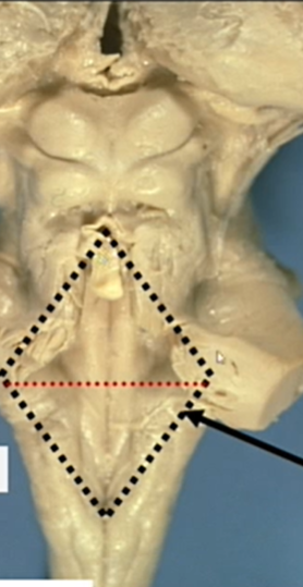
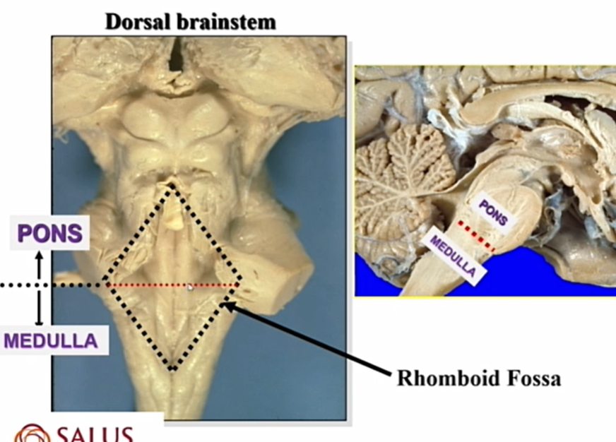
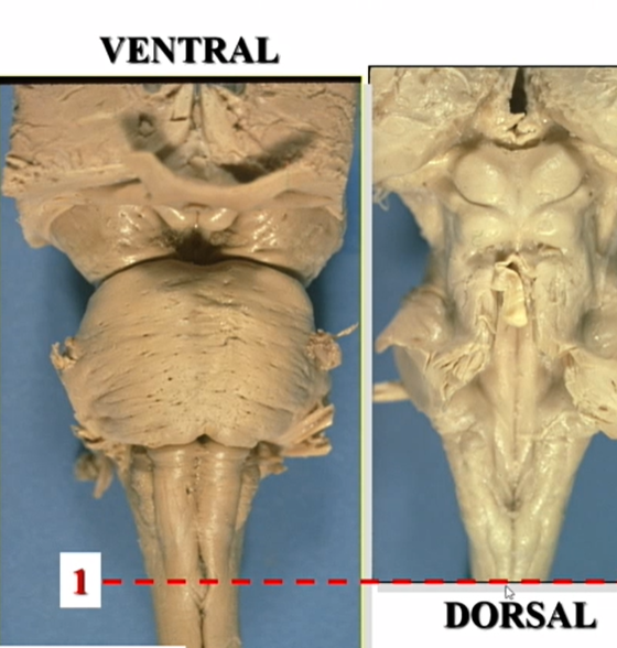
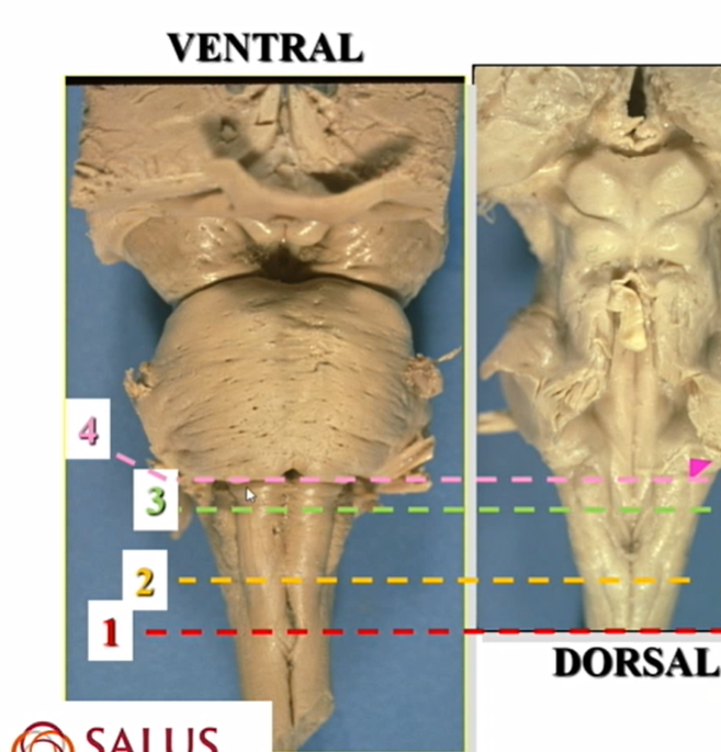
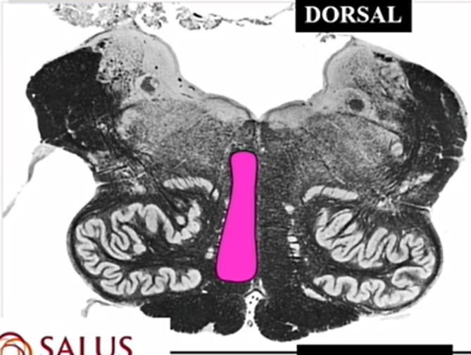
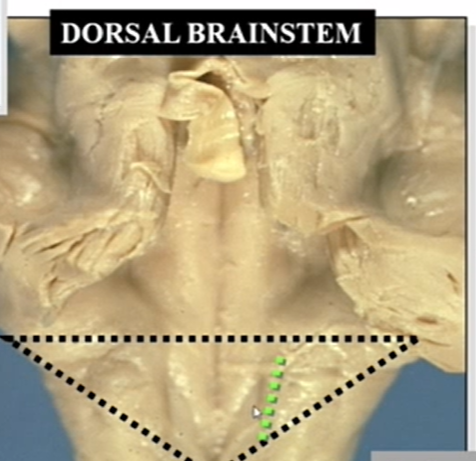
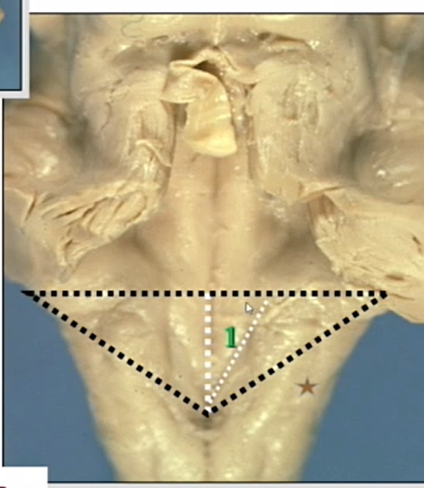
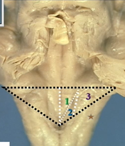
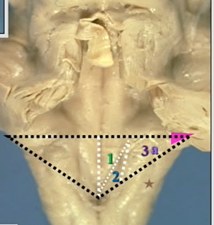
dorsal brainstem
above line is pons
below is medulla
rhomboid fossa
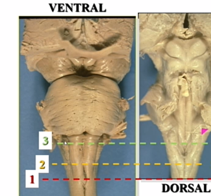
name and view
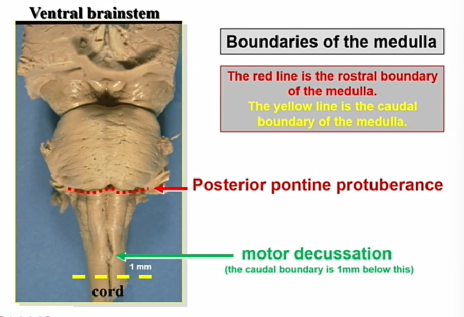
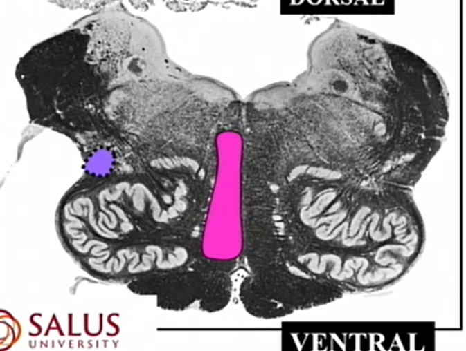
ventral brainstem
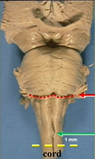
showing the rostral - red boundary of the medulla —>posterior pontine protuberance to fourth ventricle
showing the caudal - green boundary of hte medulla —> 1 mm caudal to the motor dec
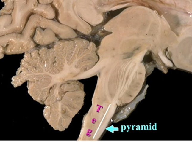
name and whats there
tegmentum
more dorsal
general sensory and reticular formation
name 1 and what travels there
pyramids
more ventral — motor
corticospinal and corticobulbars
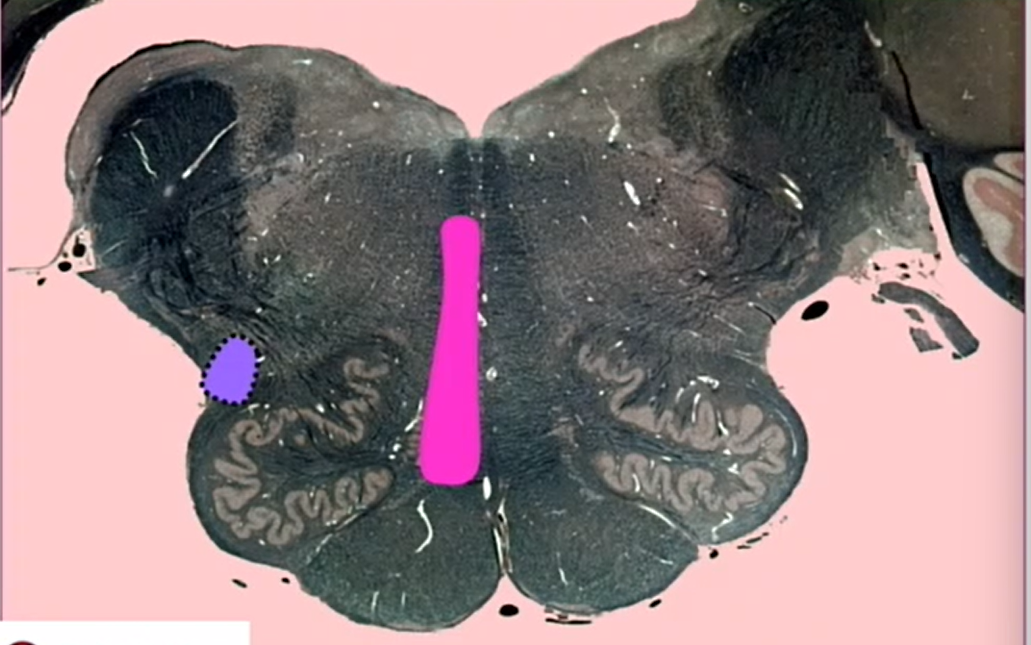
name what is separated by what line
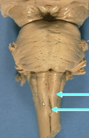
name and view
ventral brainstem
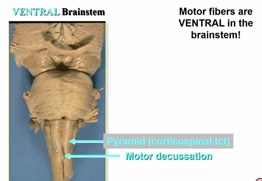
whats this cut thru
motor dec
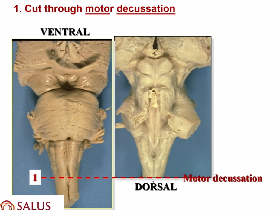
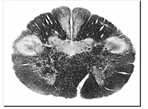
whats this a cut of
motor decussation in medulla
diagonal opening on bottom - bc fibers cross
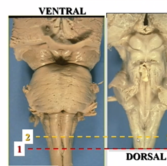
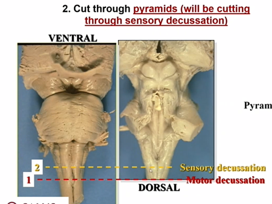
whats 2 cutting thru
sensory dec
cut thru bumps that house nuc gracilis
JUST BELOW OLIVES
still see pyramids
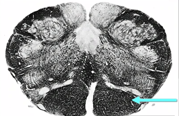
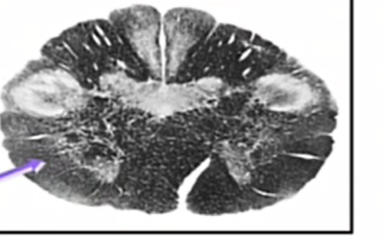
whats this a cut thru (ignore arrow)
sensory dec in closed medulla
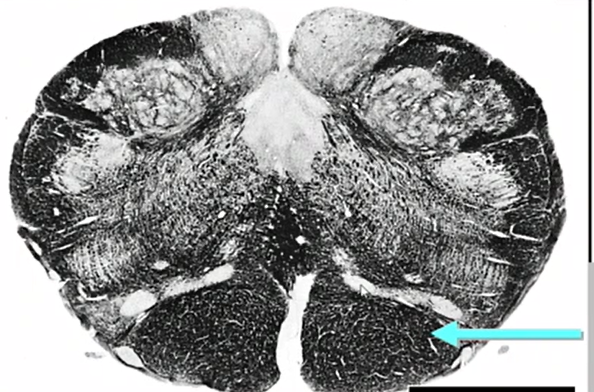
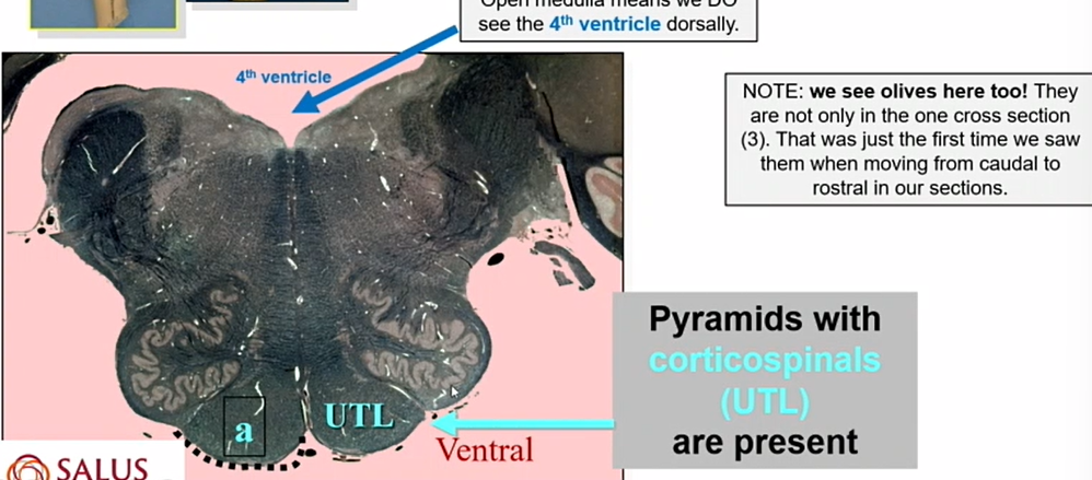
what is indicated by the blue arrow
pyramids w corticospinals (Upper extremities, trunk, lower extremities —> medial to lateral)
closed medulla so no 4th ventricle
NOT THE LATERAL CORTICOSPINALS BC WE HAVENT CROSSED YET!!
no corticobulbars bc they have already come off
name cut of 3
inferior olives
we are in the 4th ventricle — we are in rhomboid fossa
pyramids are present
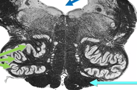
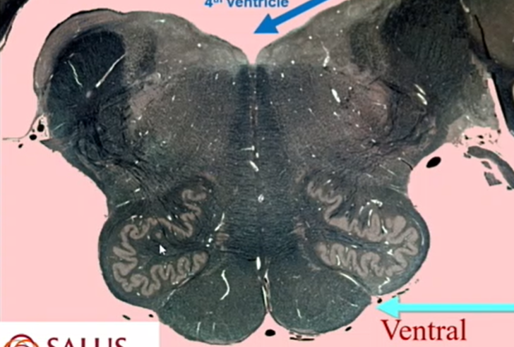
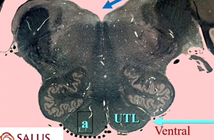
whats this a cut thru
inferior olives
name all arrows
if we see olives we are in open medulla
still no more cortical bulbars

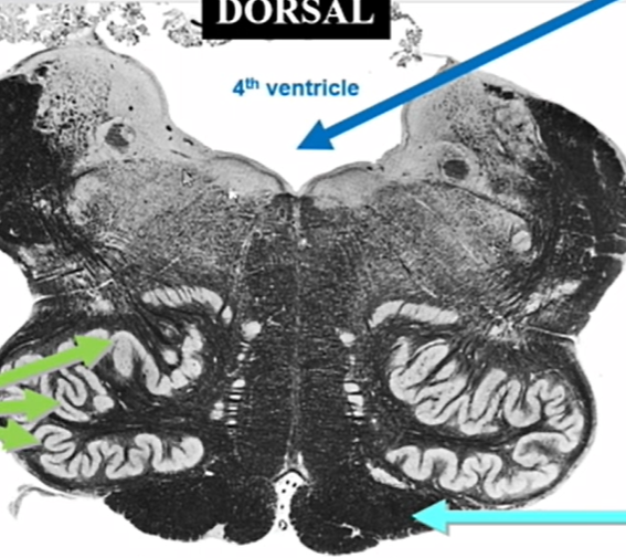
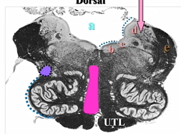
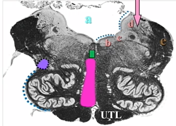
name 4 cut
highest medulla
pyramids still prseent
name the cut
highest medulla (open medulla)
name all arrows
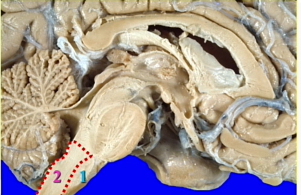
dotted line means its something you would see on gross brain - pyramid
visualize the order from rostral to caudal of medulla cuts
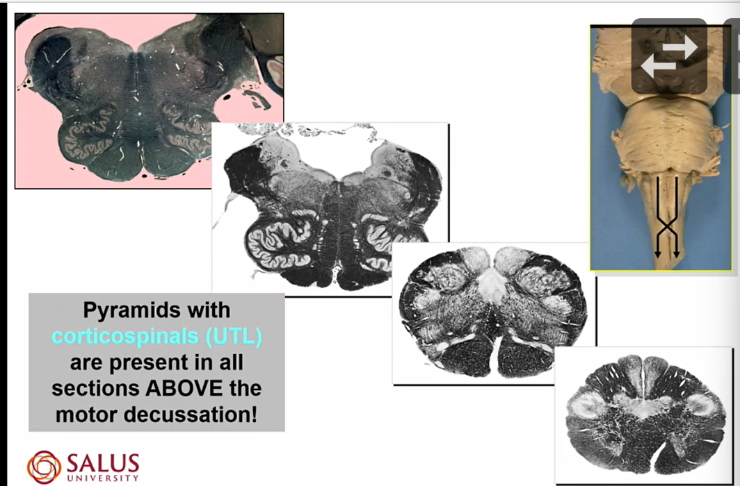
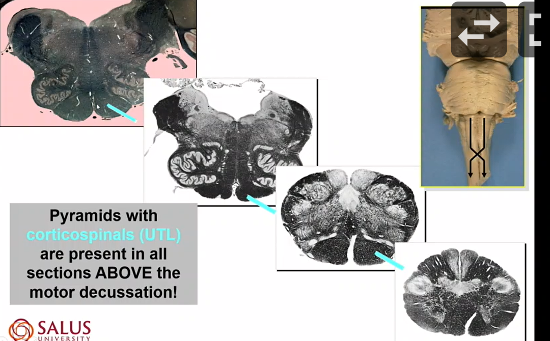
describe the corticospinals
anywhere above the crossing is corticospinals
after motor dec they are lateral corticospinal

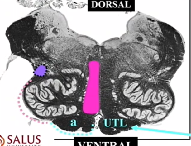
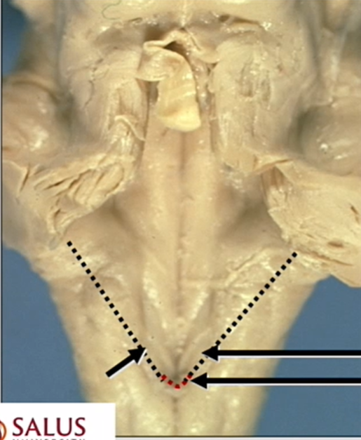
whats the dark blue dotted line
gracile tubercule - can see on gross brainstem
sensory dec
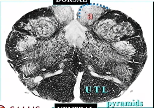
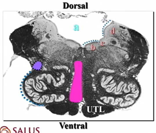
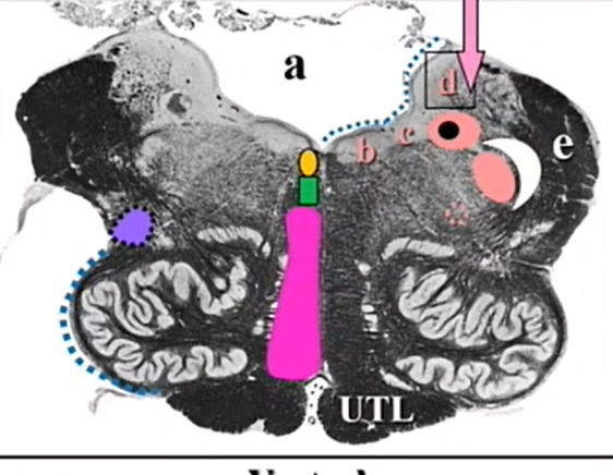
whats a
nucleus gracilis
sensory dec

dotted blue line over b
cuneate tubercle
sensory dec
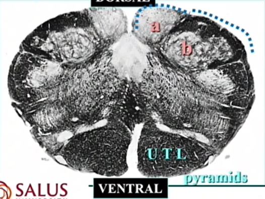
b
nucleus cuneatus

dotted hot pink
internal arcuates
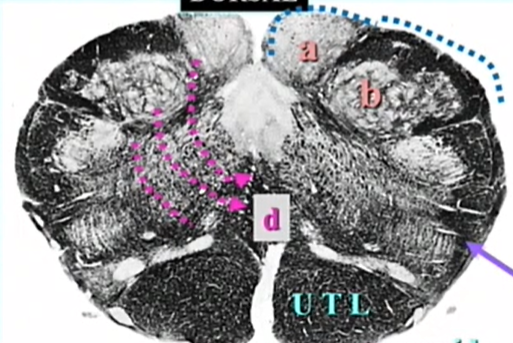

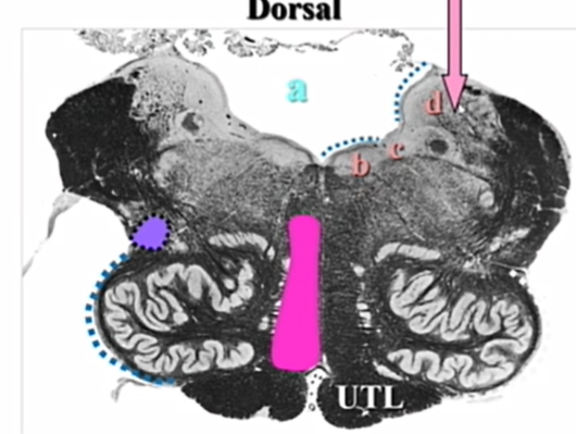
d
sensory dec

purple arrow
spinothalamic
if we see olives we are at _______ medulla
open
name
medial lemniscus
inferior olives
purple
ventral and lateral spinothalamics
inferior olives
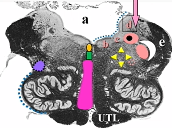
name pink and purple and view
pink - medial lemniscus
purple = ventral and lateral spinothalamic
highest open medulla
note that the spot is basically the same as inferior olives cut
name
spinothalamics
name light pink dots
inferior olive
olivary complex (nucleus)
inferior olives
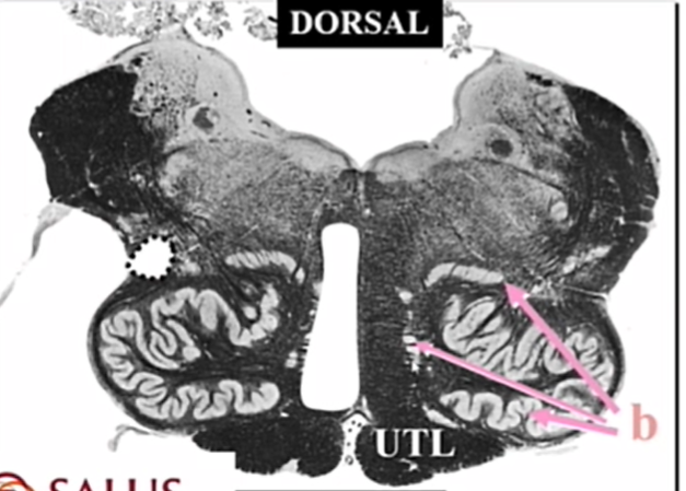
name pink arrows
main olive
medial accessory olive
dorsal accessory olive
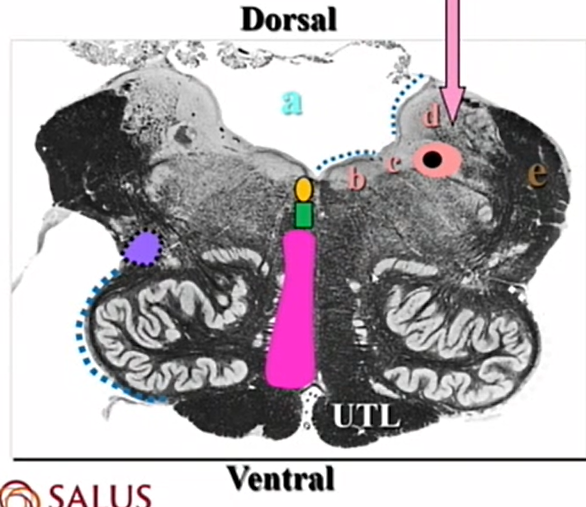
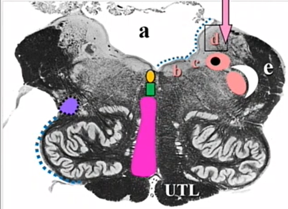
name green arrow and its purpose
sulcus limitans in medulla
separates motor (medial) from sensory (lateral)CN nuclei
name 1 and what it contains
hypoglossal trigone
contains hypoglossal (CN 12) nuc

2 adn what it contains
vagal trigone
dorsal efferent nuc
3
lateral recess
a and pink triangle
vestibular area (vestibular nuclei)
acoustic tubercle

orange square
nucleus prepositus hypoglossi
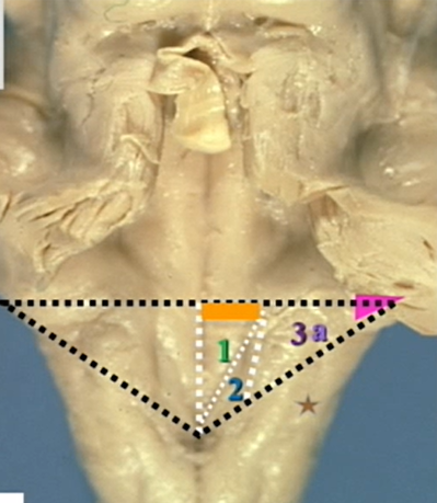
star
inferior cerebellar peduncle

a
fourth ventricle
floor of rhomboid fossa
inferior olives cut
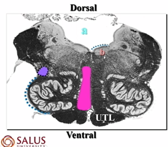
b
hypoglossal nuc
inferior olives cut

c and its type of autonomic fiber and whats it associated w
dorsal efferent nuc
associated w CN 10
parasympathetic preganglionic
inferior olives cut
d
vestibular nuc — lateral bc sensory
inferior olives cut
pink arrow
inferior vestibular nucleus
d is our vestibular nuclei — and we are lower in medulla as far as its parts go
inferior olives cut
e
inferior cerebellar peduncle
green
tectospinal
motor reflex
inferior olives cut

yellow
medial longitudinal fasciculus
how CN communicate
donut shape
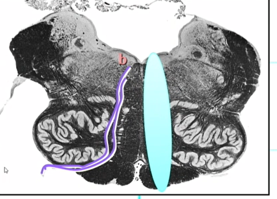
looks like an egg and you have one egg for breakfast - nucleus and tract of solitarius
nucleus = light and tract = dark and inside
pink and crescent
nucleus and tract of 5
light = pink = nucleus
tract = white = dark
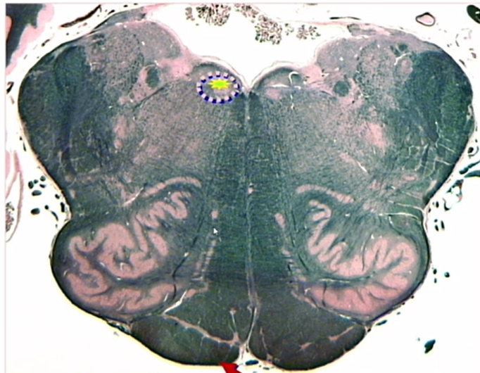
dotted pink circle and what it contains
nucleus ambiguous
CN 9, 10, 11
name yellow triangle shape and what it contains
reticular formation
contains descending autonomics
whats all a part of the reticular formation
everything left over
descending autonomics
nuc ambiguus
vomiting center
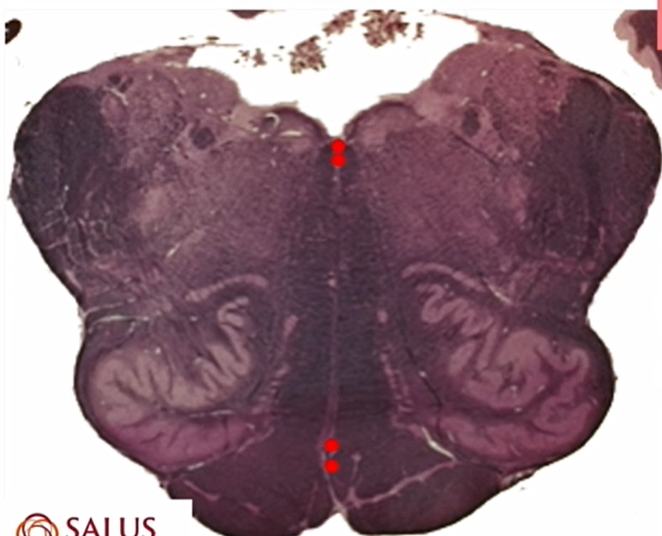
red dots
obex
red line and what its responsible for
area postrema
vomiting center
orange arrow
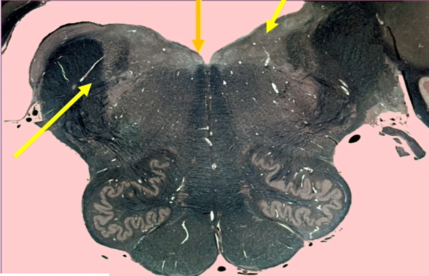
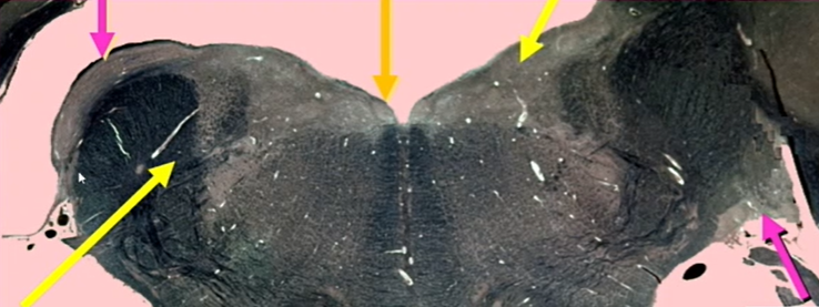
nuc prepositus hypoglossi
high medulla cut
what are the pink nuclei
tells you we are in high medulla
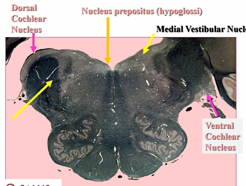
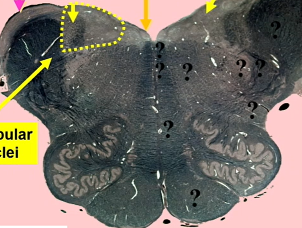
name yellow dotted area and yellow arrows
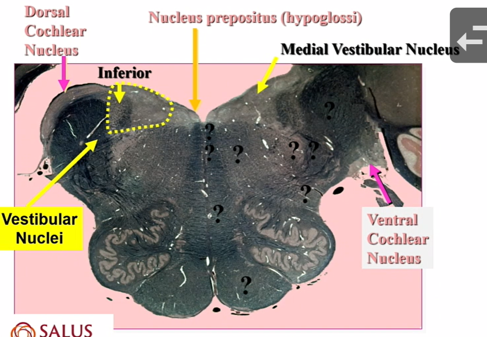
fxn of nucleus prepositus hypoglossi
gaze holding center (neural integrator) for lateral gaze
name neuron
CN 12 (LMN)
CNS bc in brainstem
lesion of hypoglossal nuc/ CN 12
paralysis is contralateral — corticospinal
tongue deviates towards sign of lesion
dark line is the CN

where do taste (7,9,10) synapse
VPM
VPL nucleus
sensory nucleus for BODY
VPM is sensory nuclei for
HEAD
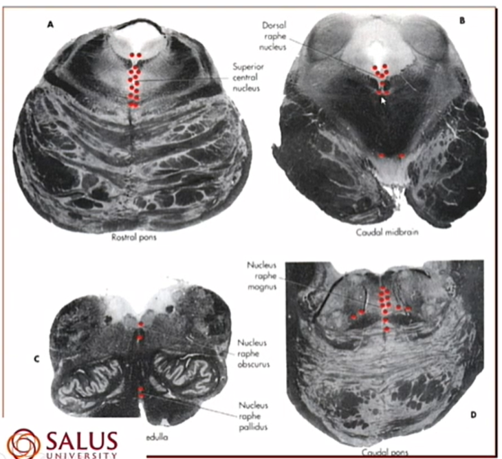
name and where its seen
Raphe nuclei
seen more in high medulla than low pons