Abdomen 3: Organs
1/124
There's no tags or description
Looks like no tags are added yet.
Name | Mastery | Learn | Test | Matching | Spaced | Call with Kai |
|---|
No analytics yet
Send a link to your students to track their progress
125 Terms
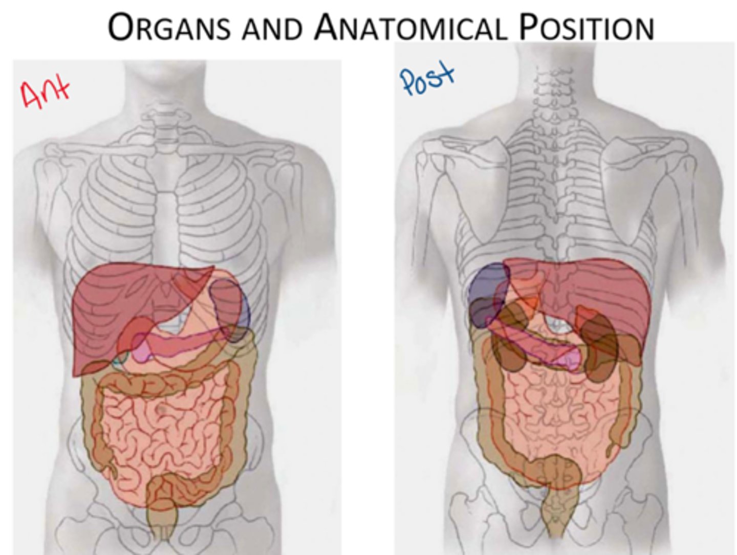
Be familiar with the anatomical position of the organs within the abdomen
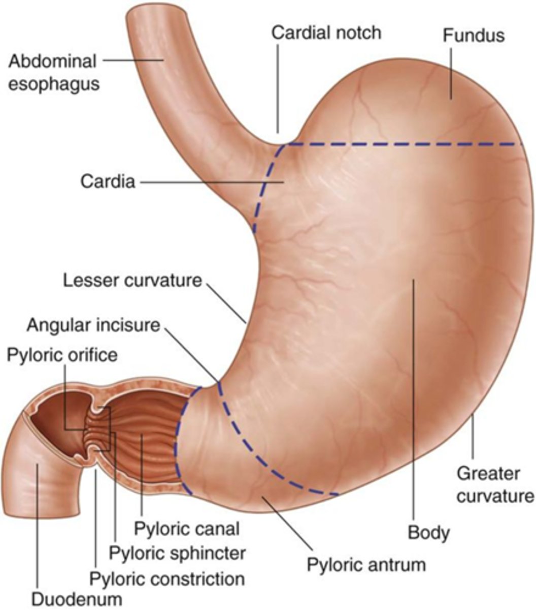
Label the parts of the stomach
What provides parasympathetics to the stomach?
vagus nerve: via anterior and posterior vagal trunks on the stomach
What is the esophagus innervated by?
vagus nerve
Where do the contents of the stomach empty to?
duodenum (first part of small intestine)

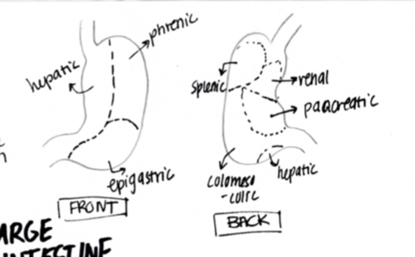
Draw the contact points of the stomach to other viscera within the abdomen
Left is anterior right is posterior
note that the pancreas is the largest organ that will contact the stomach posteriorly
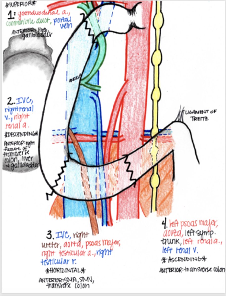
Label the parts of the duodenum
What portions of the duodenum are retroperitoneal and which are intraperitoneal?
1st and 4th portion are intraperitoneal, 2nd and 3rd portions are retroperitoneal
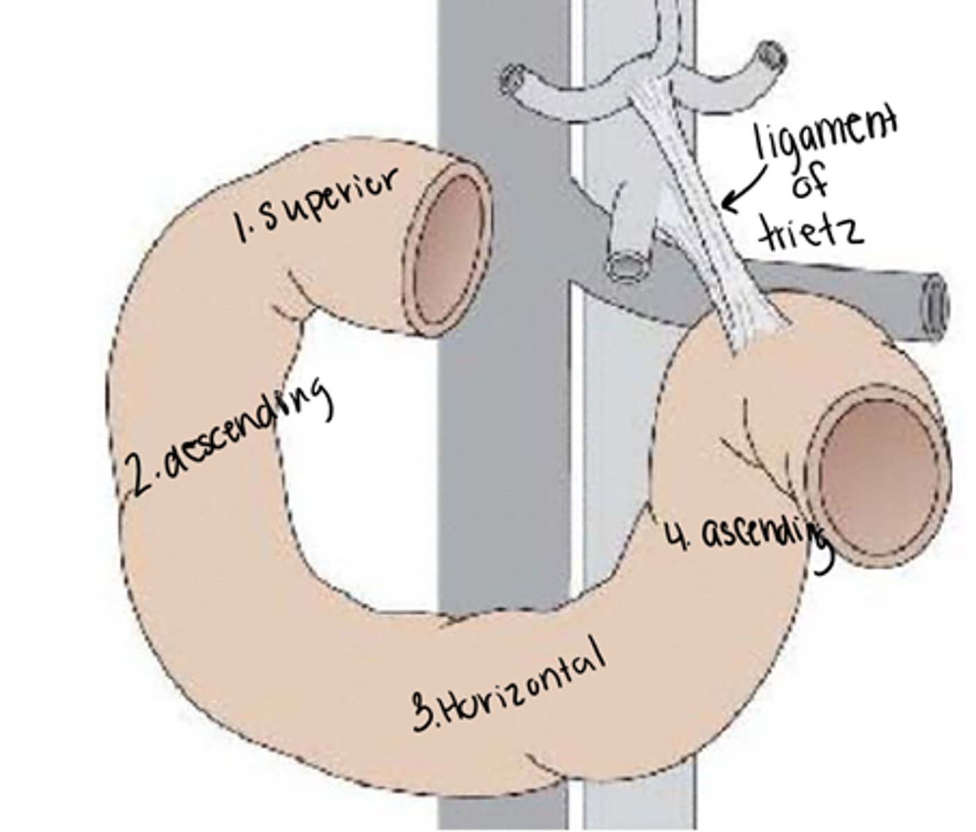
What is the ligament of trietz?
Ligament found at the transition from duodenum to jejunum (suspensory ligament)

Is the stomach intraperitoneal or retroperitoneal?
intraperitoneal
Describe the position of the duodenum within the abdomen and what it is in contact with
Anterior to inferior vena cava and aorta
Has intimate contact with the head of the pancreas (C shaped portion)
What part of the duodenum will bile and pancreatic ducts open into?
second part of duodenum (descending portion) through papilla
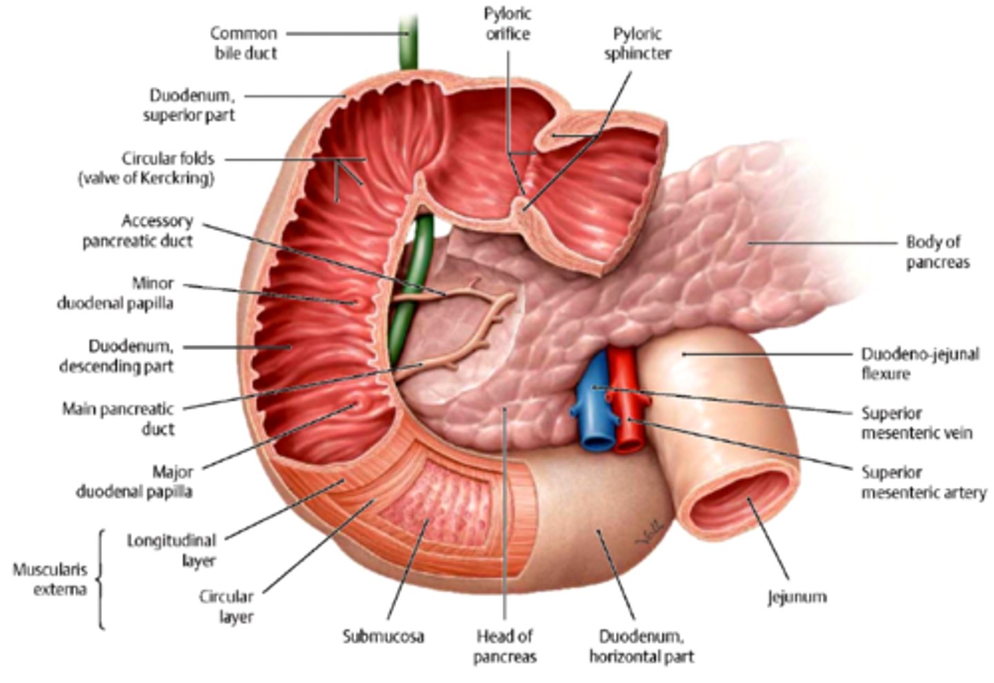
What type of secretions will come from the major papilla of the duodenum?
secretions from the main pancreatic duct and common bile duct (within 2nd portion of duodenum)

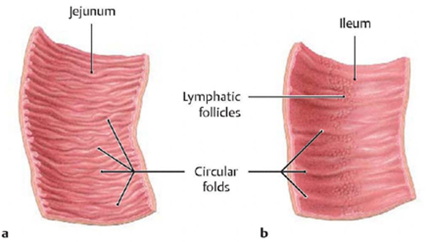
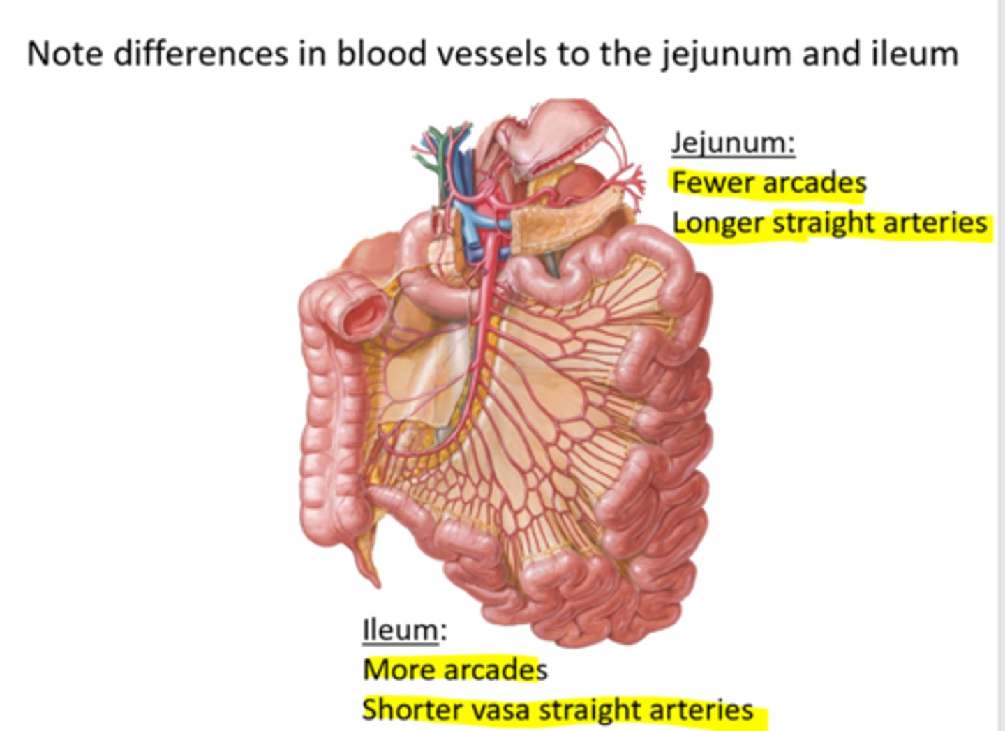
Compare and contrast the defining features of the jejunum and ileum
Jejunum is the first place that absorption will occur thus the walls will be thicker and have numerous folds
vasa recta=straight arteries
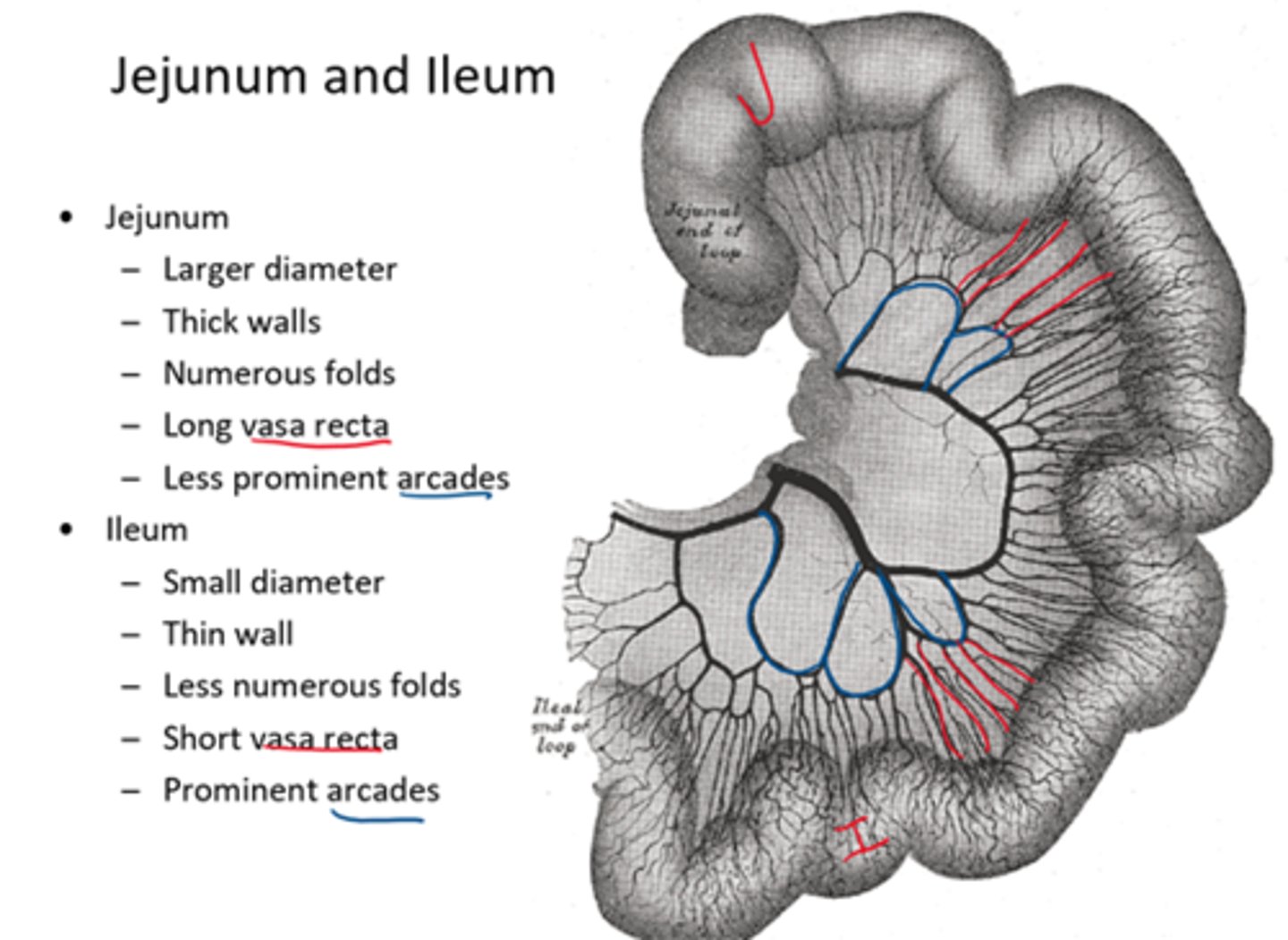
In which part of the small intestine will vasa recta be longer and which will be shorter?
Longer in jejunum, shorter in ileum
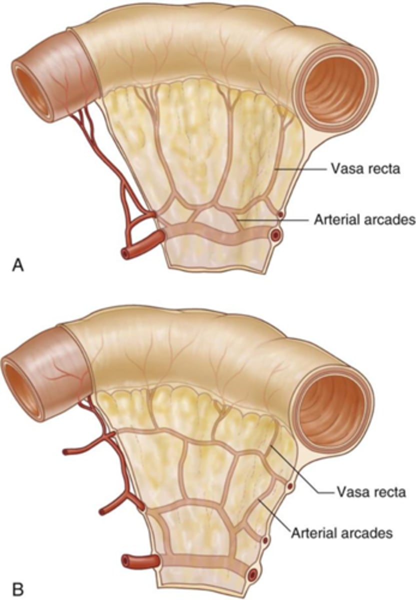
Explain the differences in folds in teh jejunum and ileum
More folds found in the jejunum than the ileum as there is more absorption that occurs in the jejunum
folds increase surface area for absorption
What is the cecum?
first part of the large intestine

What allows passage from ileum to cecum?
iliocecal valve
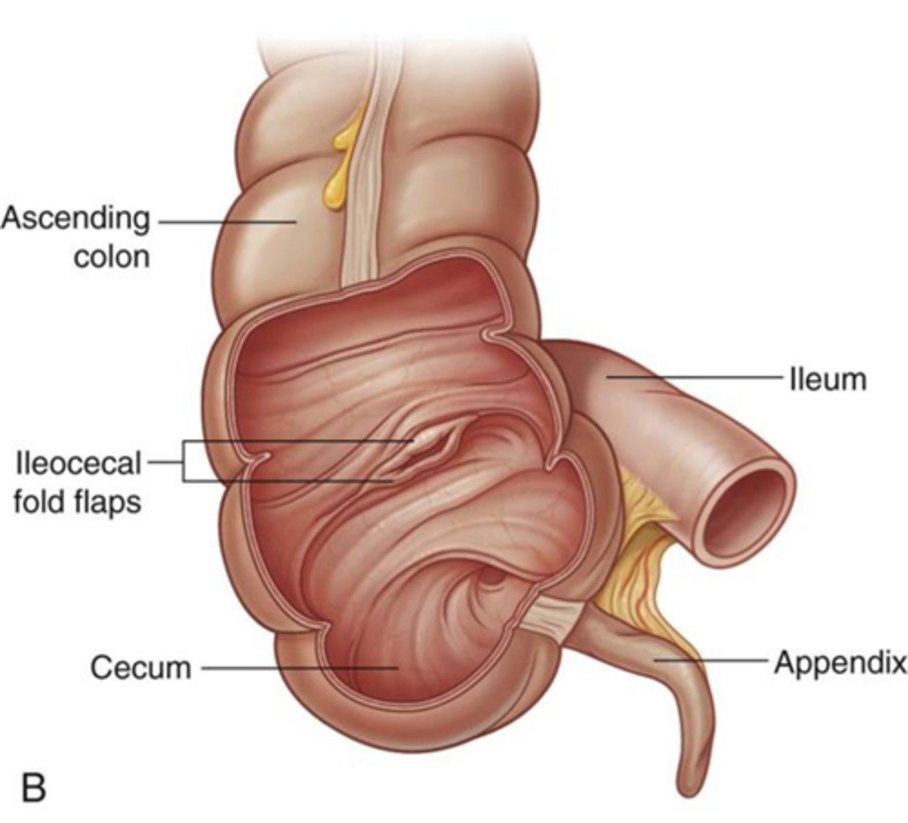
What is the appendix?
Small tail-like projection that is connected to the cecum (blind pouch that's function is unknown)

What are taeniae coli?
longitudinal muscles that aid in the process of peristalsis
If you follow it will lead you to the appendix

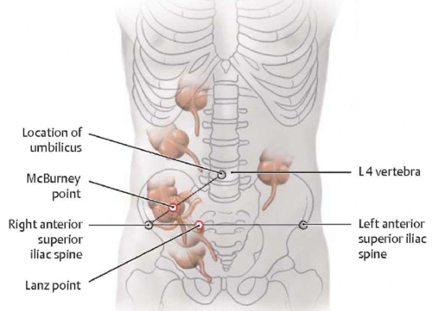
What point is used to locate the appendix?
McBurney's point: 2/3 between belly button and ASIS
Note that location is variable
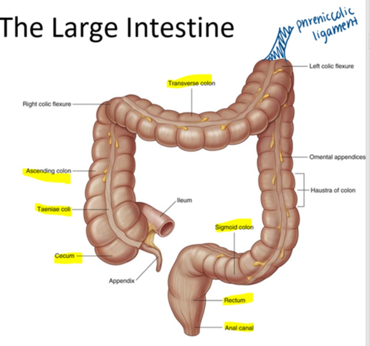
What are the characteristic features of the large intestine?
Taeniae coli
Haustra
Omental appendices

What are haustra?
"Puckers" of the large intestine which churn and constrict to move the food forward (due to taenaie coli)

Is the cecum intraperitoneal or retroperitoneal?
intraperitoneal
Is the appendix intraperitoneal or retroperitoneal?
intraperitoneal

List the parts of the large intestine and if they are intraperitoneal or retroperitoneal
Intraperitoneal: cecum, appendix, transverse colon, sigmoid colon
retroperitoneal: ascending and descending colon, and rectum

Are the jejunum and ileum intraperitoneal or retroperitoneal?
intraperitoneal
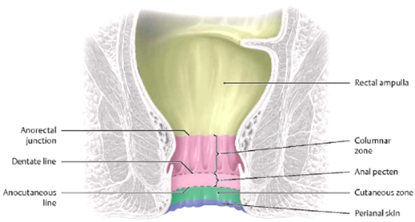
What does the pectinate line divide?
Transition between abdomen and pelvis
Above is visceral, below is somatic
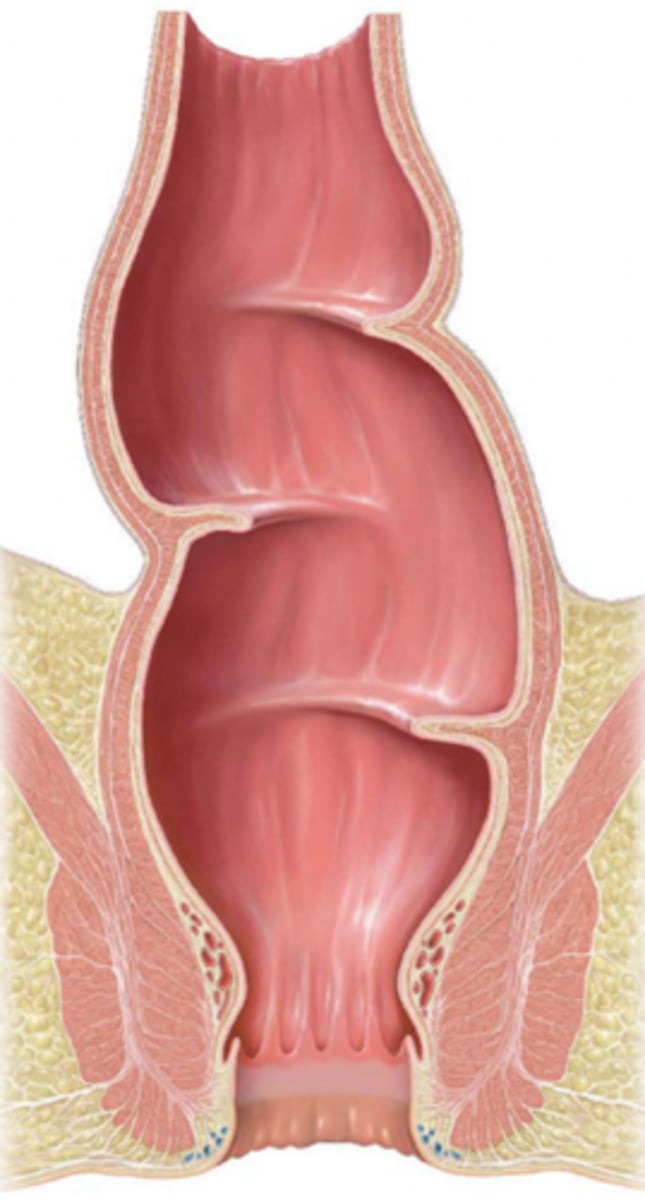
What is the function of the transverse rectal folds?
act as shelves for feces (3)
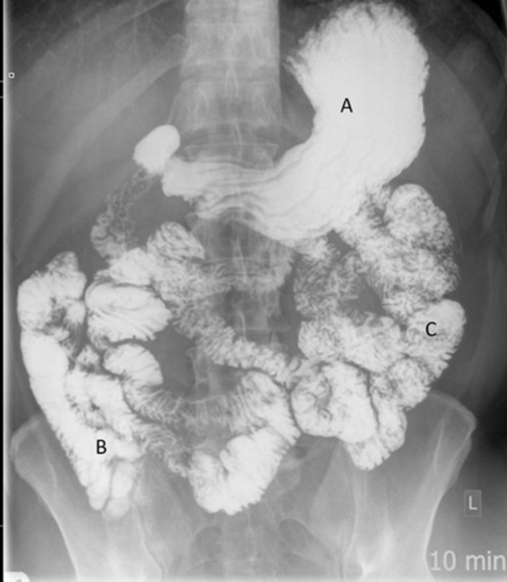
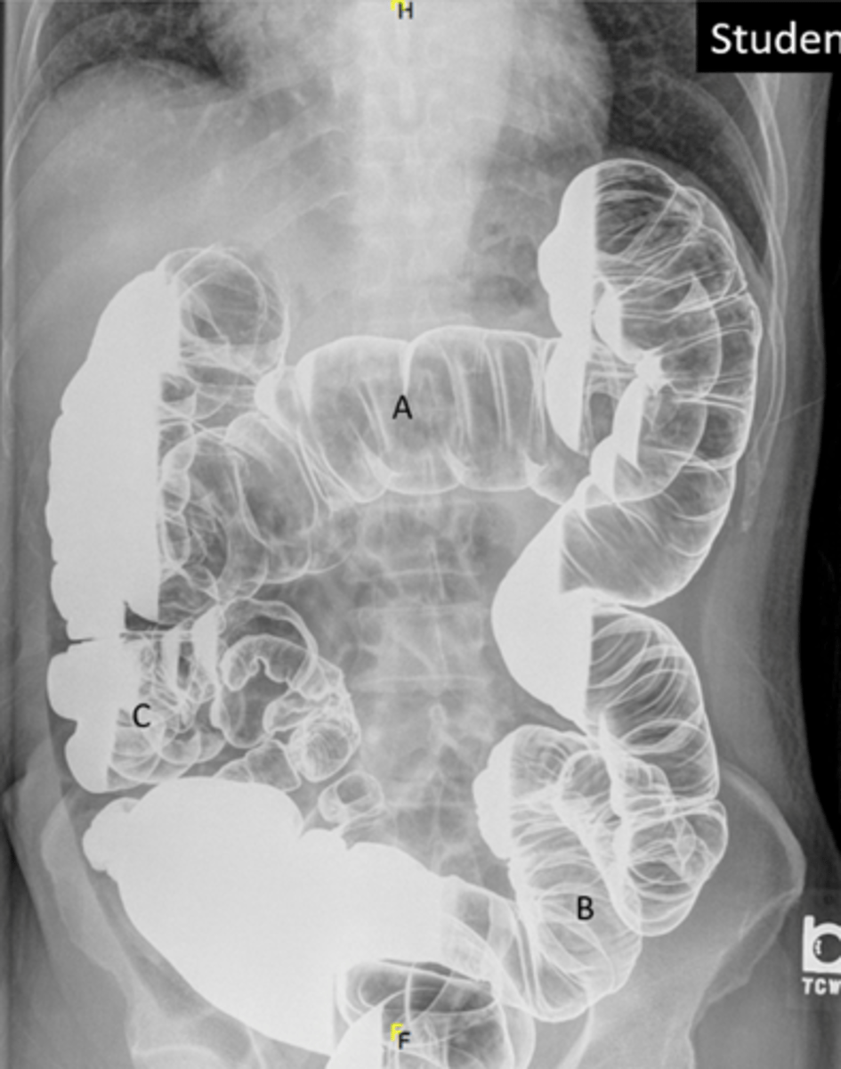
Identify the indicated structures on the following radiograph
A: stomach
B: Ileum
C: Jejunum
Identify indicated anatomy in the following radiograph
A: transverse colon
B: Sigmoid Colon
C: Cecum
F: Rectum
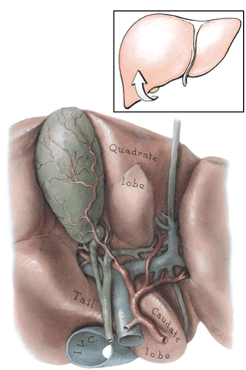
Identify the lobes of the inferior portion of the liver
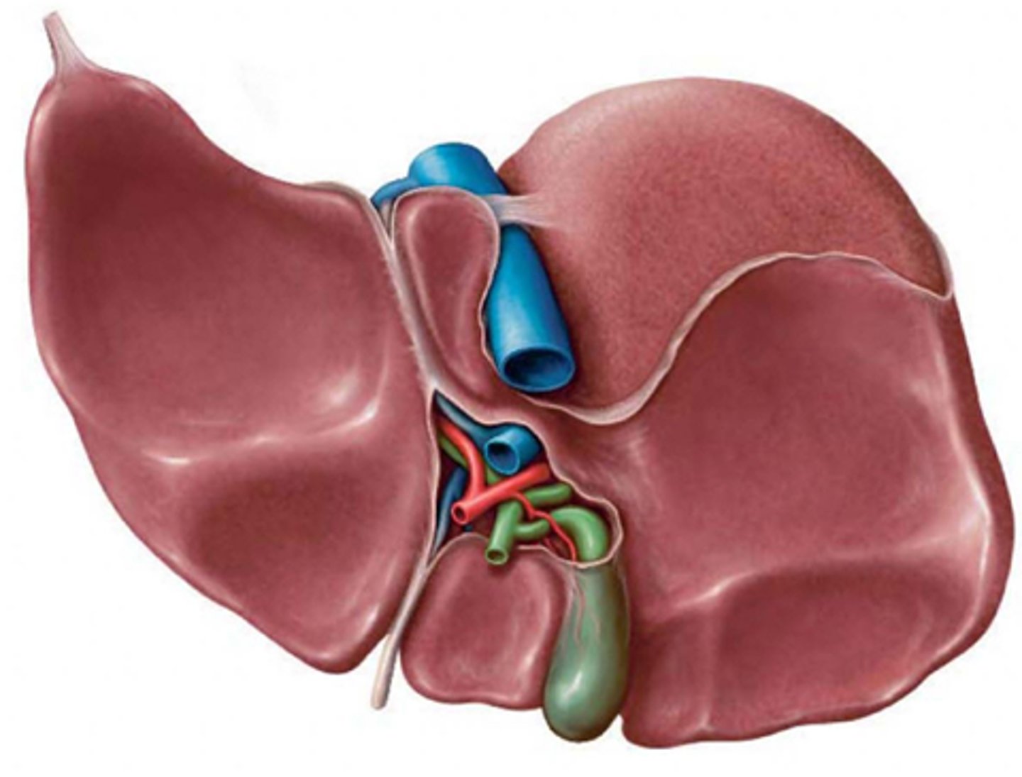
What is the porta hepatis?
Hilum of liver (transverse hepatic fissure) - contains components of hepatic artery, hepatic ducts and portal vein.
region of structures entering and exiting the liver
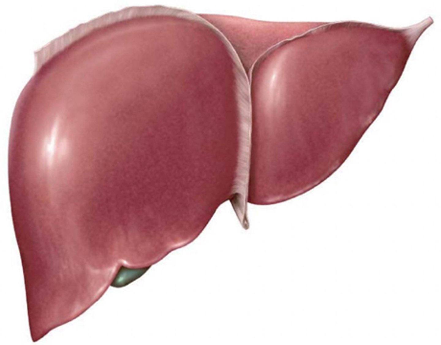
Identify the anterior lobes of the liver
Where will lymph from the bare area of the liver drain?
to inferior phrenic nodes which will drain to posterior mediastinal nodes
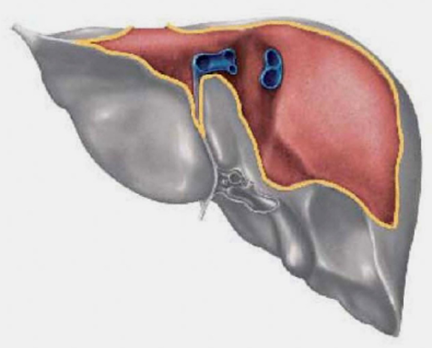
What ligament is deficient over the bare area of the liver?
coronary ligament: bare area lacks peritoneum

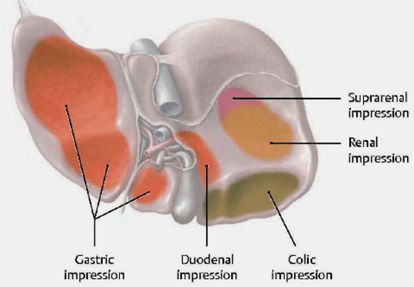
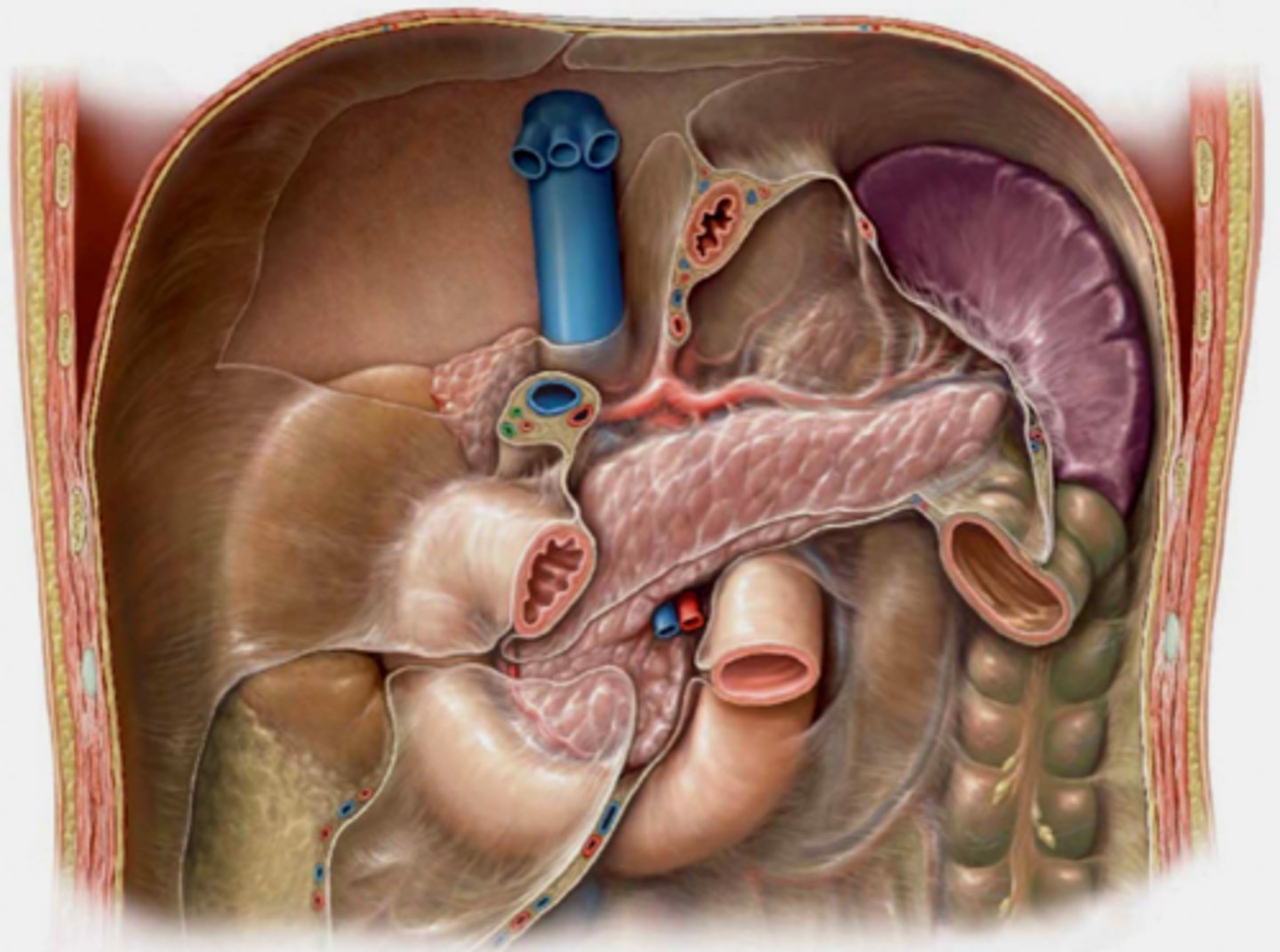
Identify impressions on the inferior portion of the liver
Note that organs in direct contact with liver can spread cancer directly to the liver
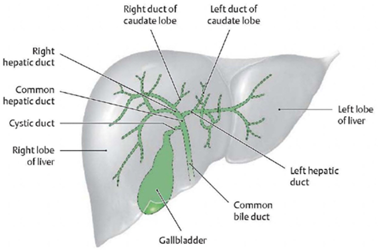
Label/draw the biliary duct system
biliary duct system
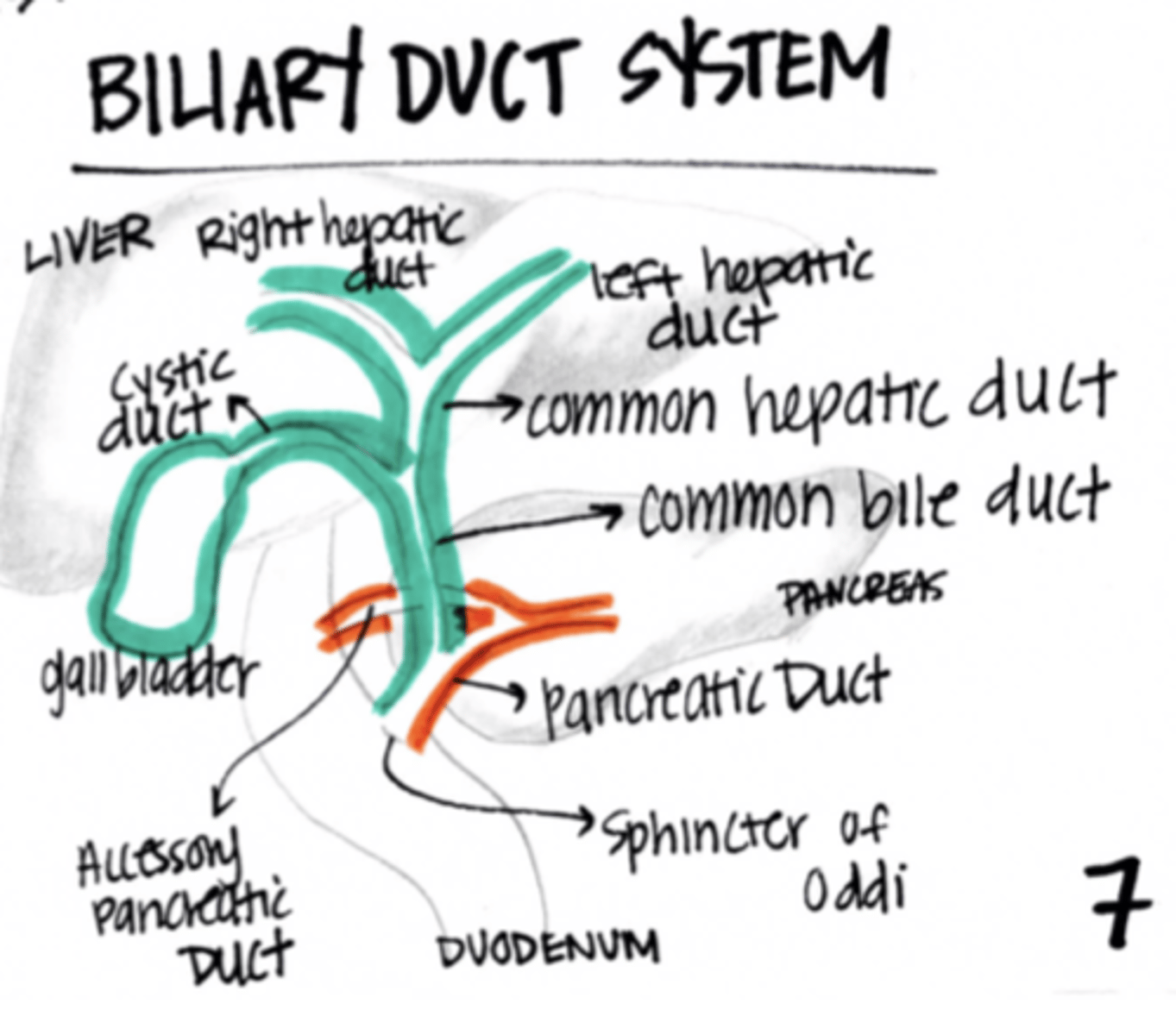
What joins to form the common bile duct?
common hepatic duct and cystic duct
What is the function of the gallbladder?
stores and concentrates bile
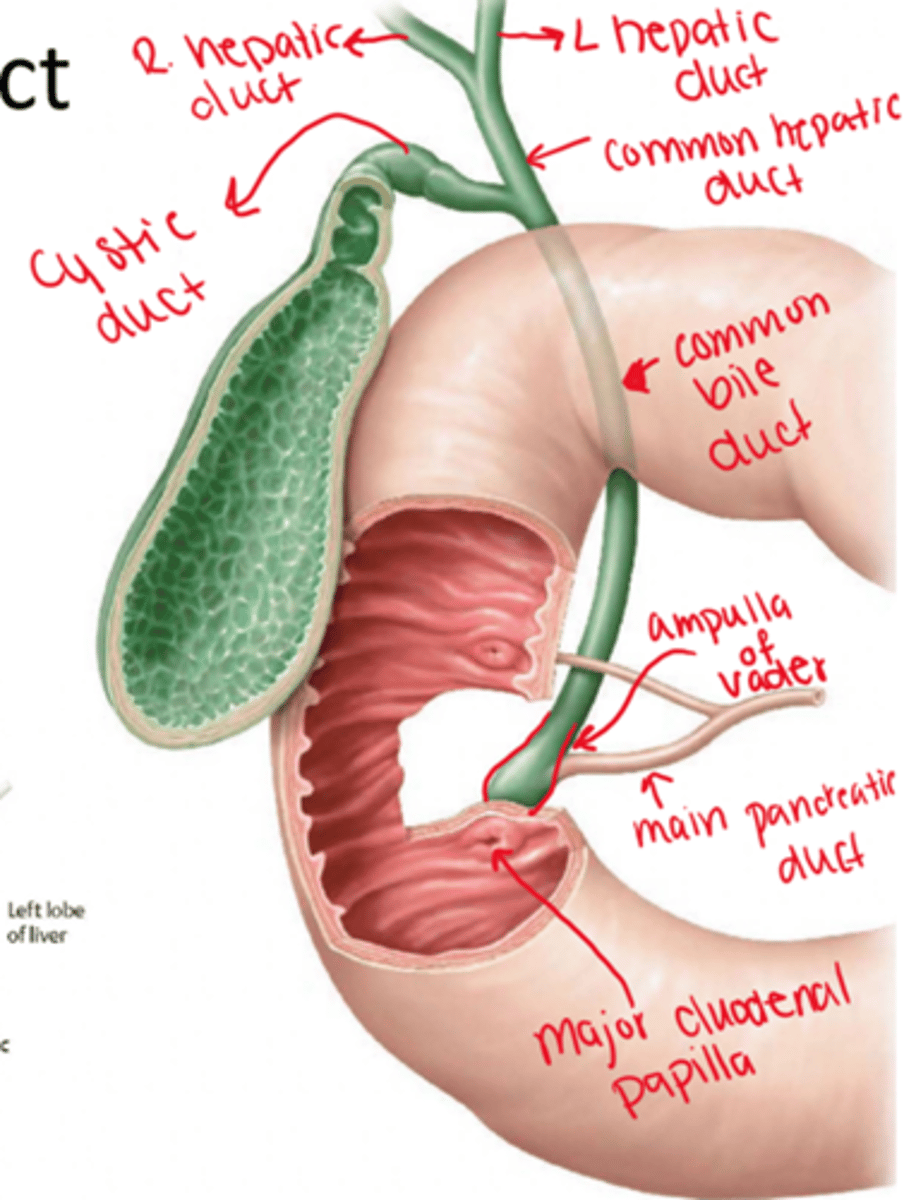
What is the ampulla of vater?
Union of the common bile duct and the pancreatic duct

What is the function of the sphincter of oddi?
Controls the flow of bile into the duodenum

Where will the ampulla of vater open to?
duodenum through the major duodenal papilla

Is the pancreas intraperitoneal or retroperitoneal?
retroperitoneal
What is another name for the ampulla of vater?
hepatopancreatic ampulla
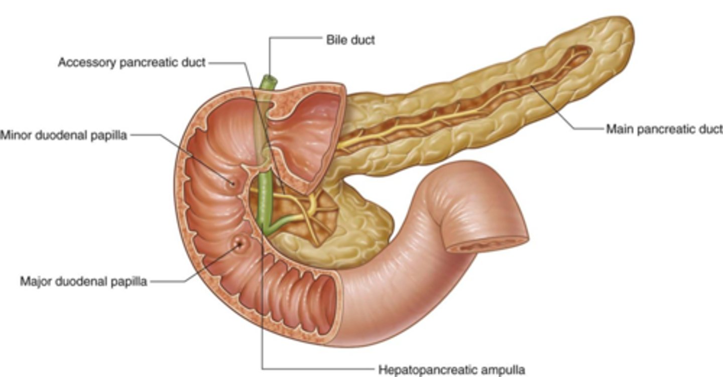
What opens to the minor duodenal papilla?
accessory pancreatic duct

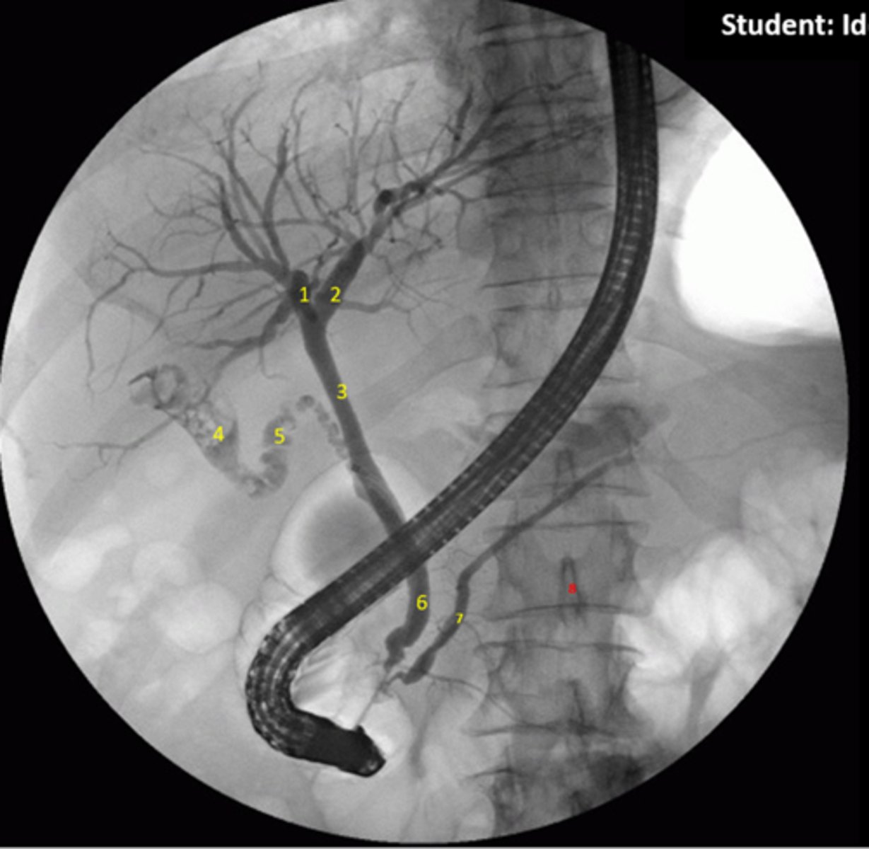
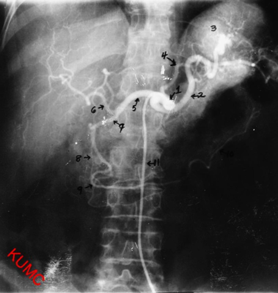
Identify indicated anatomy in the following radiograph
1.Right hepatic duct
2.Left hepatic duct
3.Common hepatic duct
4.Gallbladder
5.Cystic duct
6.Common bile duct
7.Pancreatic duct
8.Spinous process
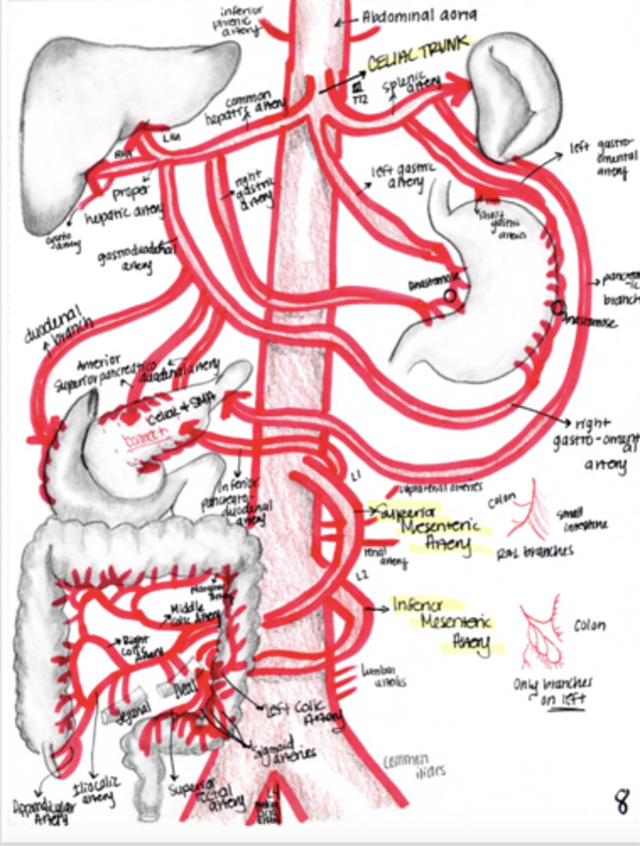
What are the main supplies of blood to the gut and what vertebral levels are they seen?
Celiac trunk: forgut (L1)
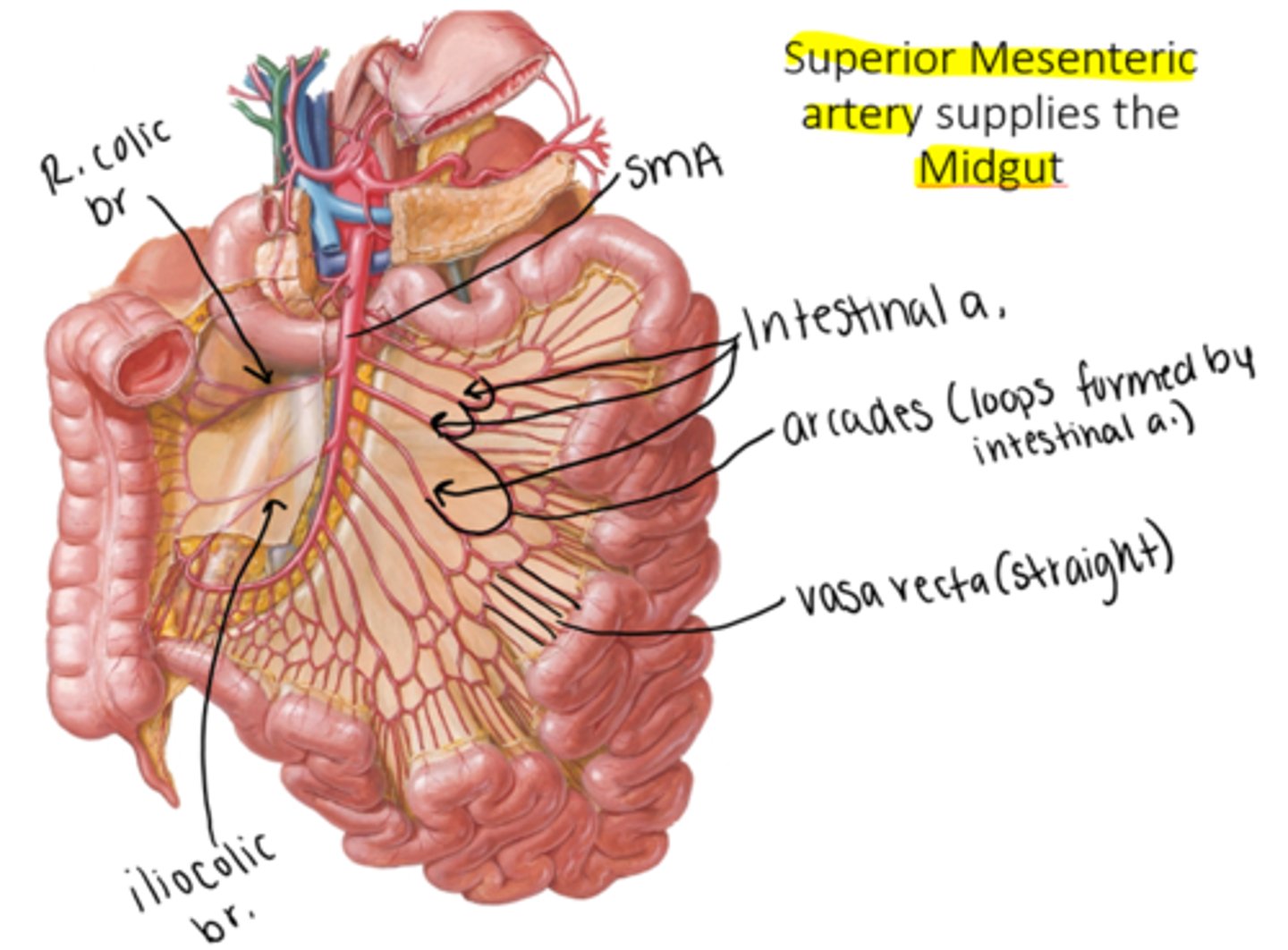
Superior Mesenteric artery: midgut (L1)
Inferior Mesenteric artery: hindgut (L3)
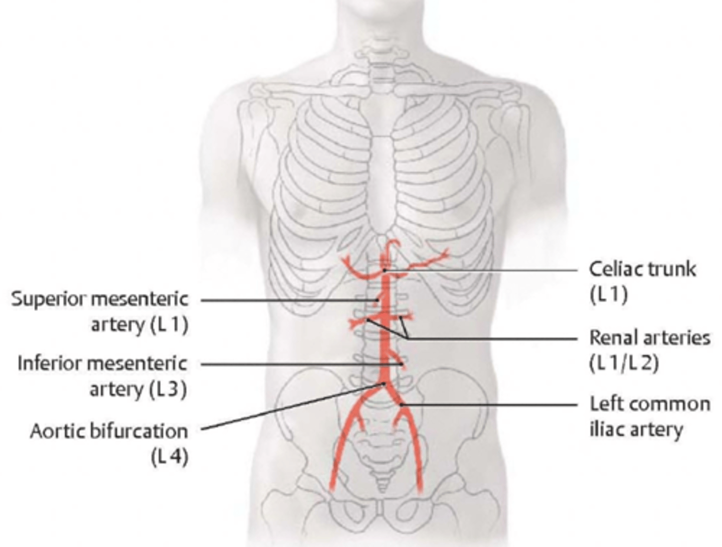
At what vertebral level will the aorta bifurcate?
L4

At what vertebral level are the renal arteries seen?
L1-L2

What is included in the foregut?
stomach, spleen, liver, and gallbladder
will be supplied by the celiac trunk

What is included in the midgut?
distal duodenum, small intestine, ascending colon, and 2/3 of the transverse colon
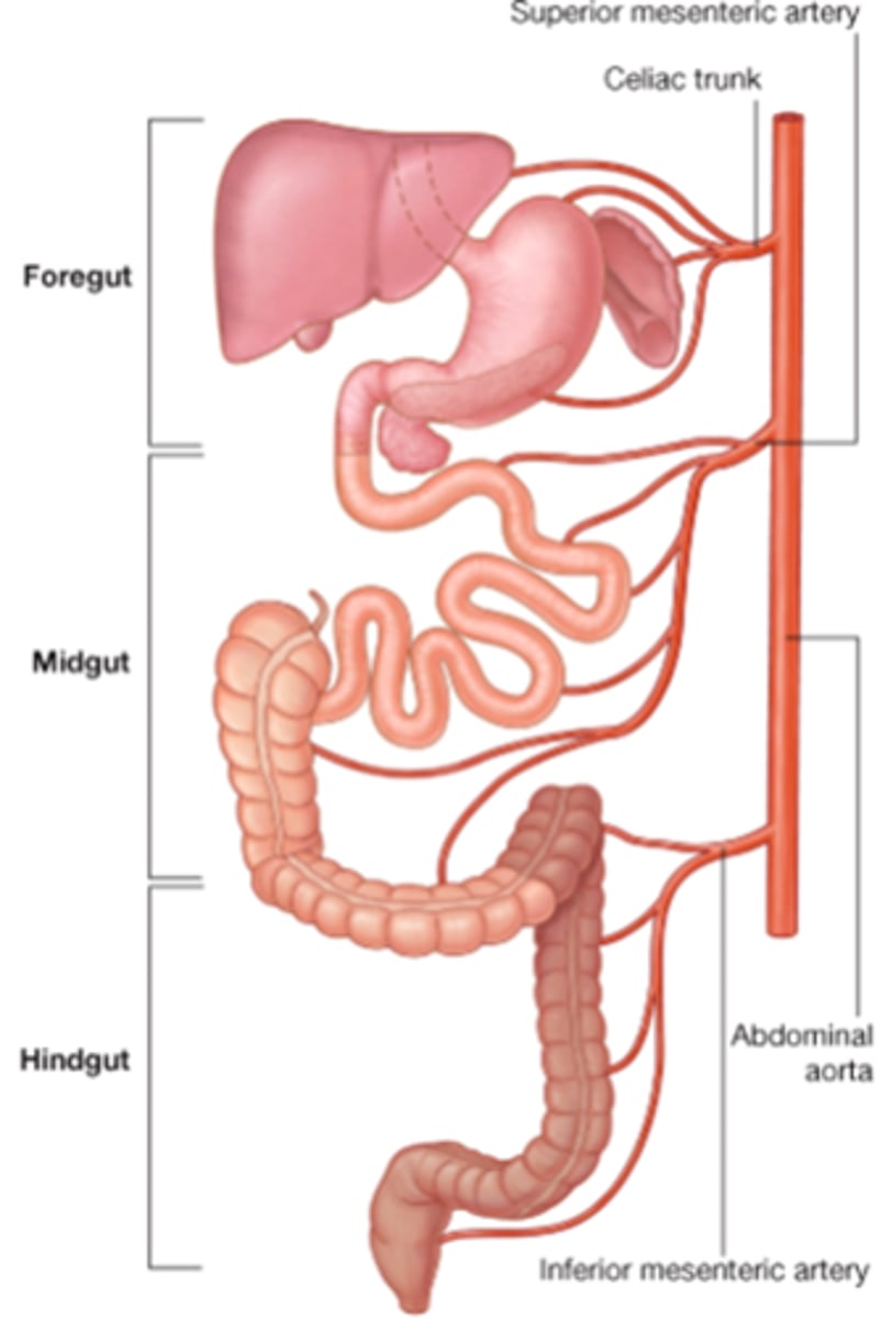
What is included in hindgut?
1. left half of the one-third of transverse colon
2. descending colon
3. sigmoid colon
4. rectum
5. superior part of the anal canal

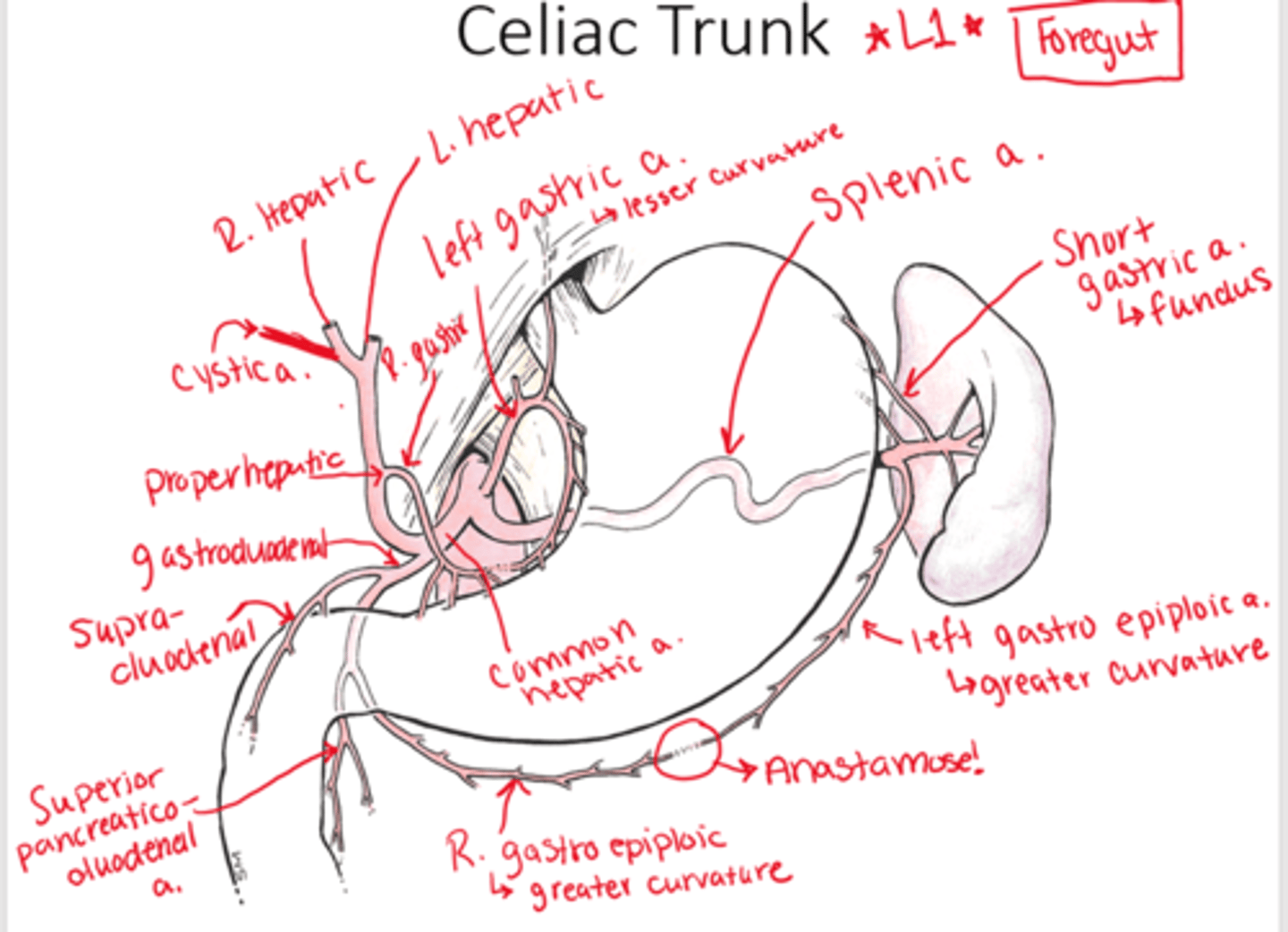
Label the branches of the celiac trunk
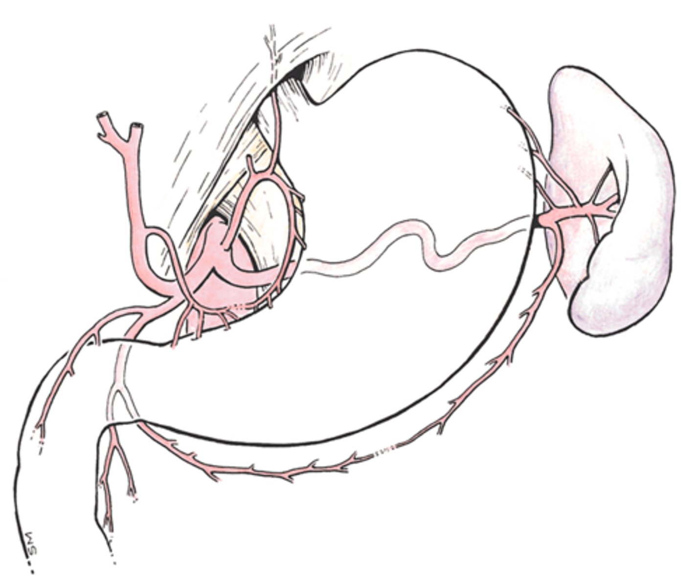
Where does the celiac trunk originate and what does it supply?
L1: supplies foregut
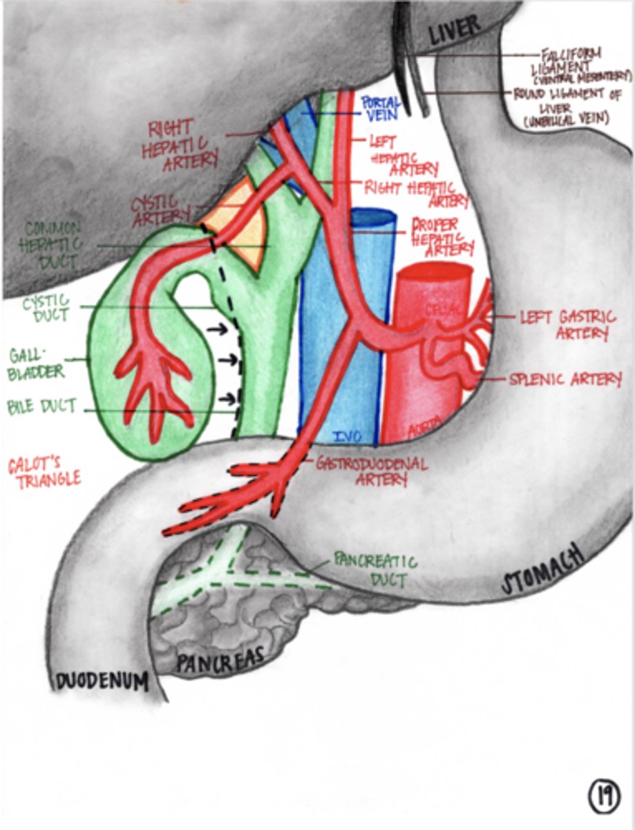
Draw where you would do Pringle's maneuver? What is in Calot's triangle?
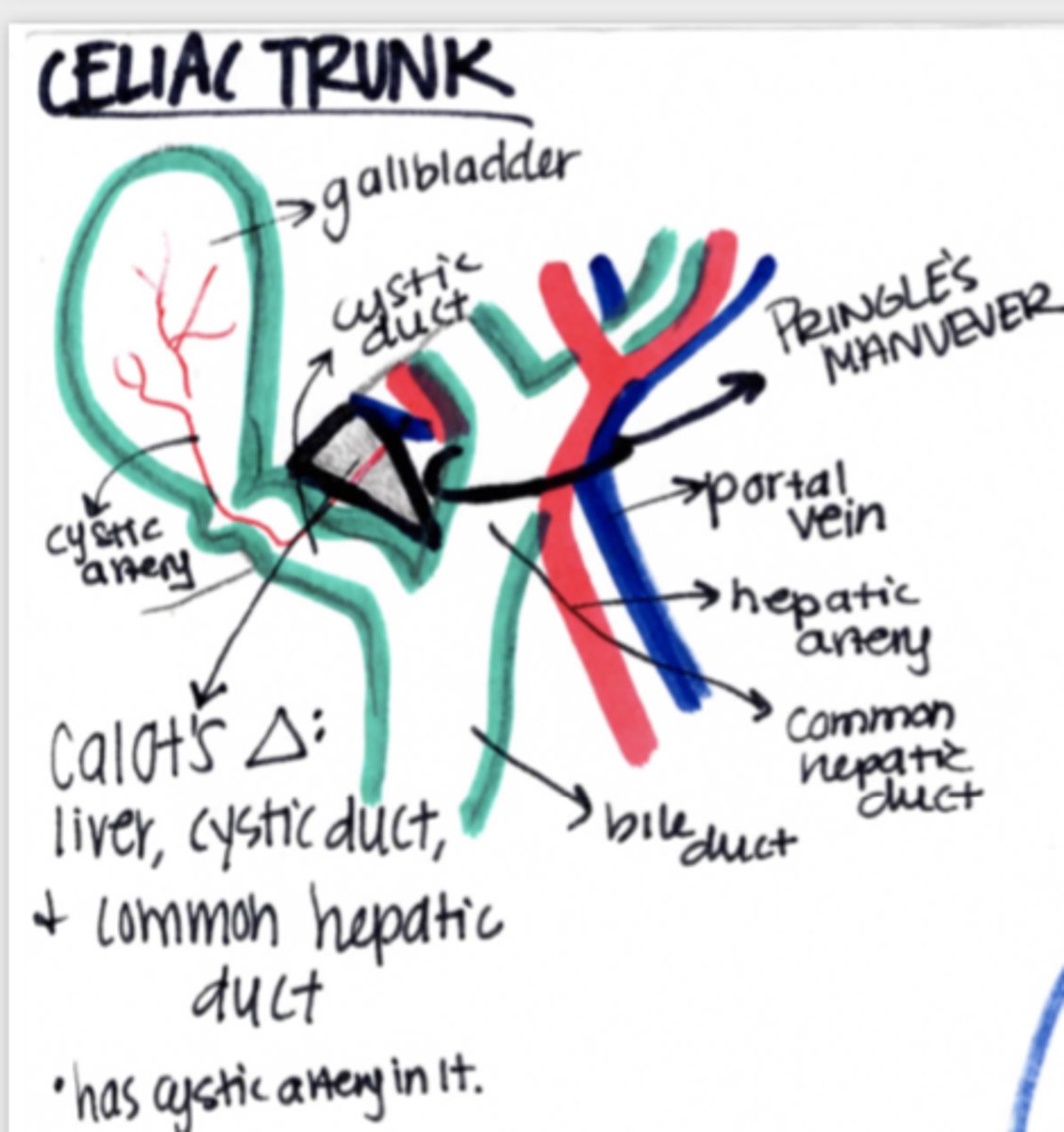
Where will anastamoses between foregut and midgut be seen?
between inferior and superior pancreaticoduodenal arteries!!
allows anastamoses between SMA and celiac trunk
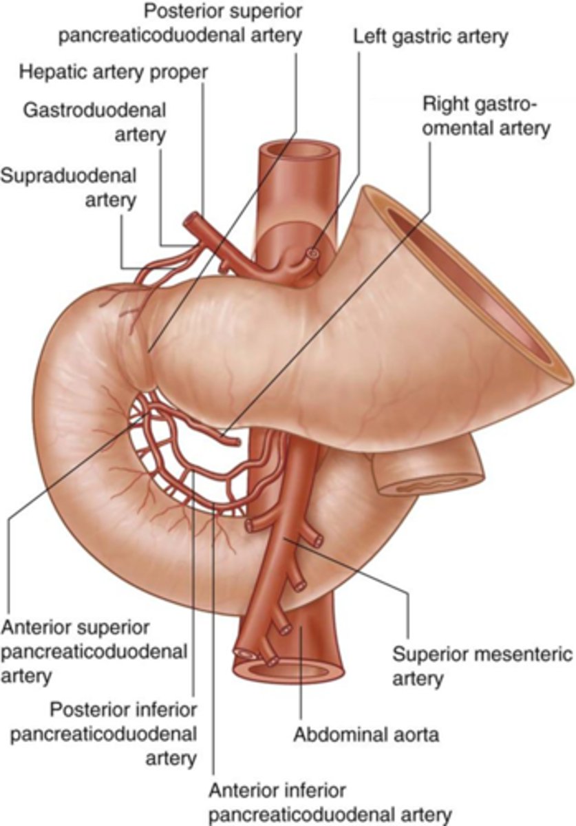
Identify the blood supply to the duodenum
Note that the supraduodenal artery also supplies the head of the pancreas

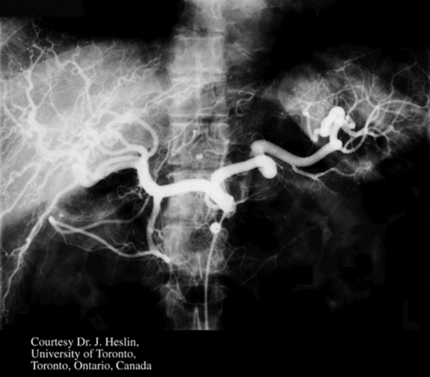
Identify pertinent anatomy in the following radiograph
What is the portal triad?
hepatic artery proper, portal vein, common bile duct
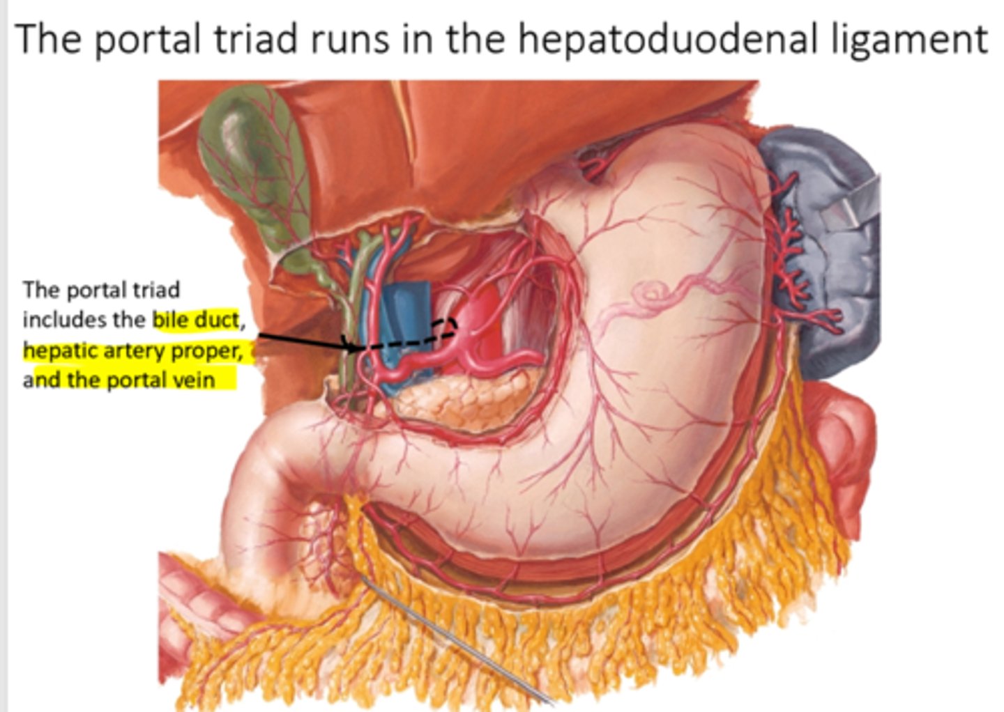
Where is the portal triad located?
within the hepatoduodenal ligament

Explain the orientation of the structures within the portal triad
Common bile duct is the most lateral structure of the portal triad
Proper hepatic artery is medial
Portal vein is posterior to other two structures in the triad (aka: bile duct and hepatic artery proper are anterior to portal vein)

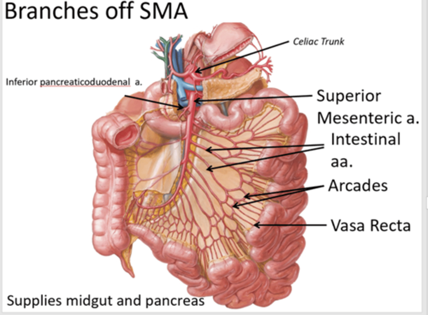
What does the superior mesenteric artery supply?
midgut

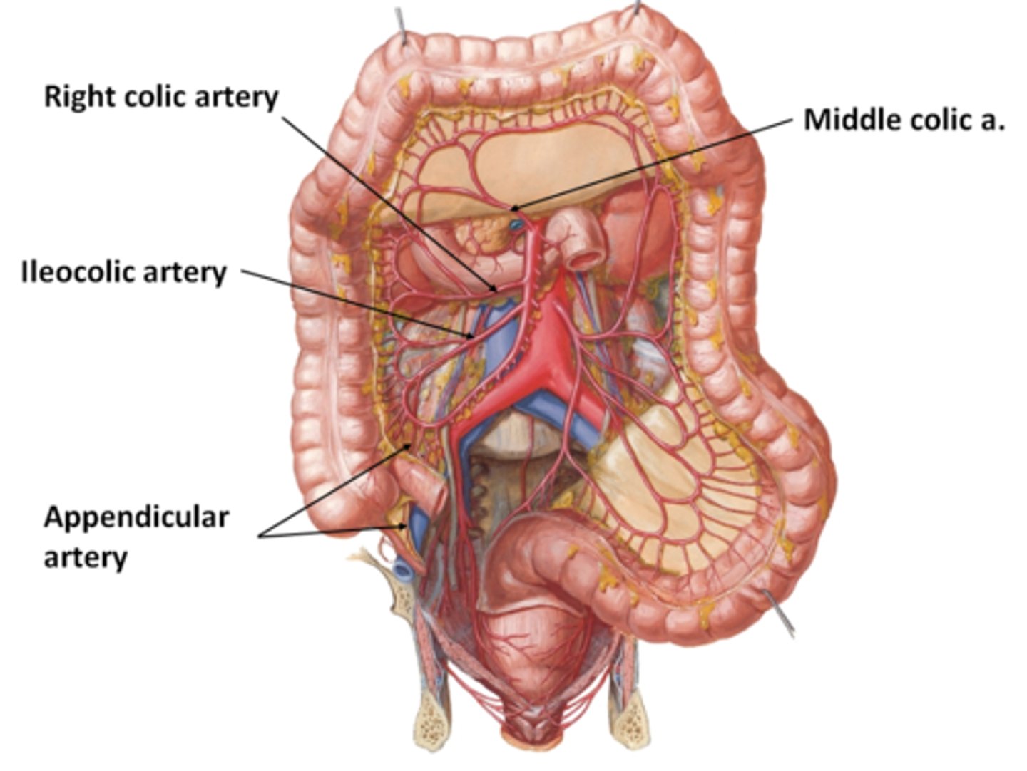
What does the right colic branch of the superior mesenteric artery supply?
ascending colon

What does the iliocolic branch of the superior mesenteric artery supply?
cecum
What branch of the SMA will supply the pancreas?
inferior pancreaticoduodenal artery
What are the differences in the vessels to the jejunum and ileum from the SMA?
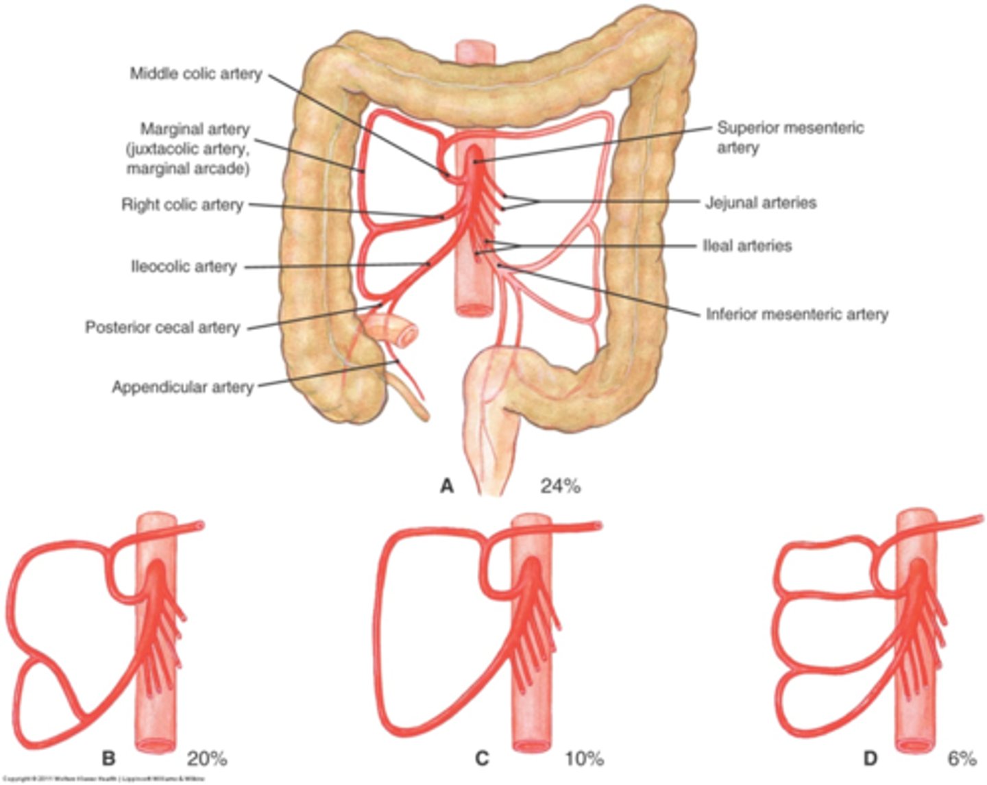
Identify branches of SMA and note what they supply
Ileocolic supplies cecum, also gives off appendicular artery to appendix
Right colic supplies ascending colon
Middle colic goes to the right colic flexure
How are the branches of the SMA named?
based on where they are going not where they originate
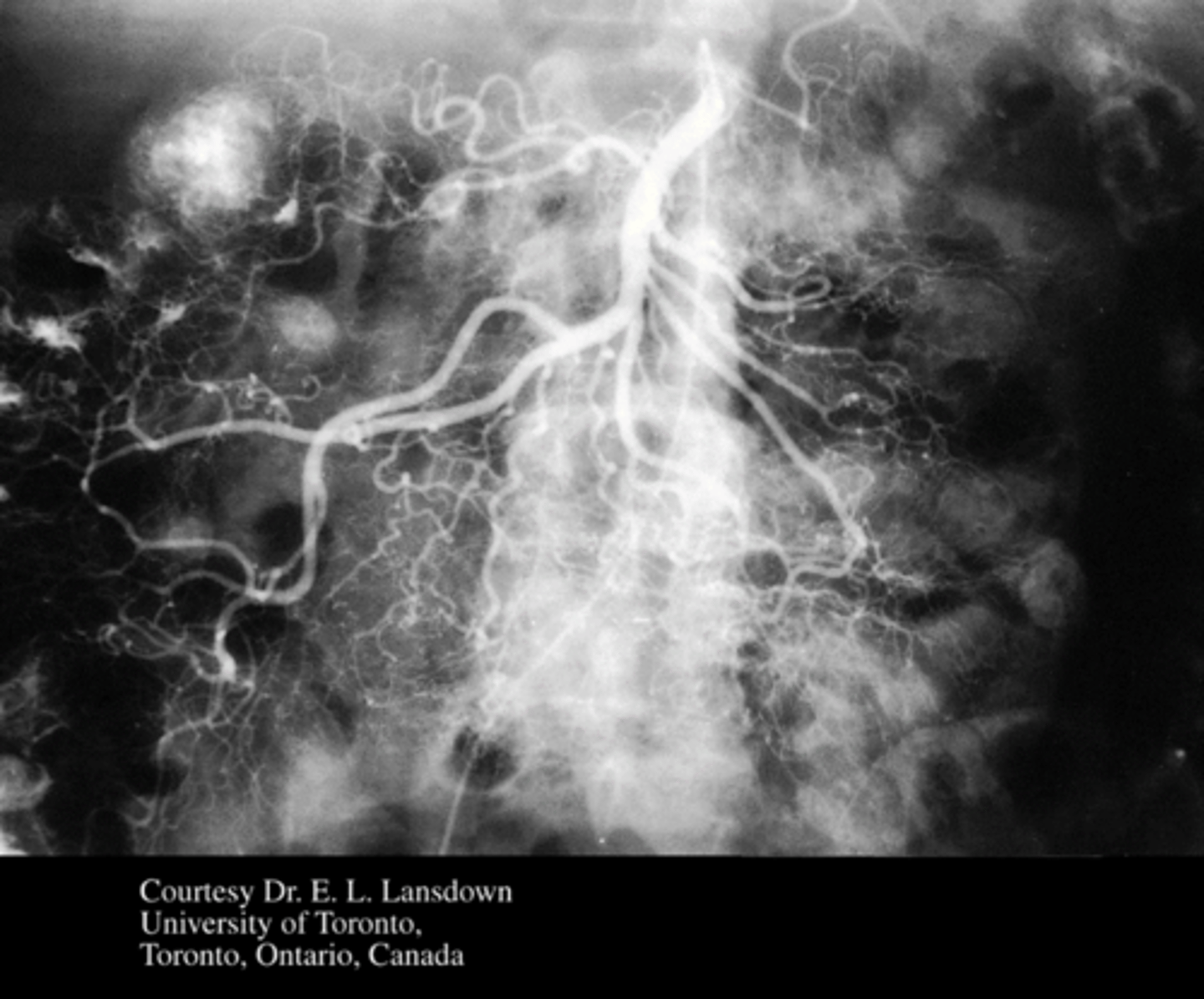
Identify pertinent anatomy in the following radiograph
SMA
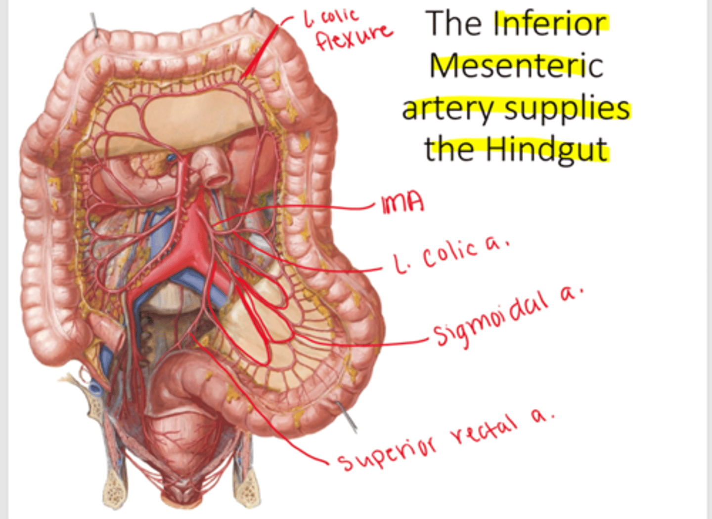
What does the inferior mesenteric artery supply?
hindgut: from left colic flexure to rectum

What is the most medial branch of the IMA?
superior rectal artery
Identify pertinent anatomy in the following radiograph
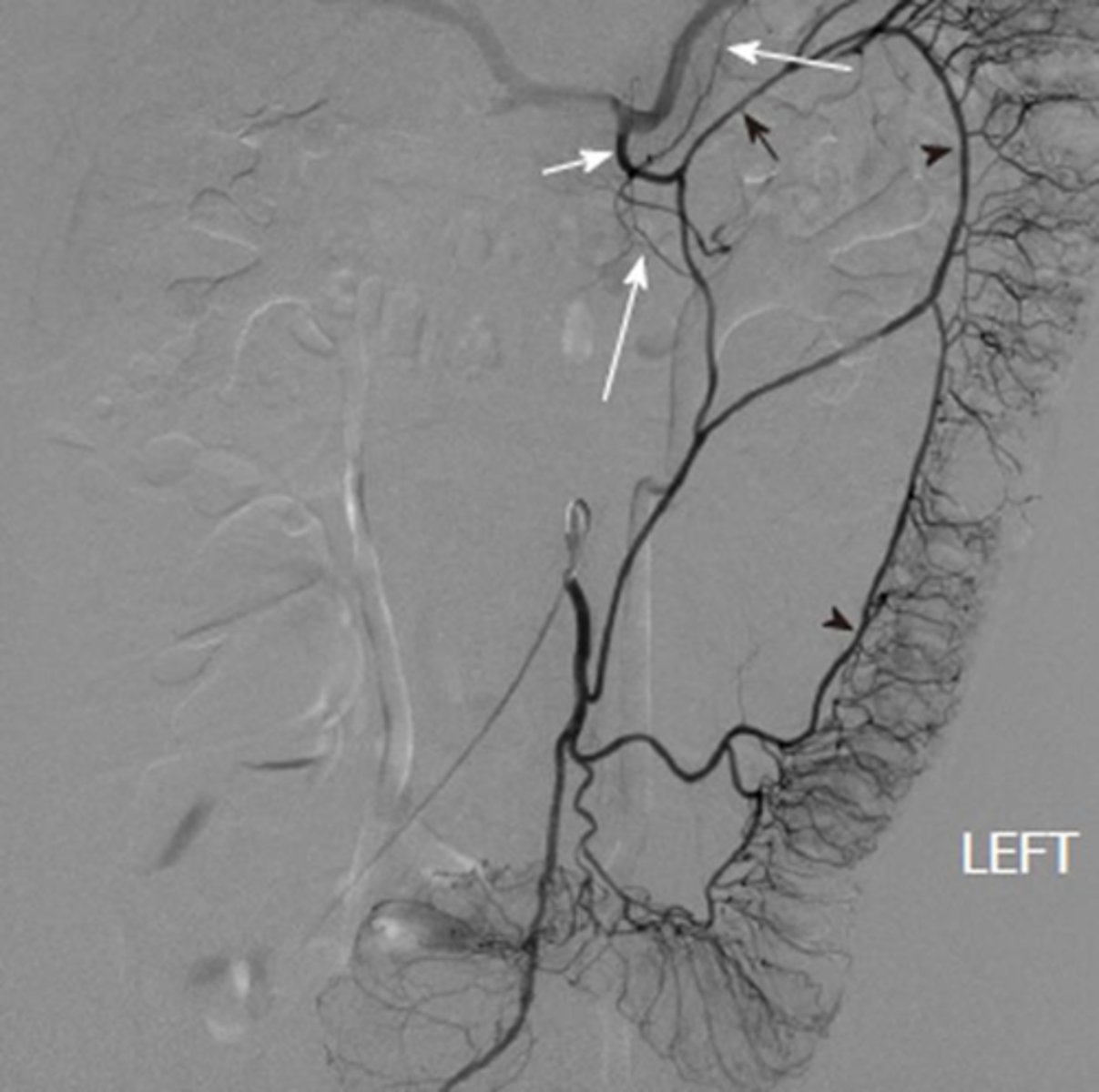
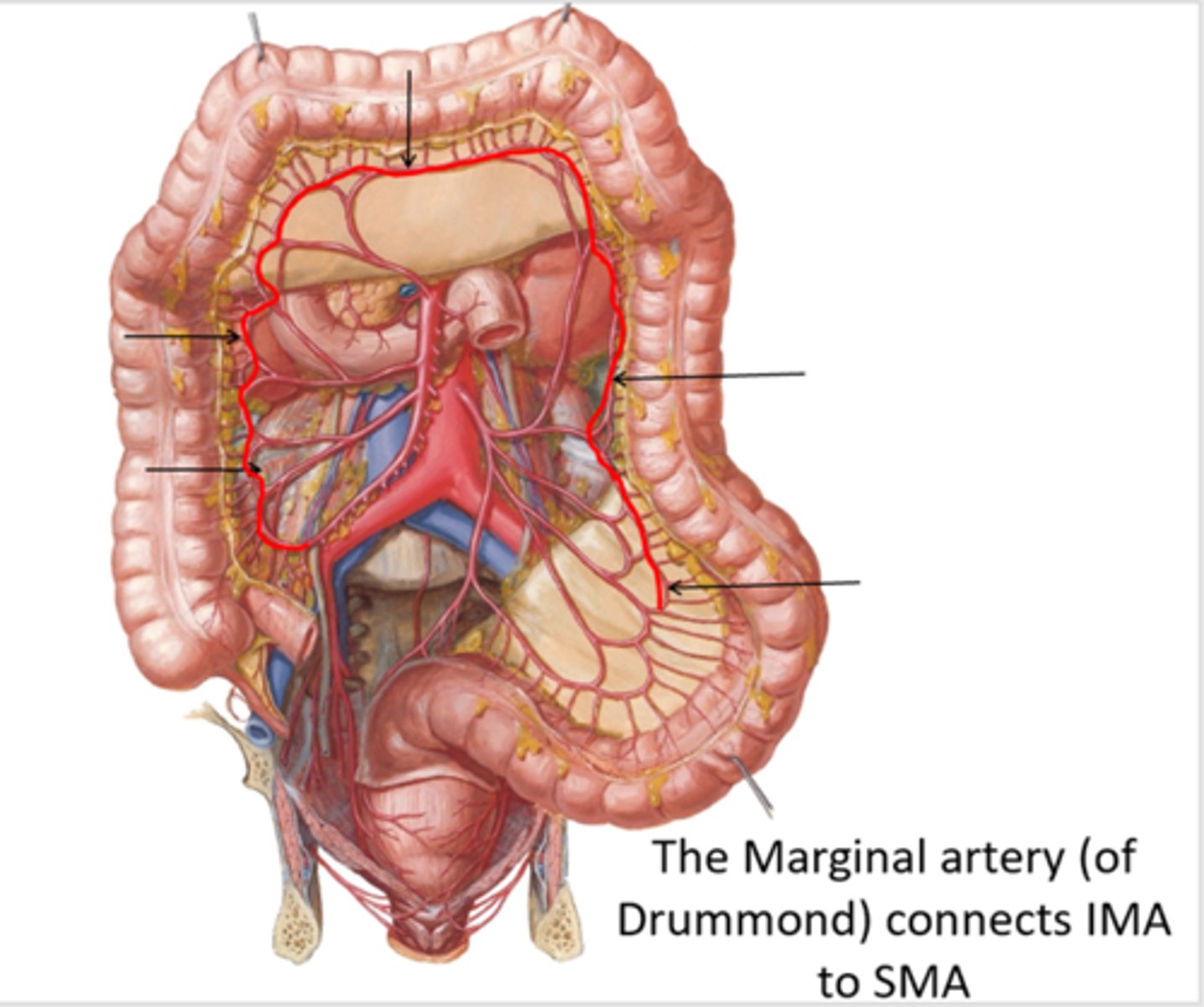
What is the anastamoses between the midgut and hindgut?
Marginal artery of drummond: connection between IMA and SMA
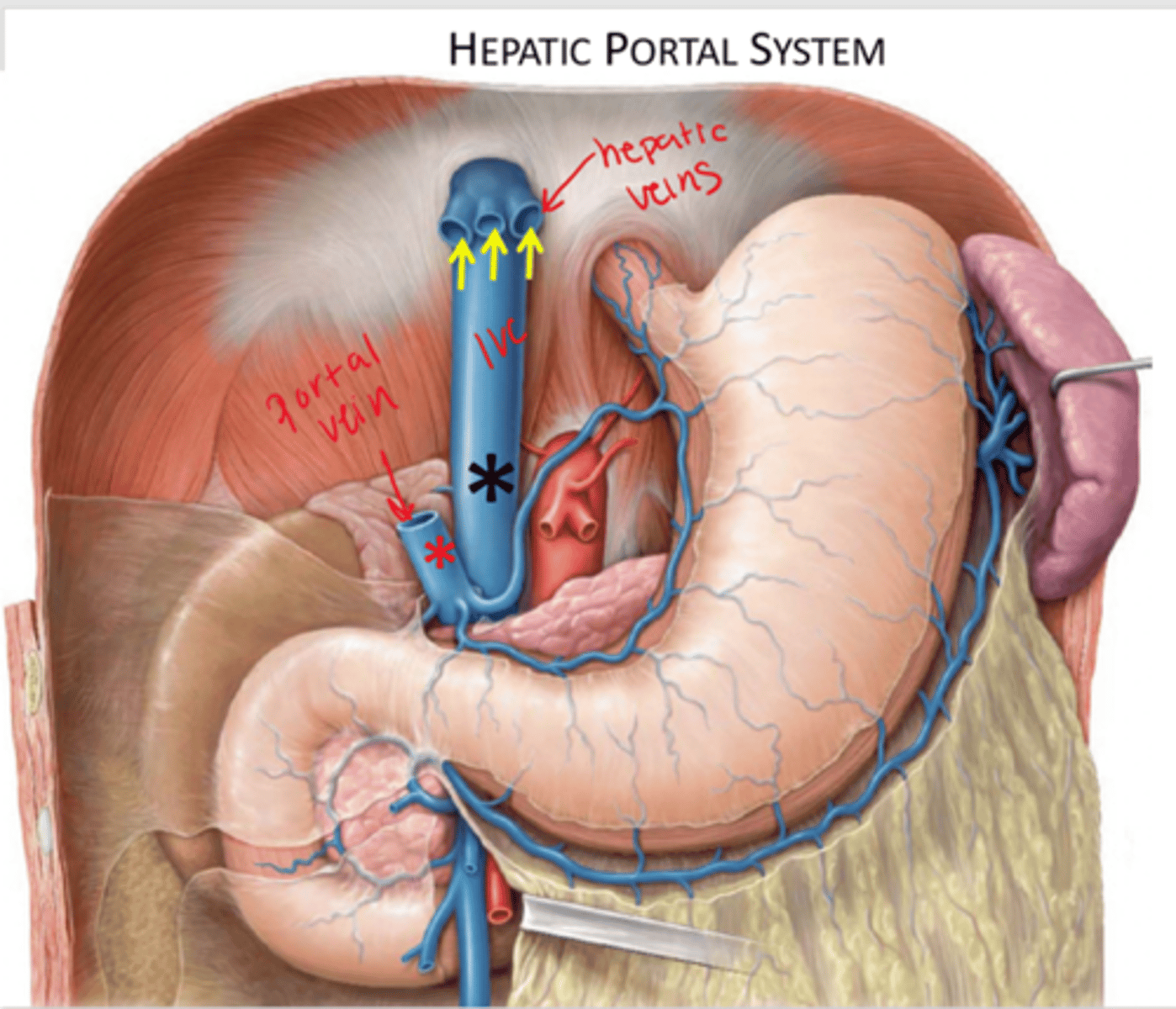
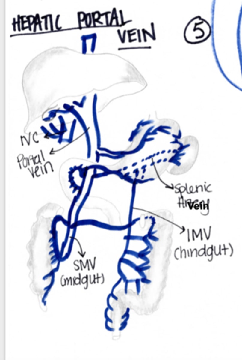
Where do all the veins from the portal system drain?
portal vein then into the liver

Where will veins in the caval system drain?
inferior vena cava: bypasses the liver and heads straight back to the heart

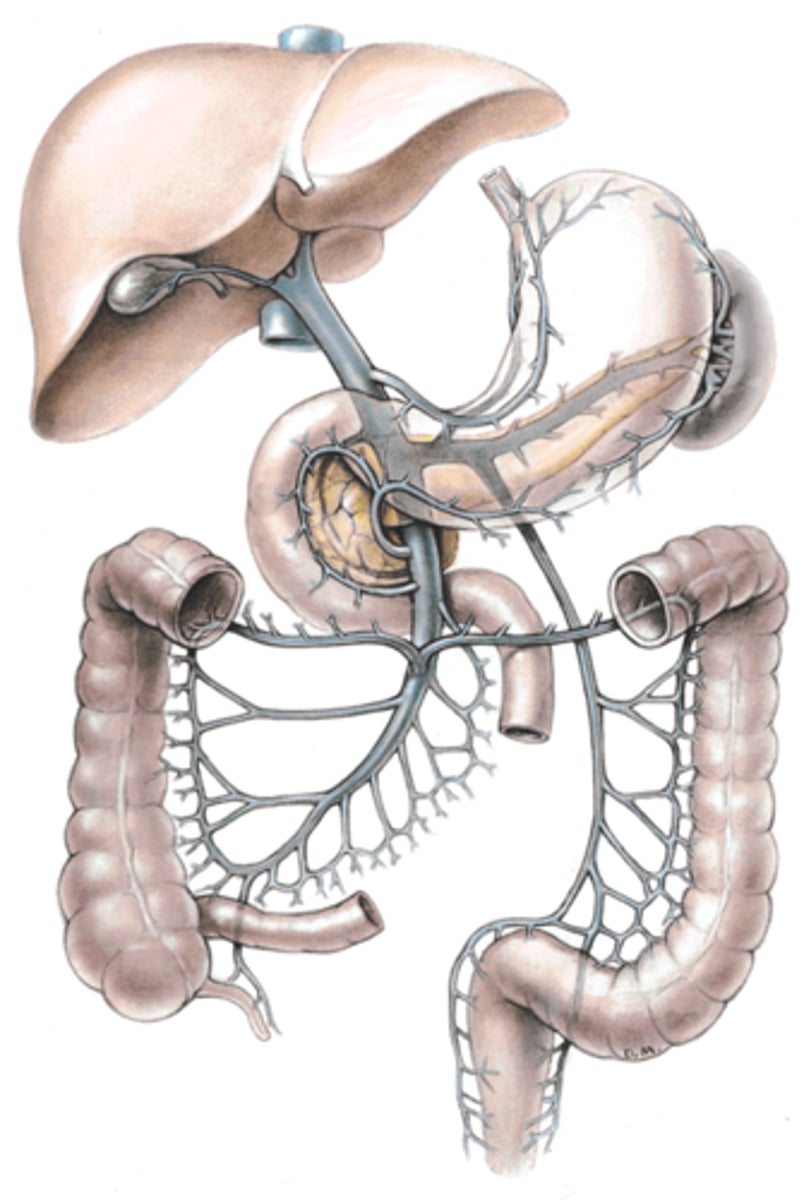
What forms the portal vein?
The union of the superior mesenteric and the splenic veins

What is carried to the liver in portal venous system?
nutrient rich blood from GI tract
broken down RBCs fromt he spleen for metabolism or storage
What are the two capillary beds utilized by the portal venous system?
Utilizes 2 capillary beds
1st capillary bed is in the gut tube
2nd capillary bed is in the liver
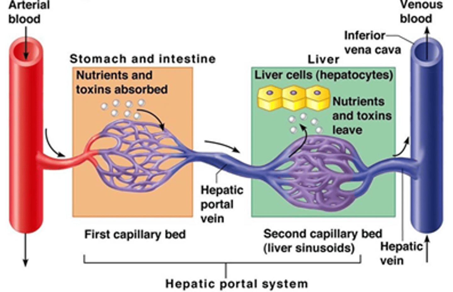
What is carried by the portal and hepatic veins?
Portal vein: carries nutrient rich blood from GI to liver
hepatic vein: carries filtered blood from liver back to the IVC
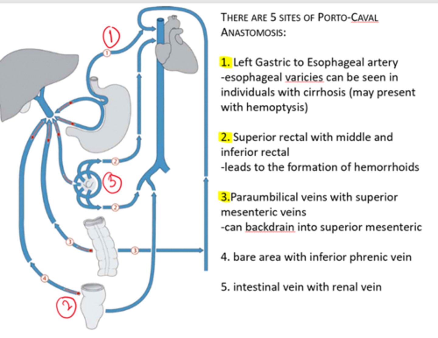
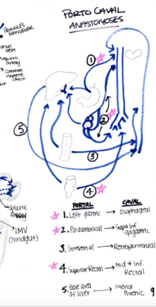
What are the sites of porto-caval anastamosis?
Note that the three highlighted can exhibit increased pressure in patients with liver cirrhosis
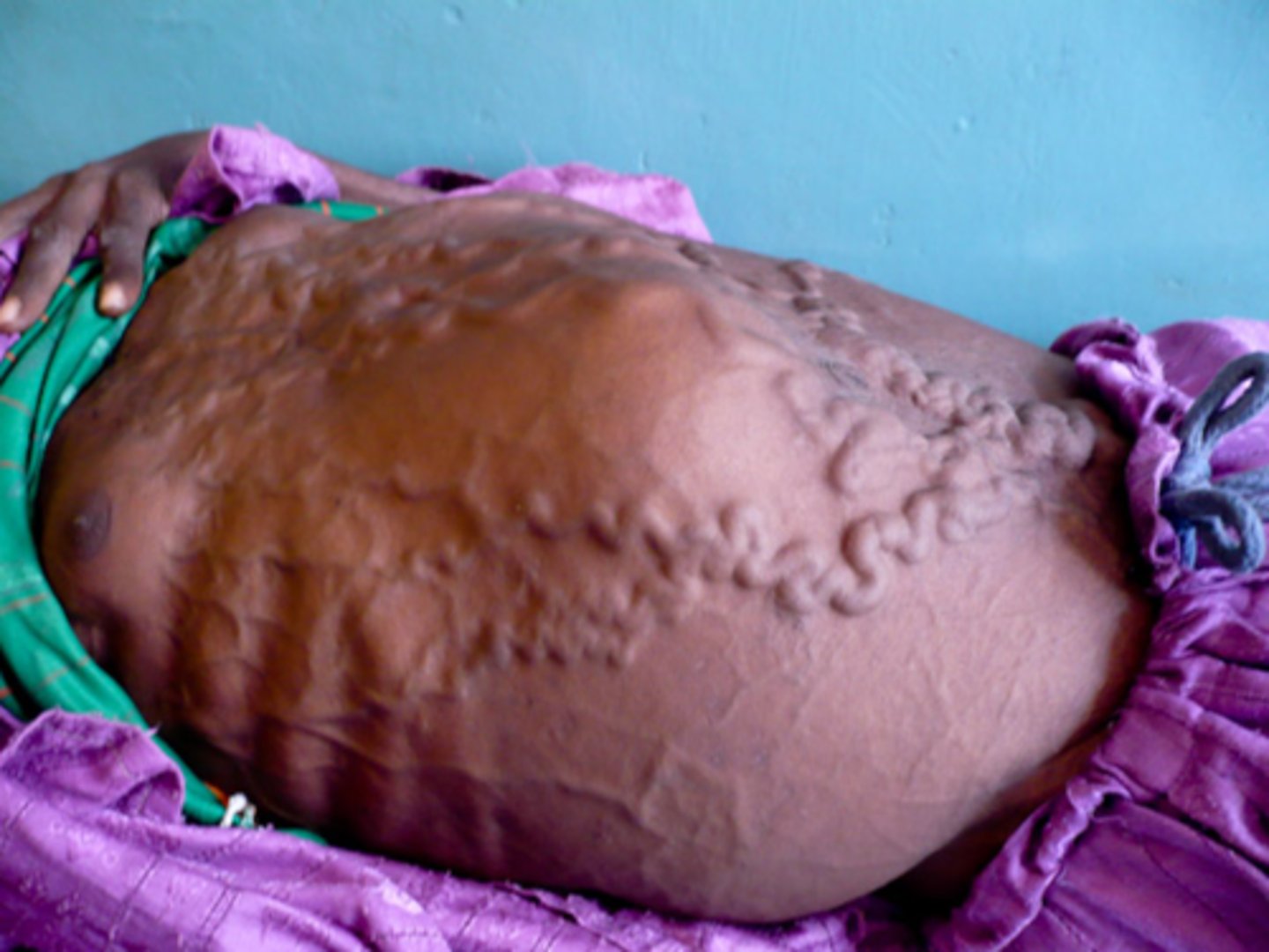
What is shown in the following image?
Caput medusa
portal vein affected is paraumbilical vein: cannot drain into portal system, thus they drain into the superior, inferior, and thoracoepigastric veins
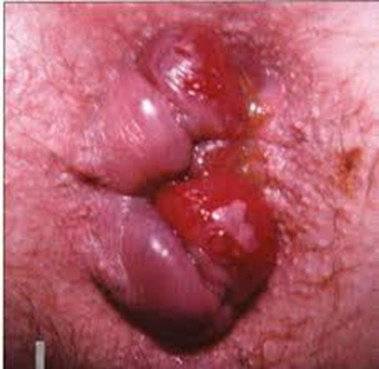
What is shown in the following image?
Hemmrhoids
Backing up of blood into the inferior rectal vein
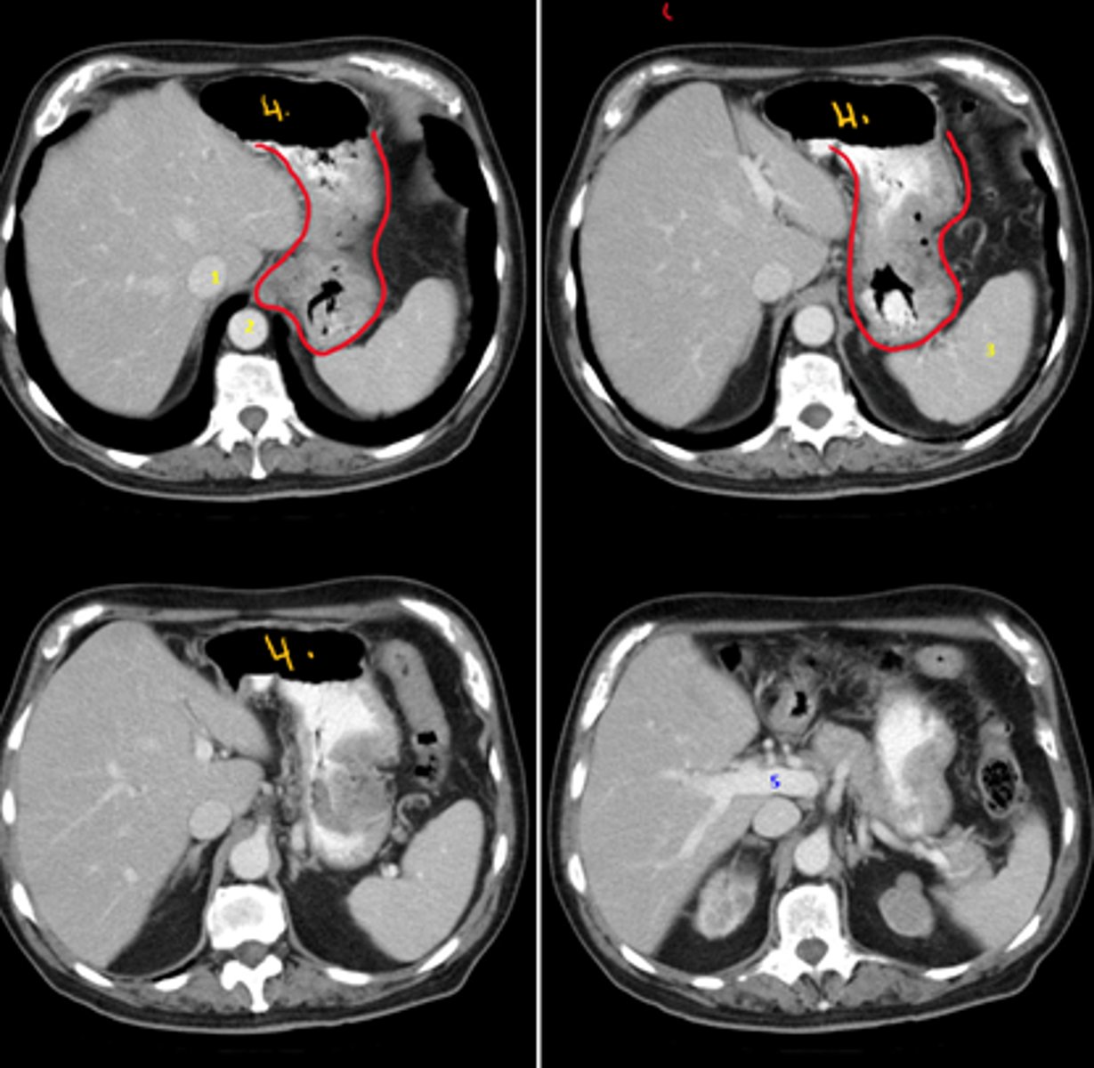
Idedntify the indicated structures on the following radiograph
1. IVC
2. Descending Aorta
3. Spleen
4. Stomach
5. Portal vein (bigger than proper hepatic artery)
growth shown in red: gastric cancer
What structures are at risk of spread of the following growth?
Structures it will spread: celiac lymph nodes to liver, local to liver and spleen, venous drainage to liver (thus the liver has two methods of spread from the stomach)
gastric cancer

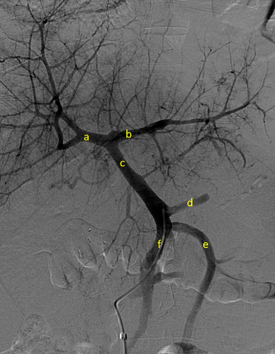
Identify pertinent anatomy of the following radiograph
Identify the pertinent anatomy in the following radiograph
A: Right portal vein
B: Left portal vein
C: Portal Vein
D: Splenic Vein
E: Inferior mesenteric vein
F: Superior mesenteric vein
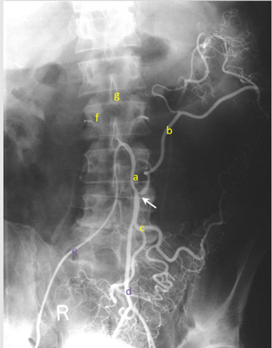
Identify pertinent anatomy of the following radiograph
1. Celiac Trunk 2. Splenic artery 3. Spleen 4. Left Gastric 5. Common Hepatic 6. Proper Hepatic 7. Gastroduodenal 8. Pancreaticoduodenal (don't have to be able to identify for test!) 9. Right gastroepiploic 10. Left gastroepiploic
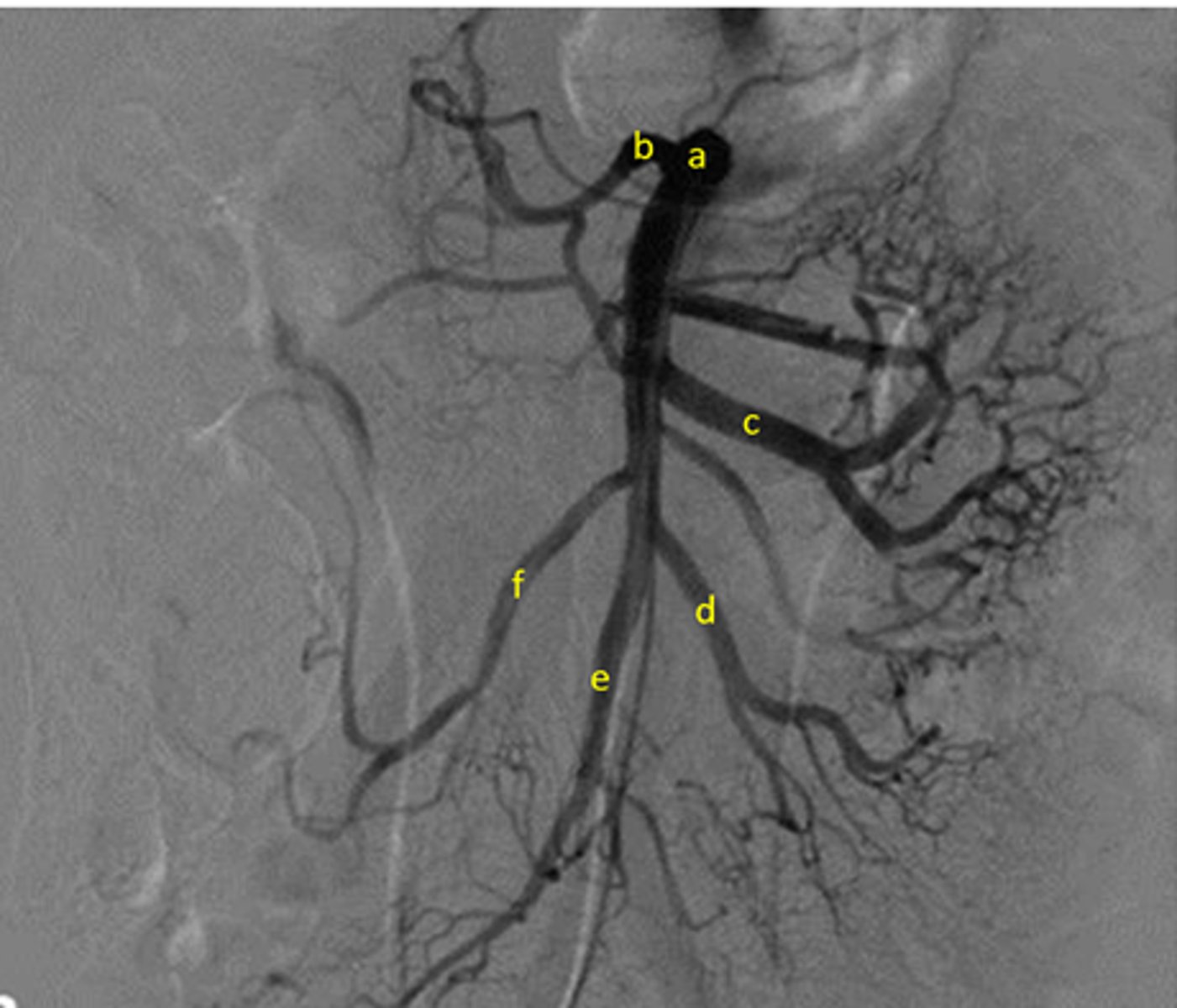
Identify pertinent anatomy of the following radiograph
A: SMA
B: Middle Colic
C:Jejunal arteries
D: Ileal arteries
E: Iliocolic
F: Right colic
Draw/identify blood supply to the abdomen. Where are the SMA, IMA, celiac trunk? What are the branches?
Draw as much as you know!
What are the important portal-naval anastomoses?
What is the venous drainage of the abdomen?
Where can the esophagus be compressed?
1. arch of aorta
2. left main bronchus
3. the diaphragm
Where is the esophagus?
begins at the level of the cricoid cartilage and the CV6 and traverses the diaphragm at TV10. ends at the TV11 by joining the stomach
situated left of midline between trachea and vertebral column. passes anterior to aorta before traversing diaphragm. then emerges left of midline and is continuous with cardiac portion of stomach.
What is the vasculature and inn. of the esophagus?
vasculature - arteries arise from the inferior thyroid, descending aorta, left gastric artery and the inferior phrenic; veins enter the inferior thyroid, azygos, hemiazygos, and gastric veins
innervation - skeletal muscle is supplied by the vagus; autonomics are derived from the vagus and sympathetic branches from the thoracic part of the sympathetic chain
What are the 2 functional sphincters assoc. with the stomach?
cardiac sphincter - located at the opening of the esophagus into the stomach
pyloric sphincter - located at the opening of the stomach into the duodenum
What are gastric ulcers?
results from erosion of the lignin got the stomach; can erode through the walls and damage adjacent; if a major artery is affected significant blood loss and death can result
What is Barrett's esophagus?
replacement of the esophageal epithelium with gastric epithelium because of chronic reflux
What mechanisms should prevent reflux of stomach acid into esophagus?
1. lower esophageal sphincter
2. folds of gastric mucosa, forming a "seal"
3. angle fo the cardiac orifice
4. diaphragm - right crus
What are duodenal ulcers?
most commonly along the posterior wall of this segment; complete ulceration of the wall will result in peritonitis and damage adjacent organs