bio 153l eyes
1/66
There's no tags or description
Looks like no tags are added yet.
Name | Mastery | Learn | Test | Matching | Spaced |
|---|
No study sessions yet.
67 Terms
rectus
straight
oblique
slanting
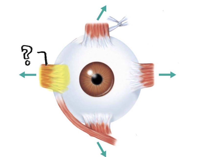
lateral rectus
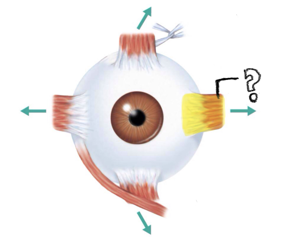
medial rectus
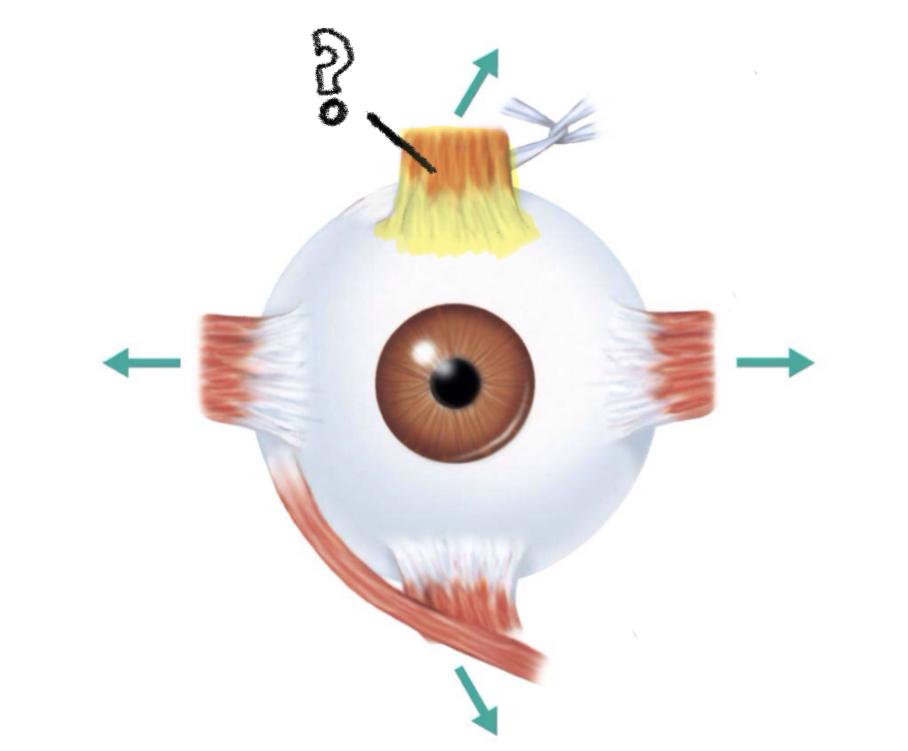
superior rectus
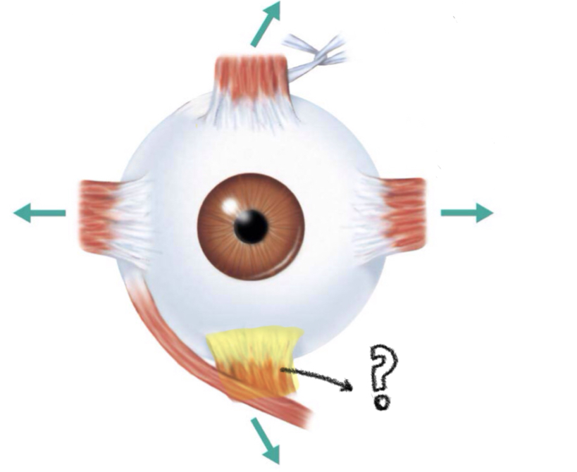
inferior rectus
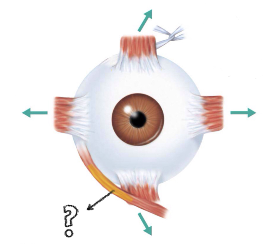
superior oblique
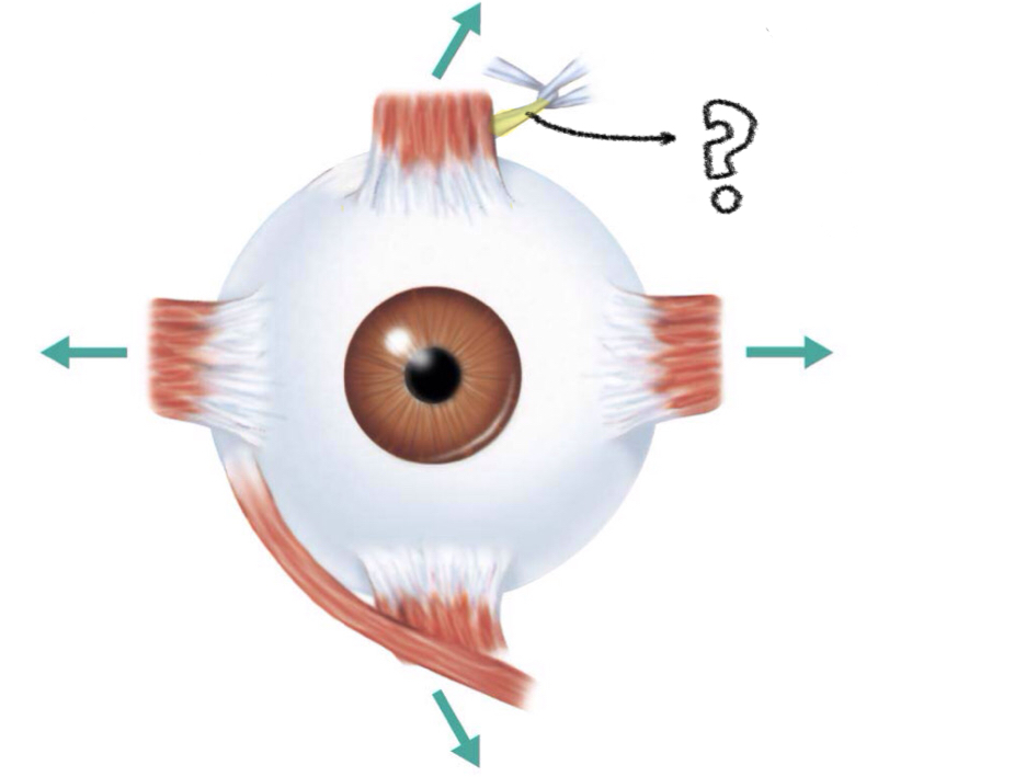
inferior oblique
moves eye laterally
action of lateral rectus
VI (abducens)
controlling cranial nerve of lateral rectus
moves eye medially
action of medial rectus
III (oculomotor)
controlling cranial nerve of medial rectus
elevates eye and turns it medially
action of superior rectus
III (oculomotor)
controlling cranial nerve of superior rectus
depresses eye and turns it medially
action of inferior rectus
III (oculomotor)
controlling cranial nerve of inferior rectus
elevates eye and turns it laterally
action of inferior oblique
III (oculomotor)
controlling cranial nerve of inferior oblique
depresses eye and turns it laterally
action of superior oblique
IV (trochlear)
controlling cranial nerve of superior obliqe
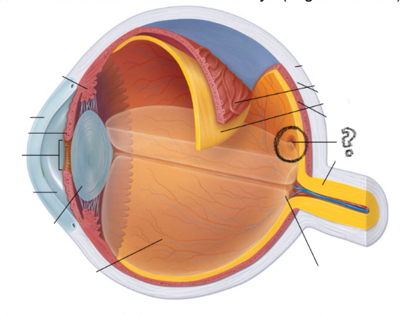
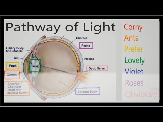
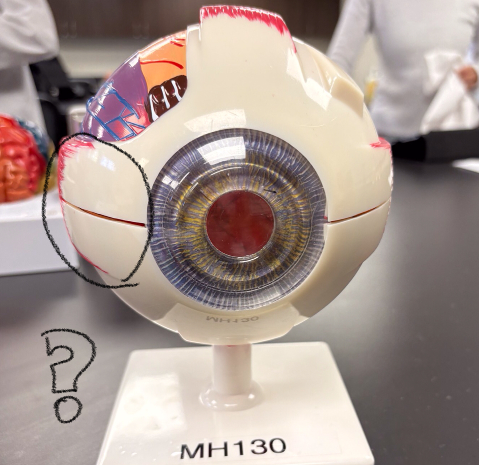
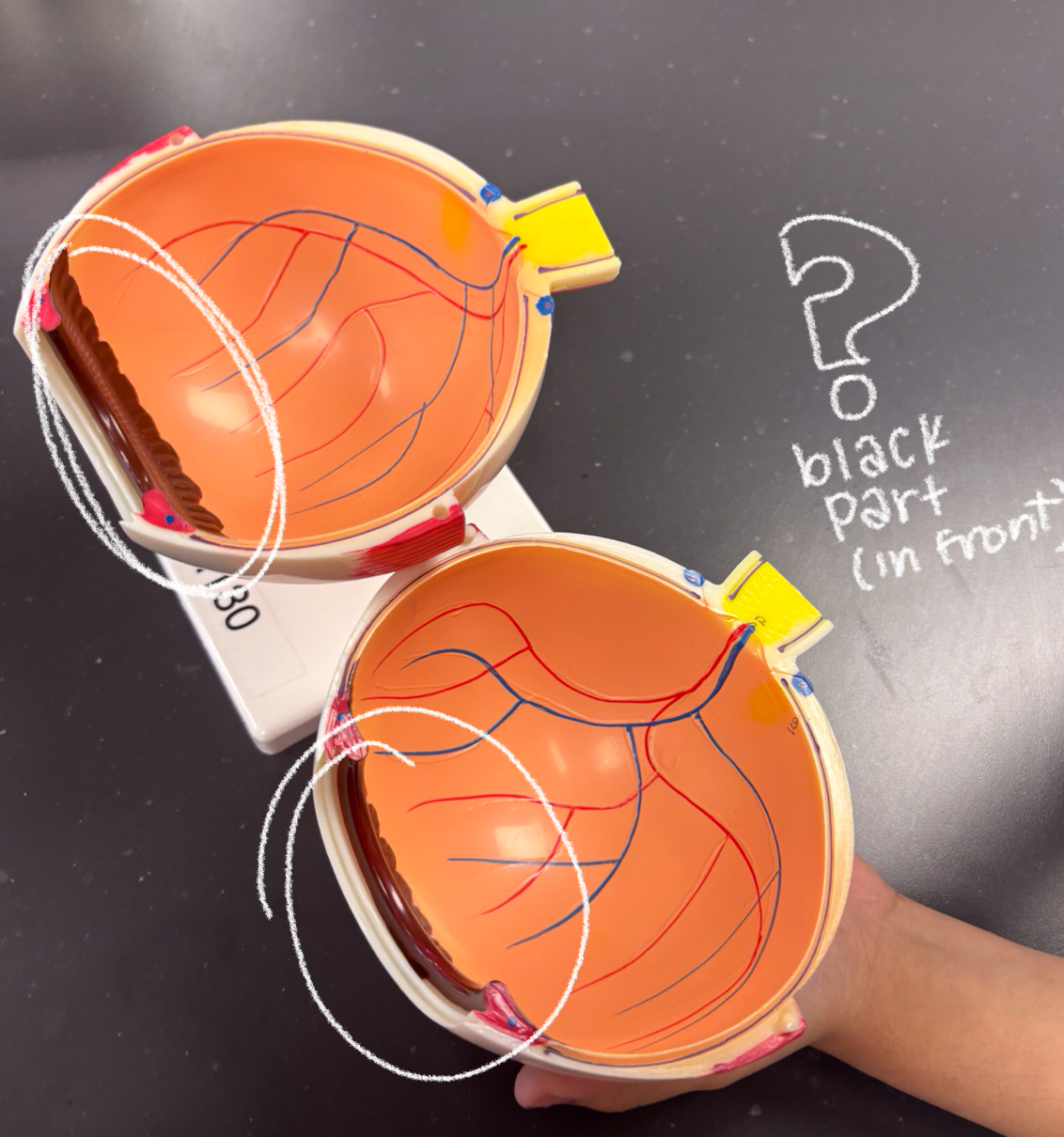
ciliary body
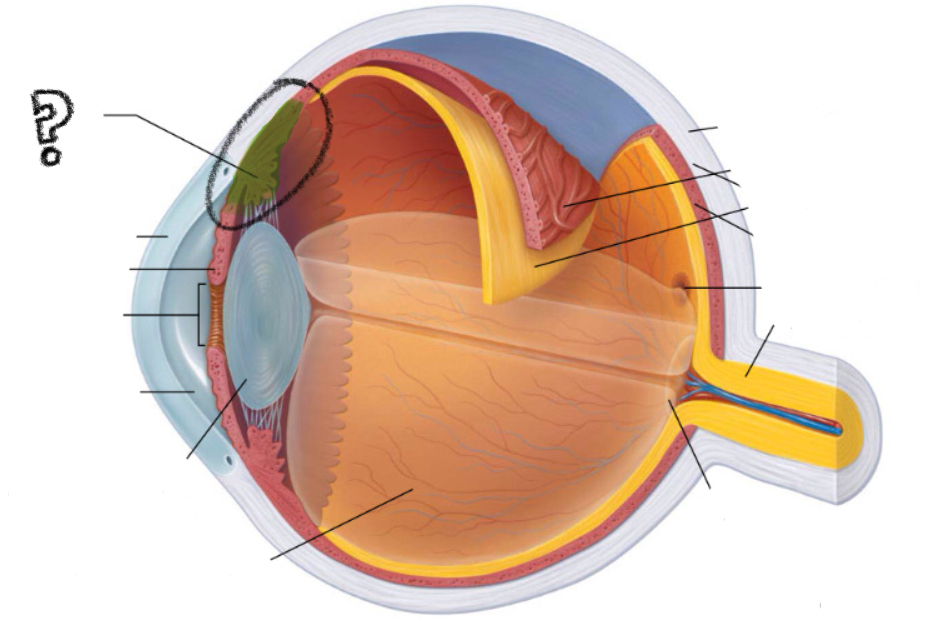
cornea
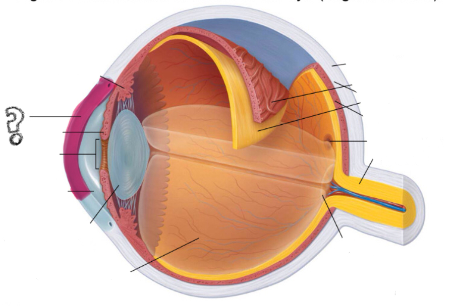
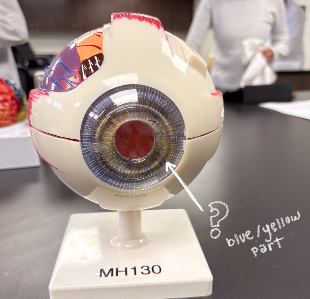
iris
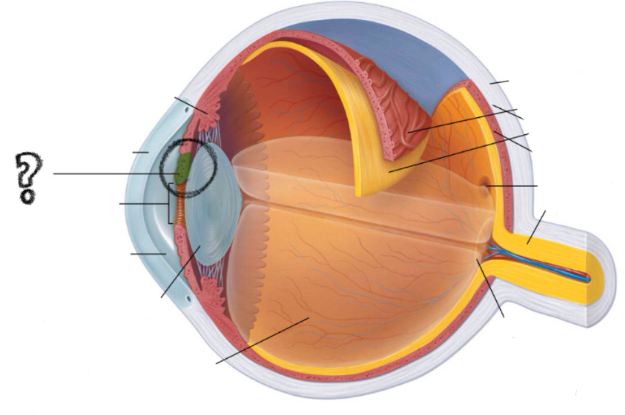
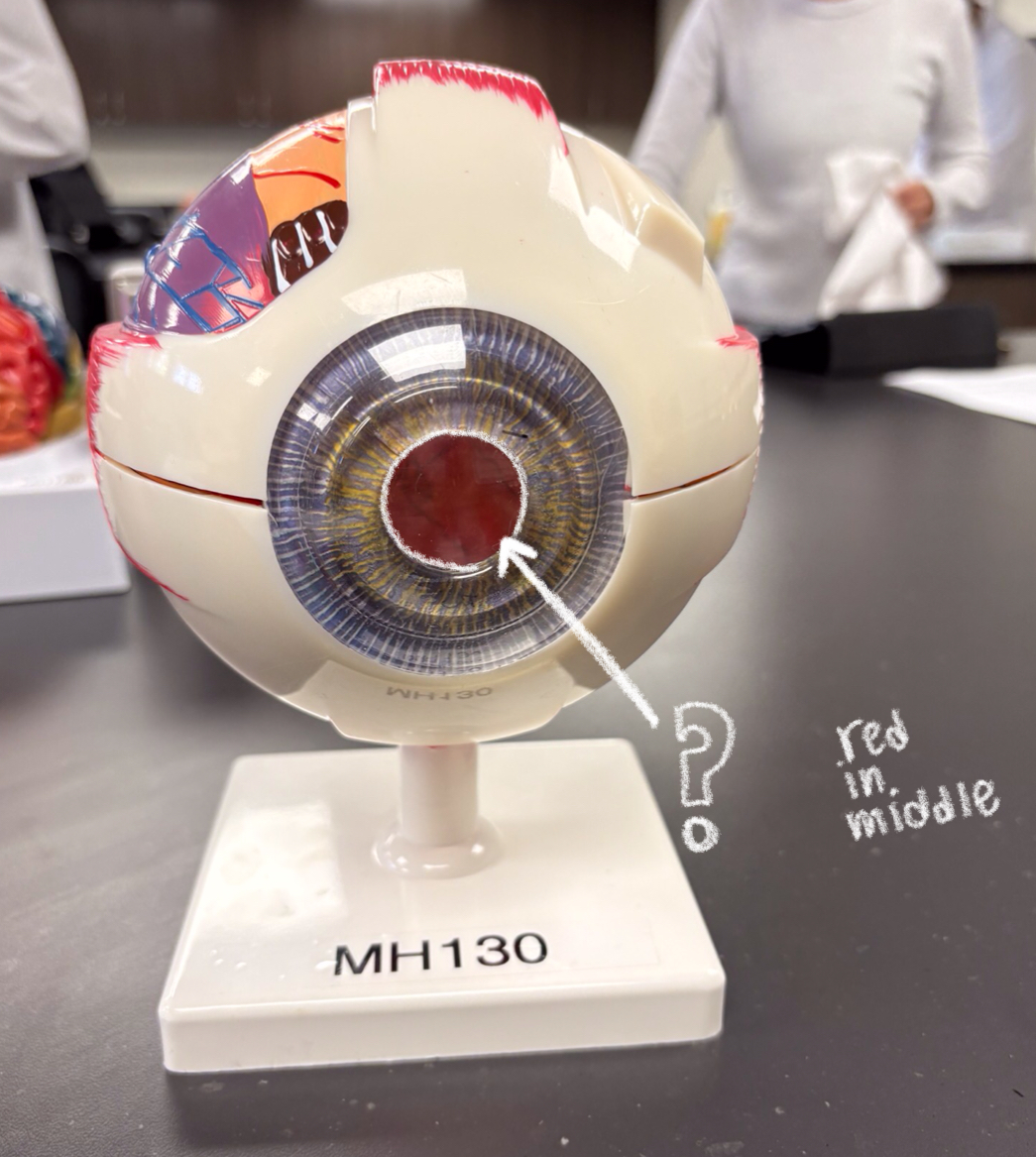
pupil
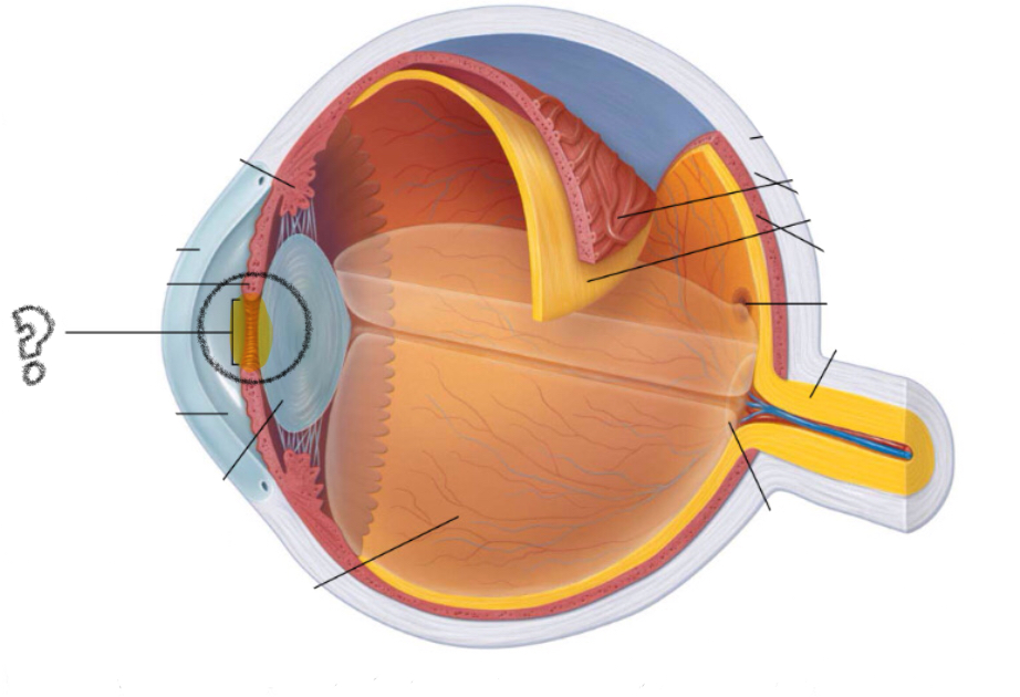
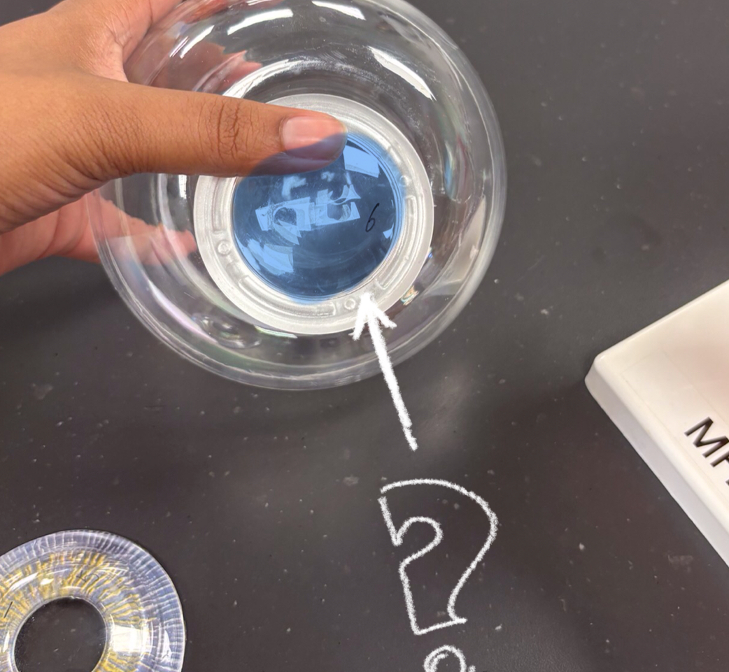
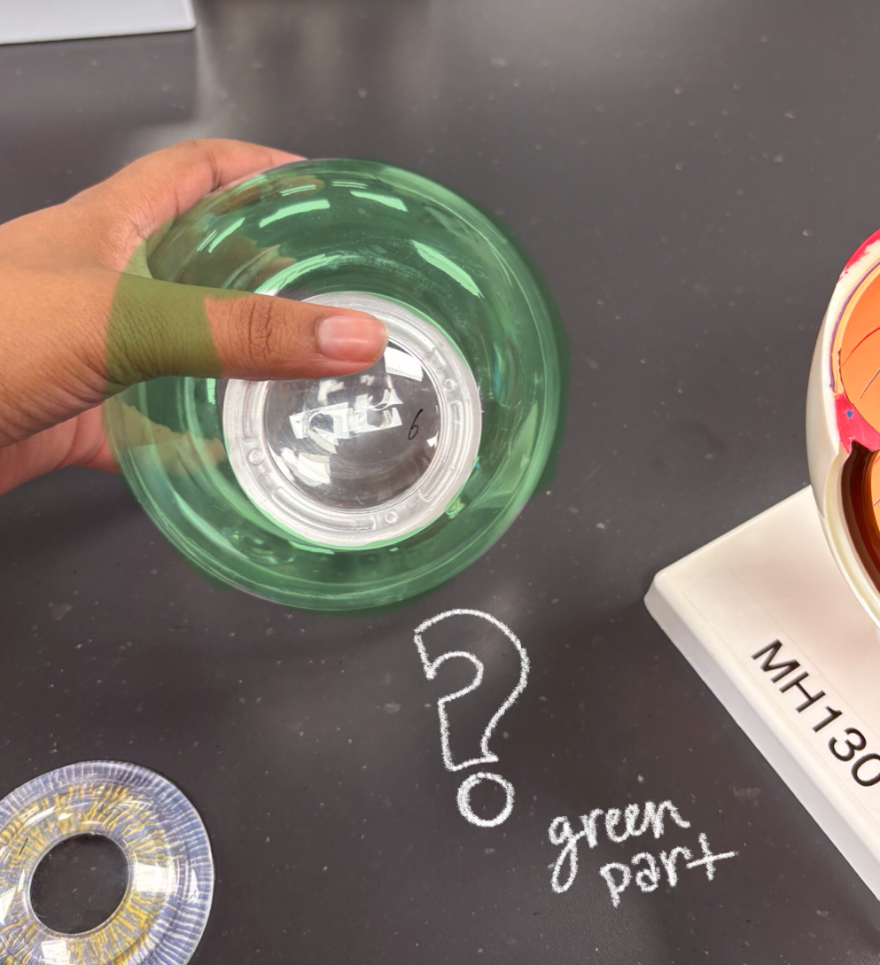
lens
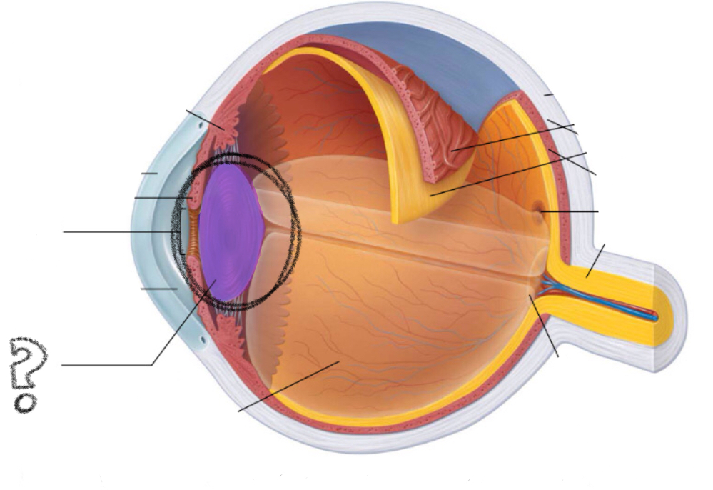
aqueous humor
i think?
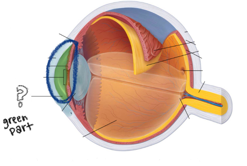
sclera
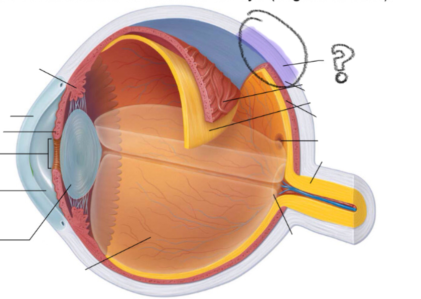
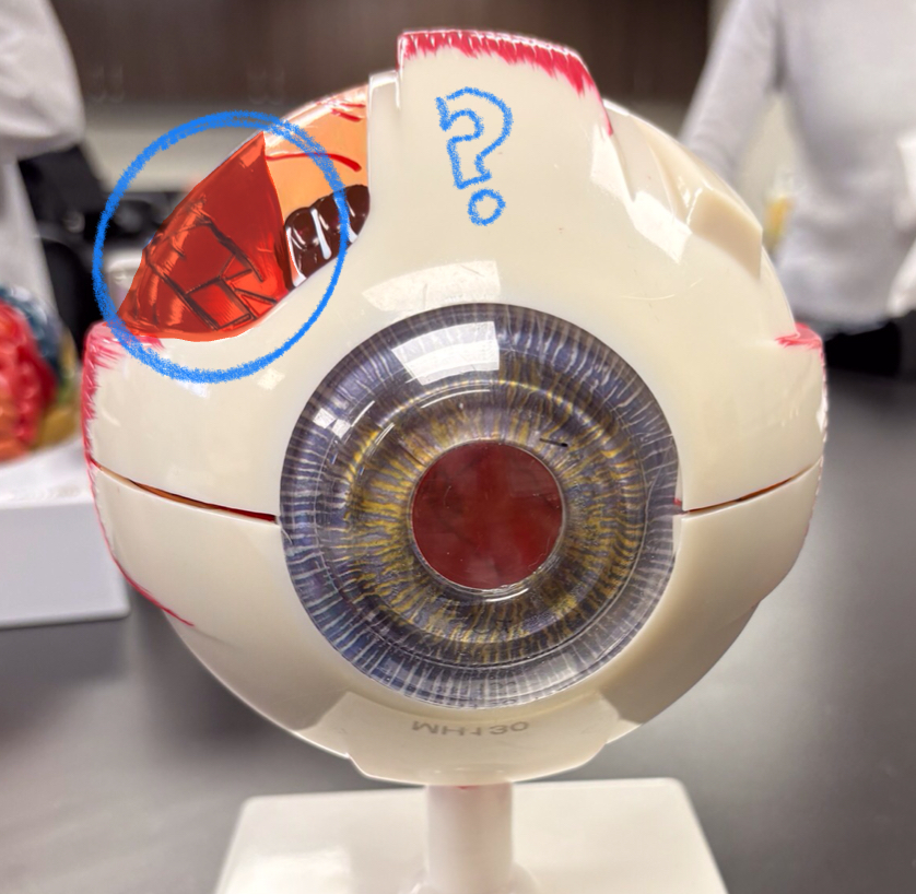
choroid
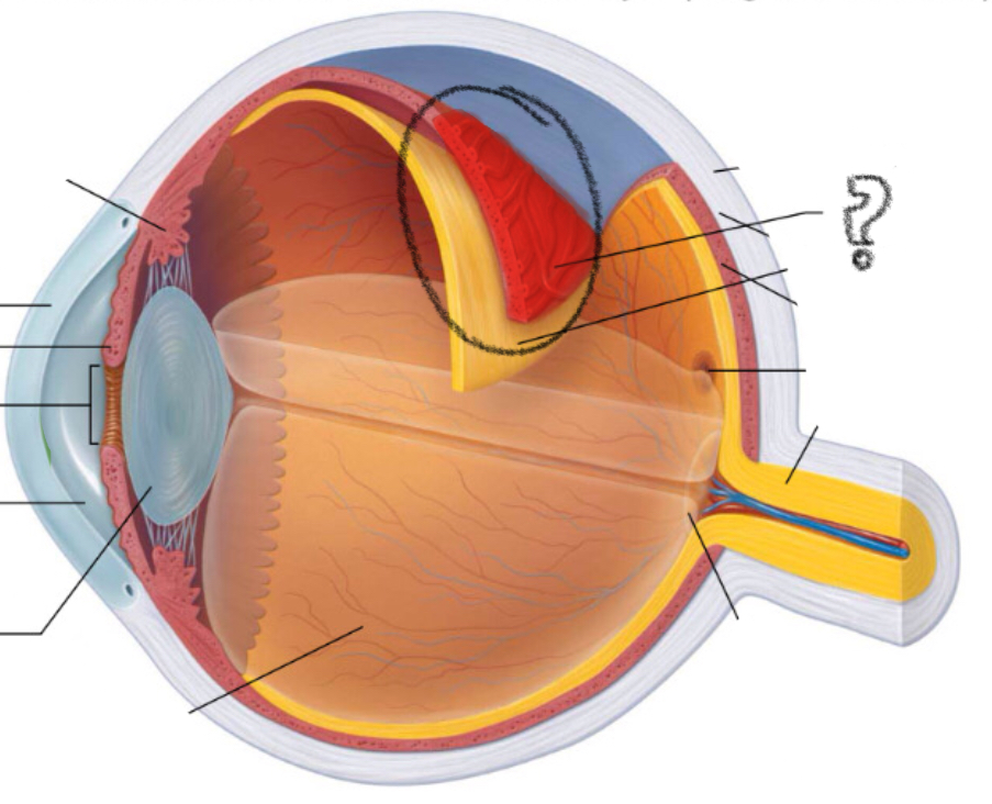
retina
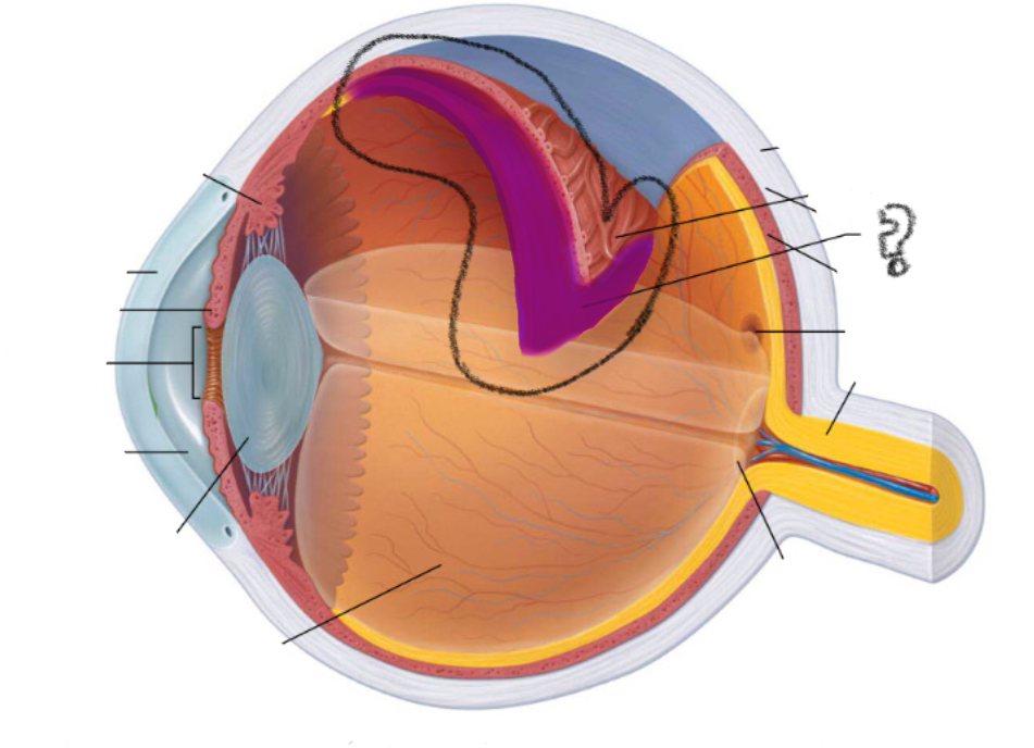
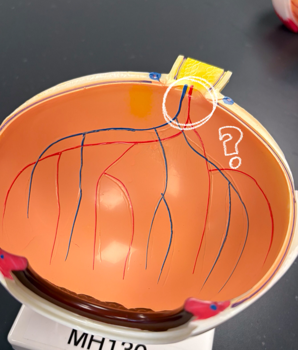
optic disc
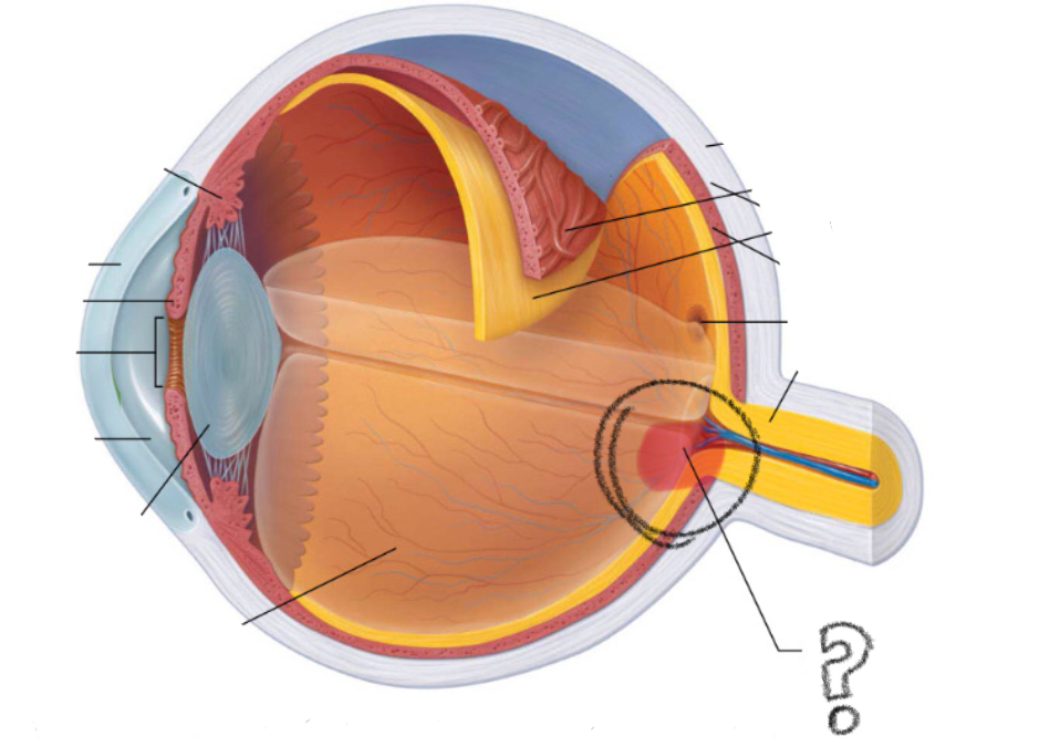
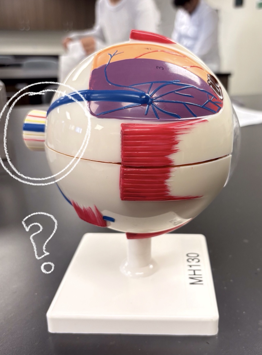
optic nerve
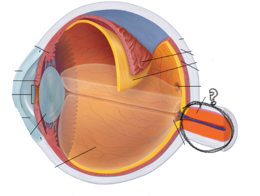
fovea centralis
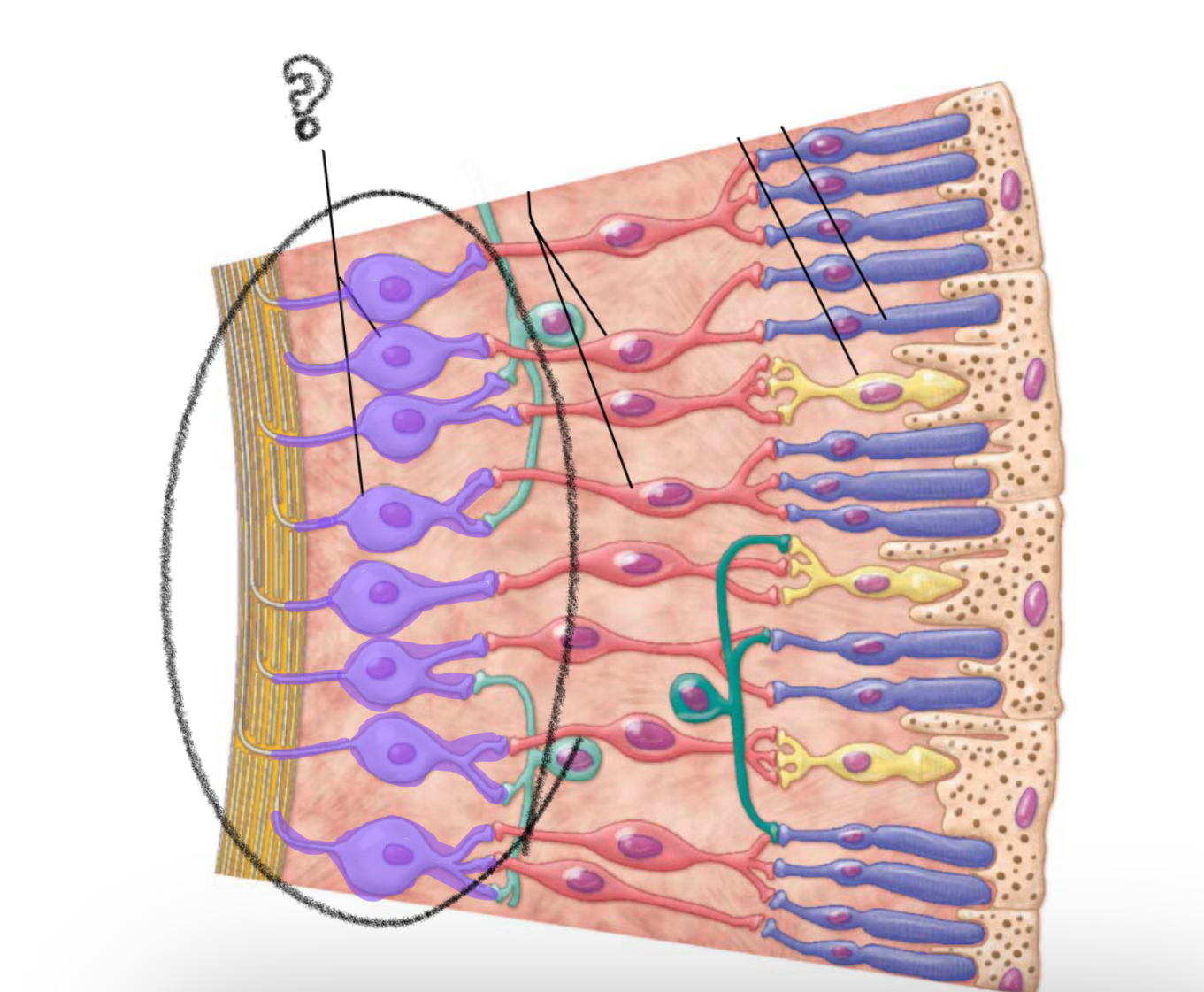
rods
photoreceptor for low light/ black and white vision
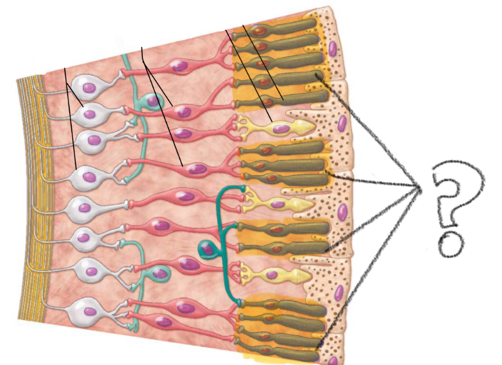
cones
photoreceptors for brighter colors/ color vision
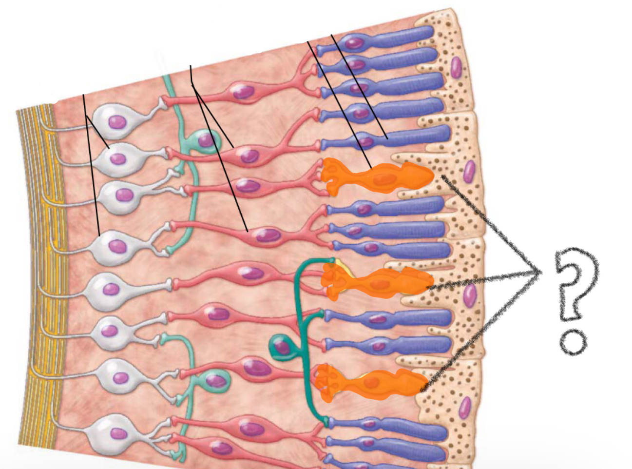
bipolar cells
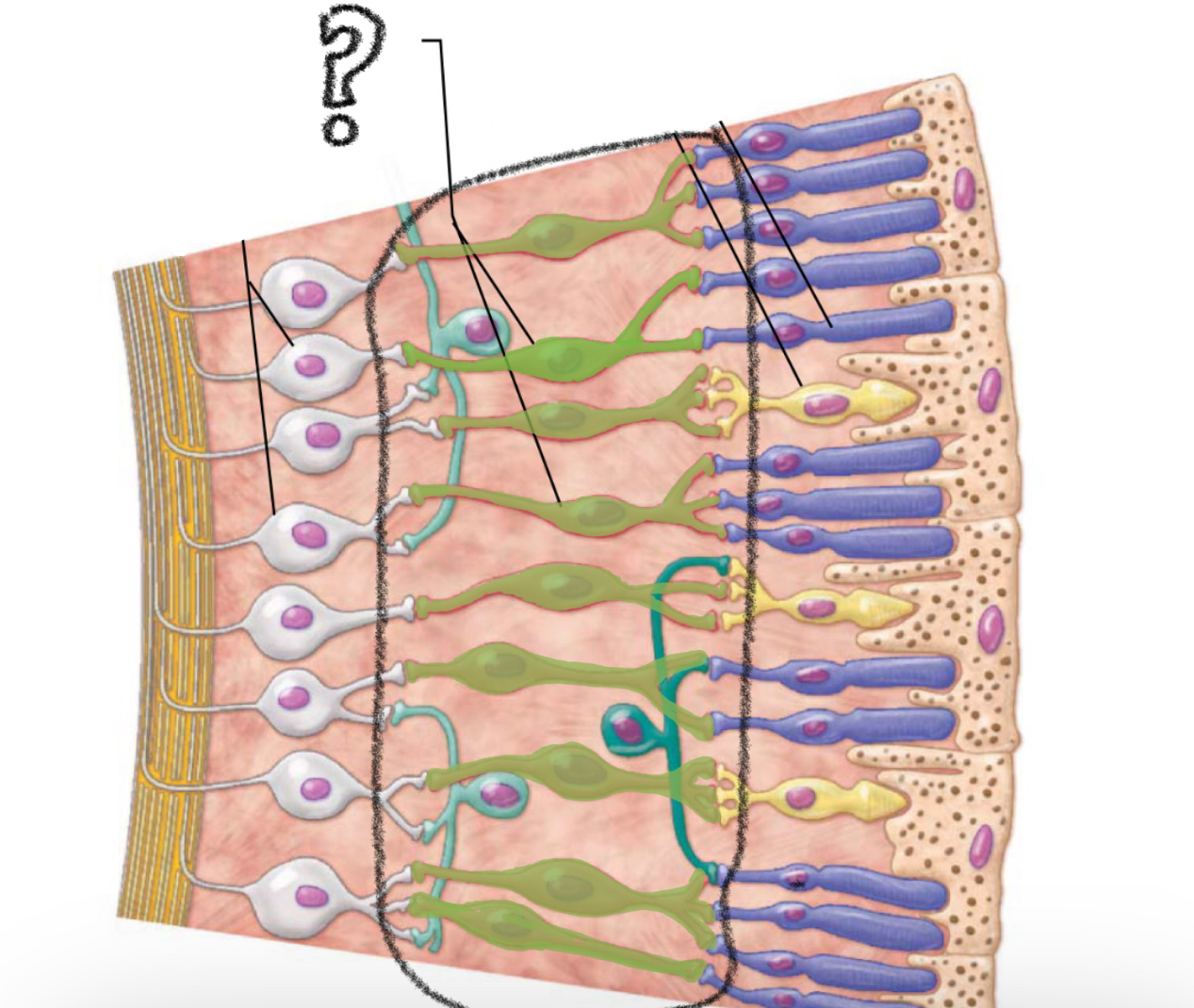
ganglion cells
(nerve cells; axons combine to form optic nerve)
light passes thru the cornea, aqueous humor, pupil, lens and vitreous humor before reaching the retina. It passes to the back of the retina( optic nerve) where it reaches the photoreceptors cells (rods and cones)
pathway for light
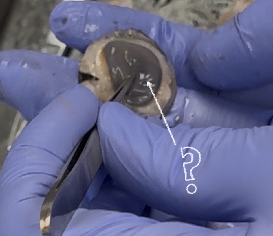
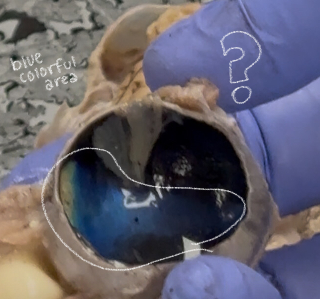
sclera (cow)
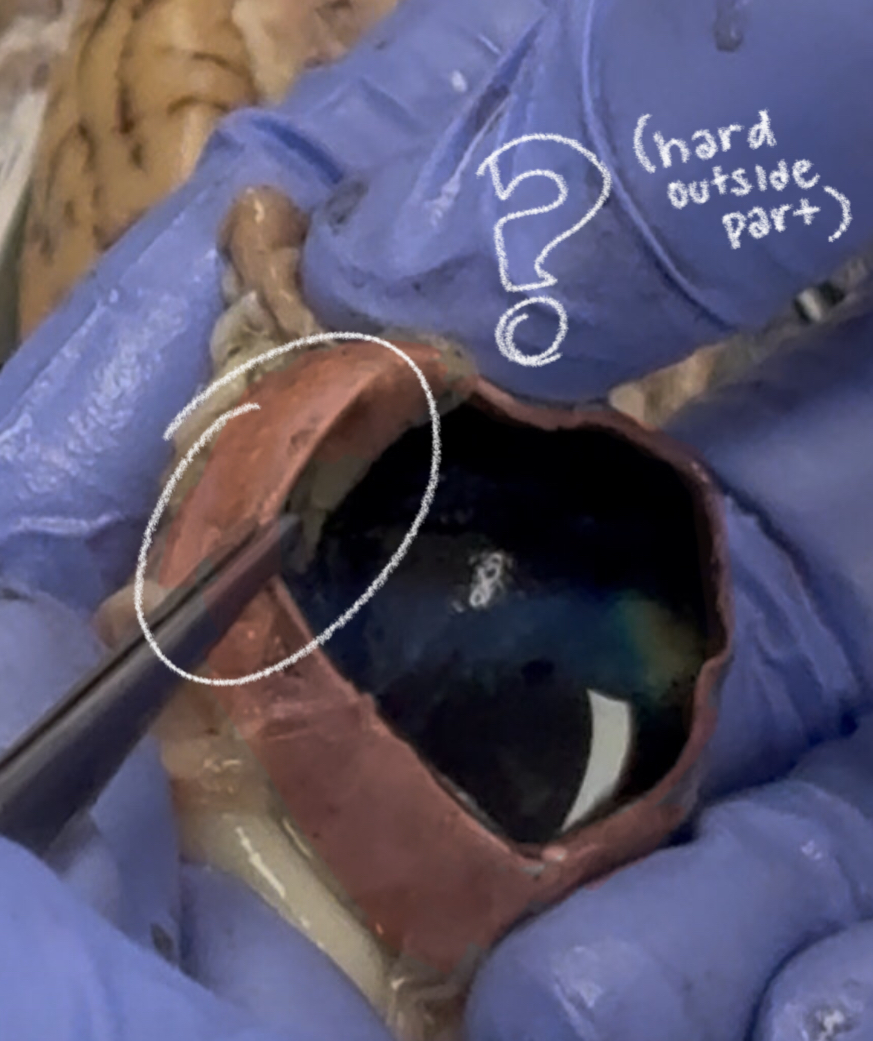
cornea (cow)
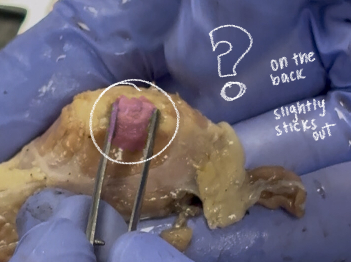
optic nerve (cow)
ciliary body (cow)
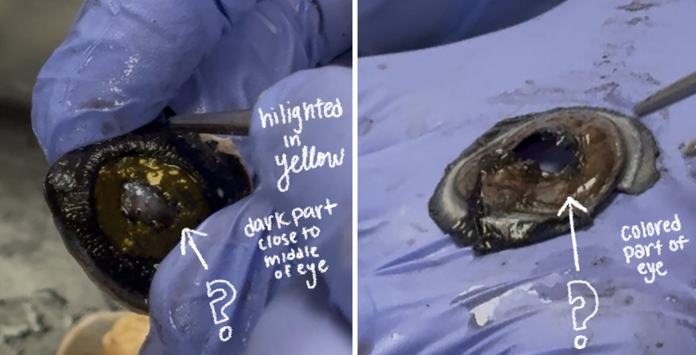
iris (cow)
pupil (cow)
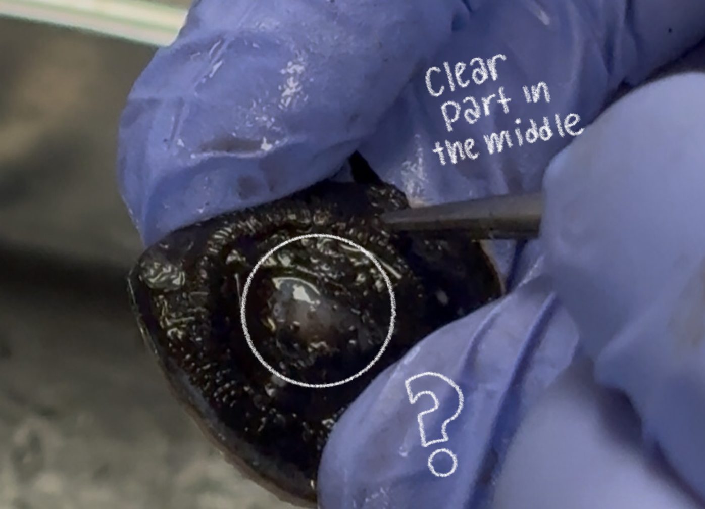
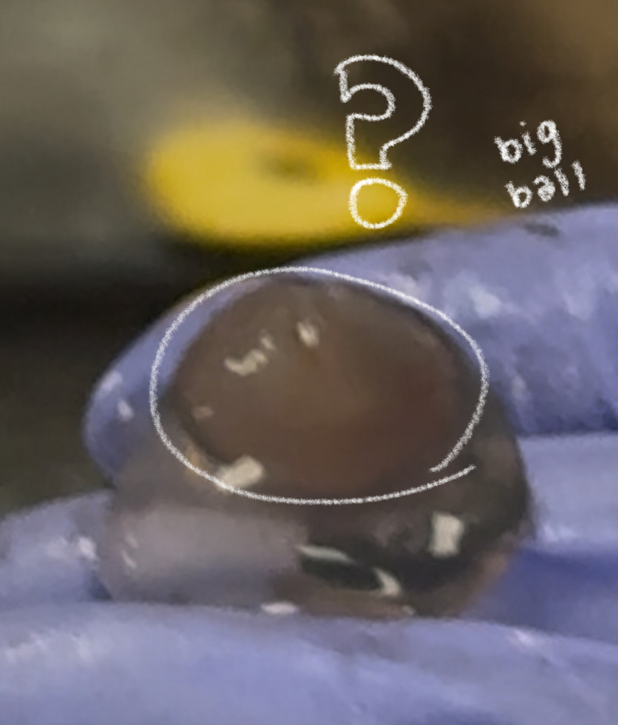
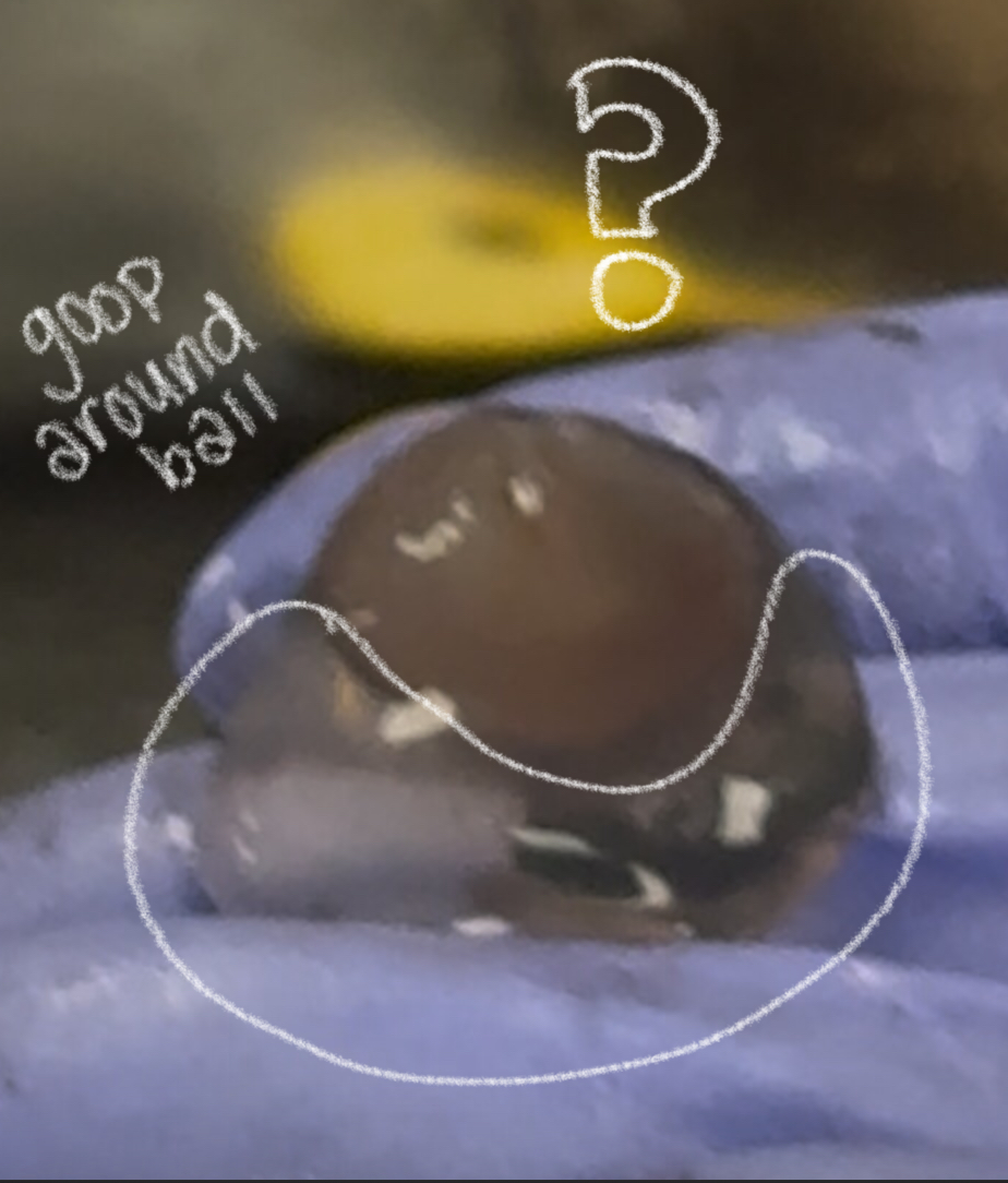
lens (cow)
vitreous humor (cow)
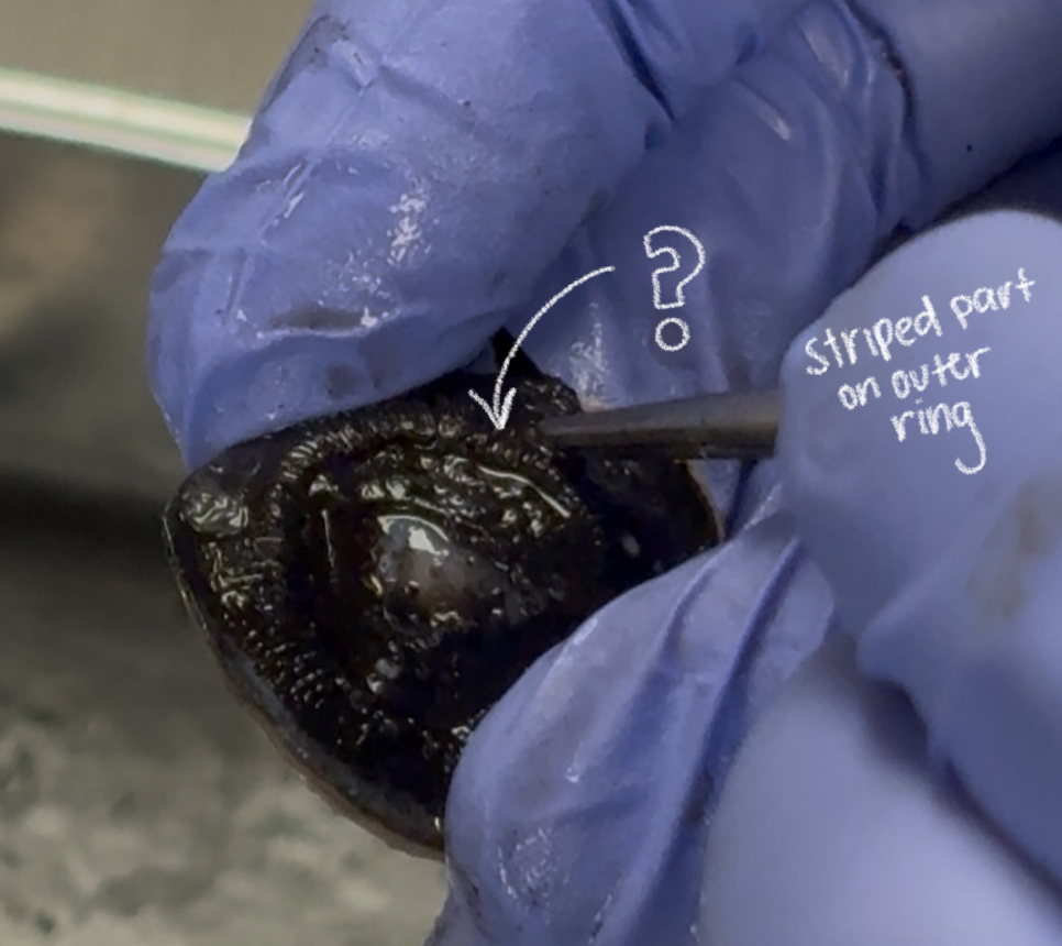
retina (cow)
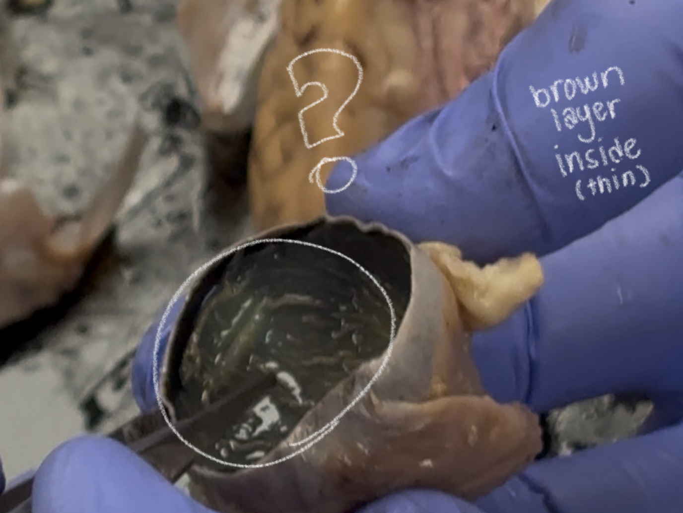
tapetum lucidum (cow)
makes cow & cat eye reflect light; improves night vision
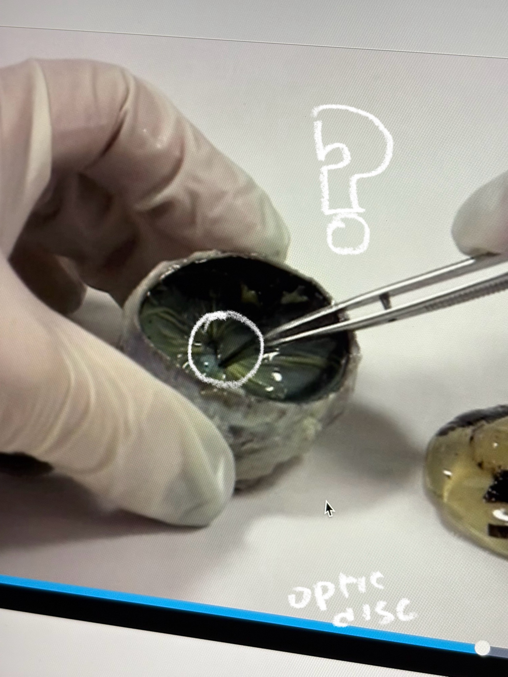
optic disc (cow)
dot in the middle??
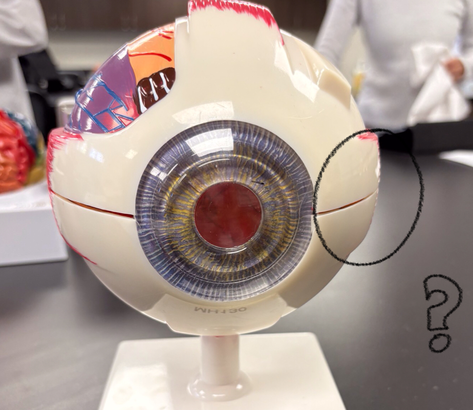
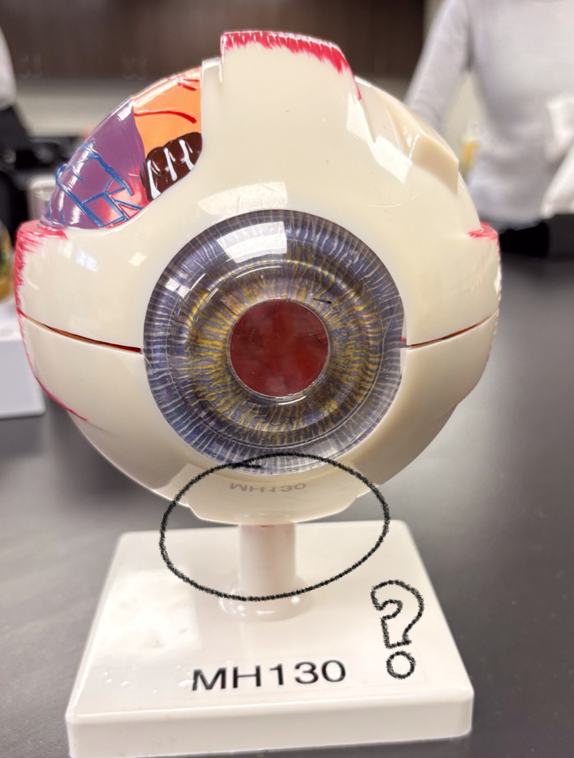
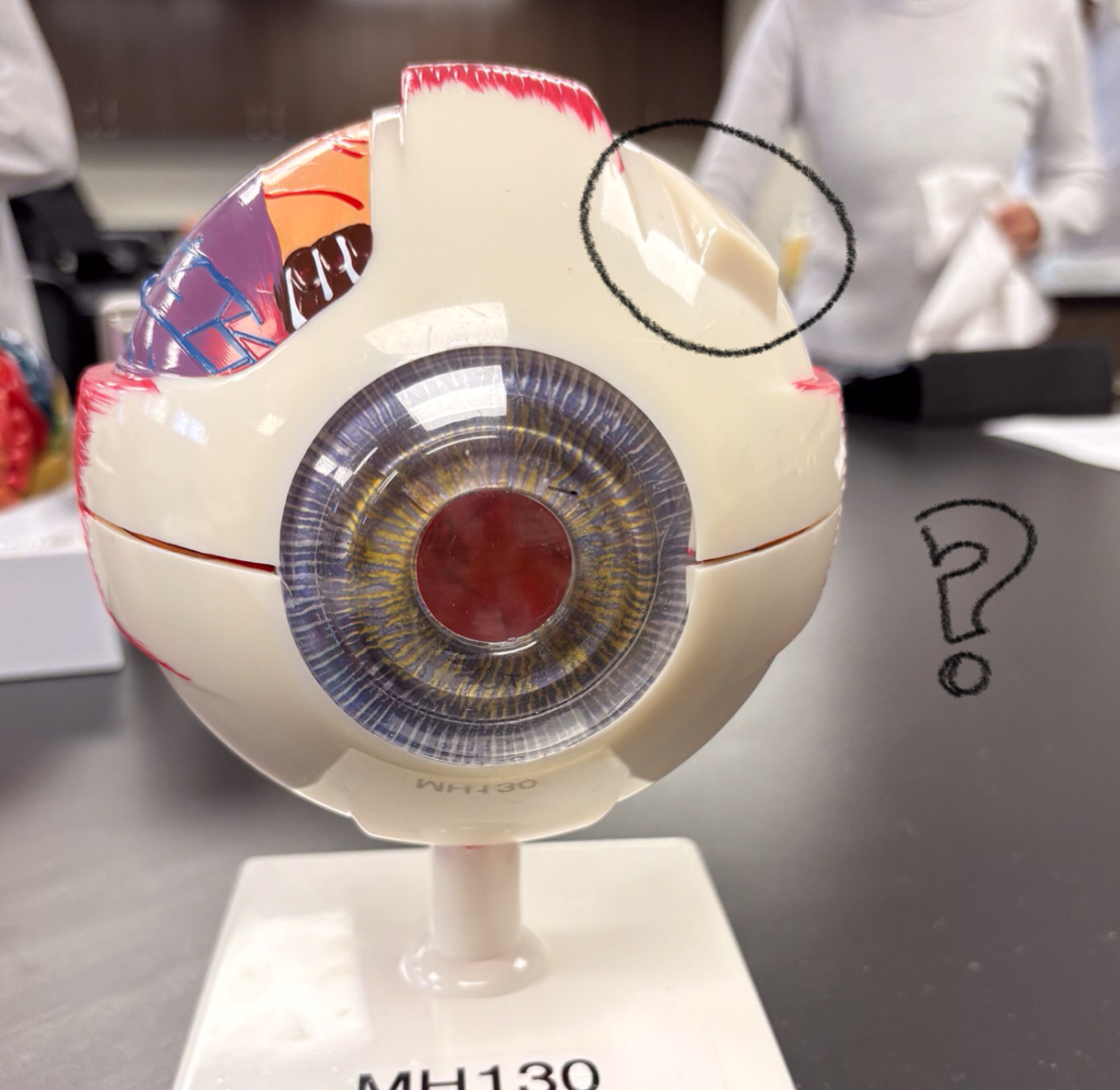
sclera model
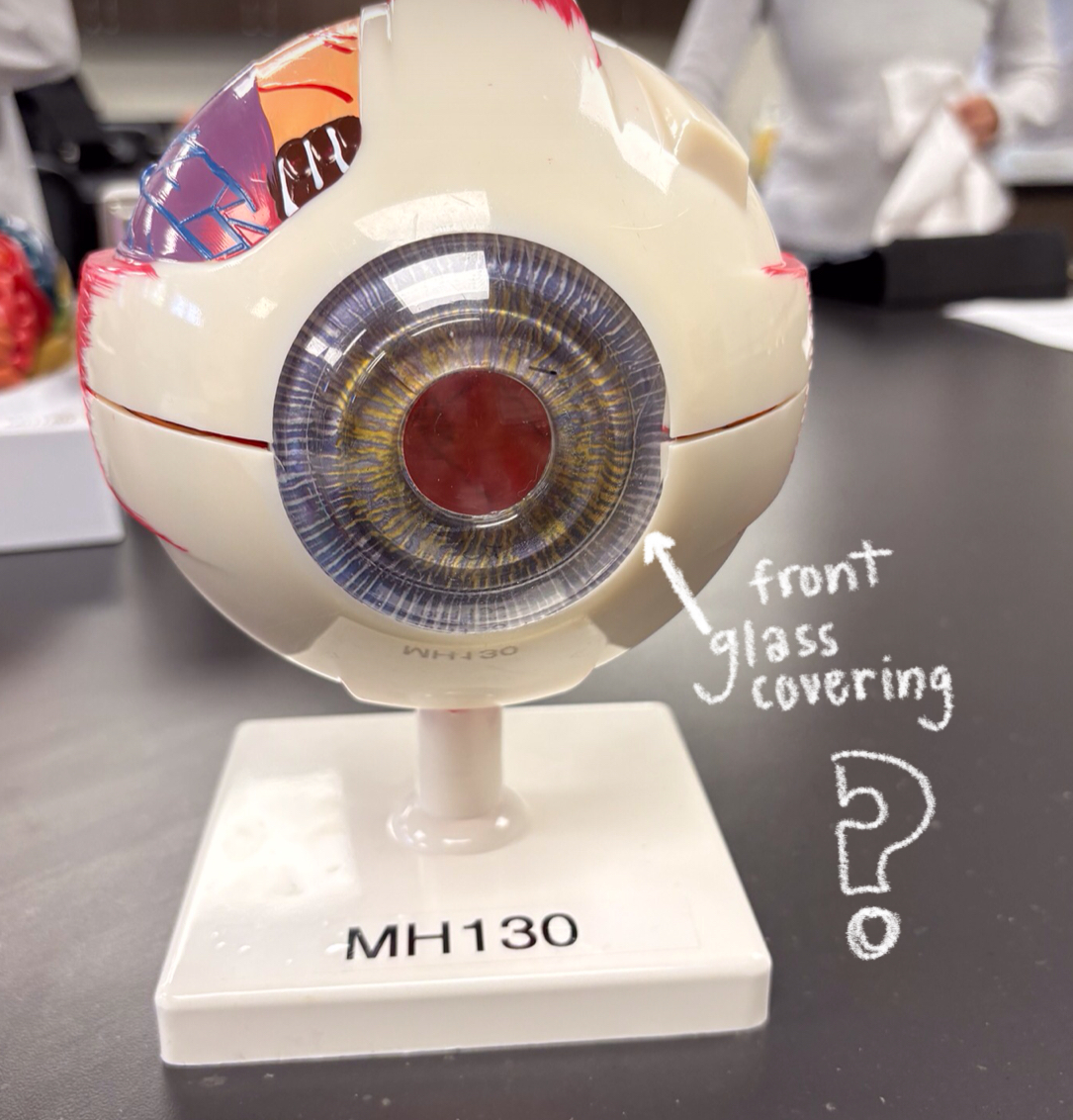
cornea model
pupil model
iris model
colored part of eye
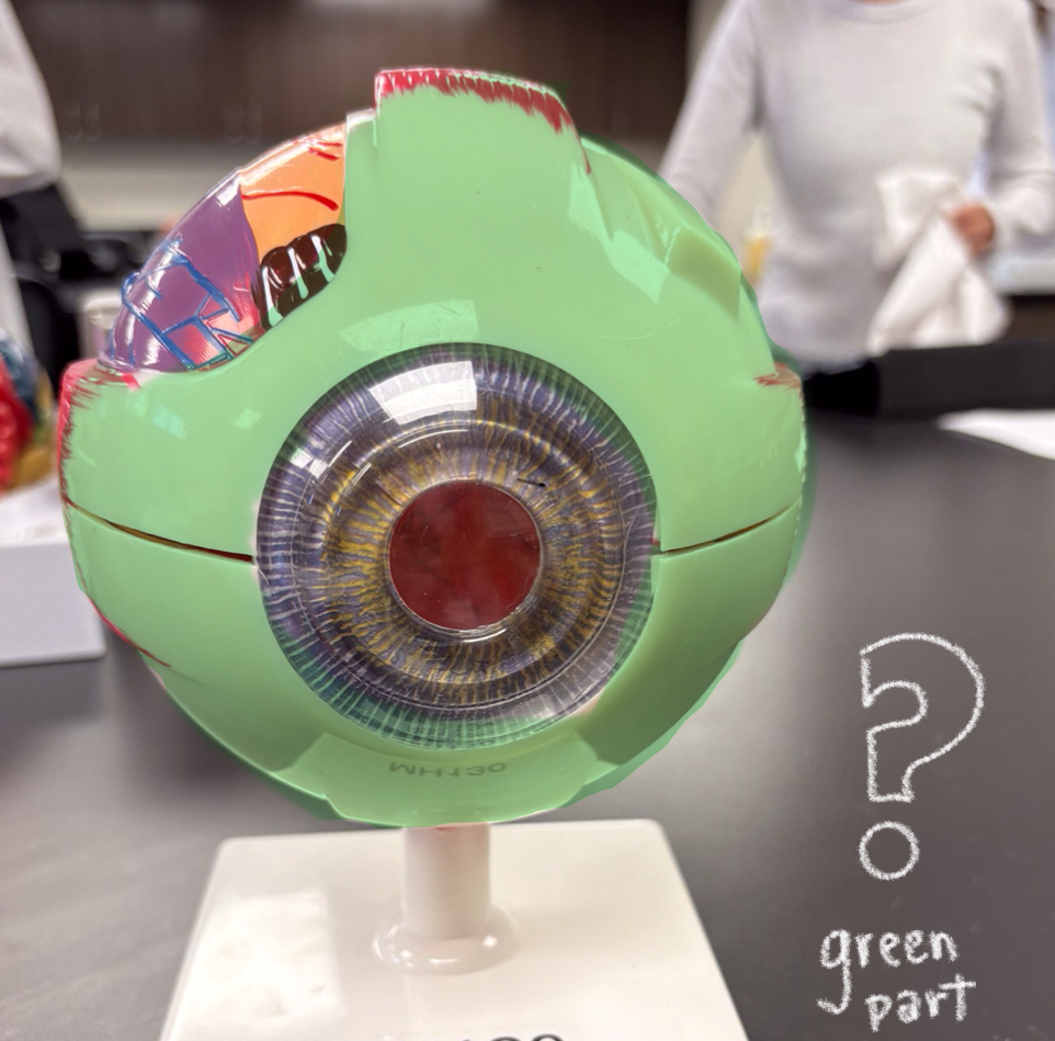
chorid model
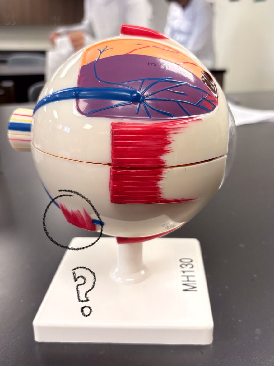
lateral rectus model
superior rectus model
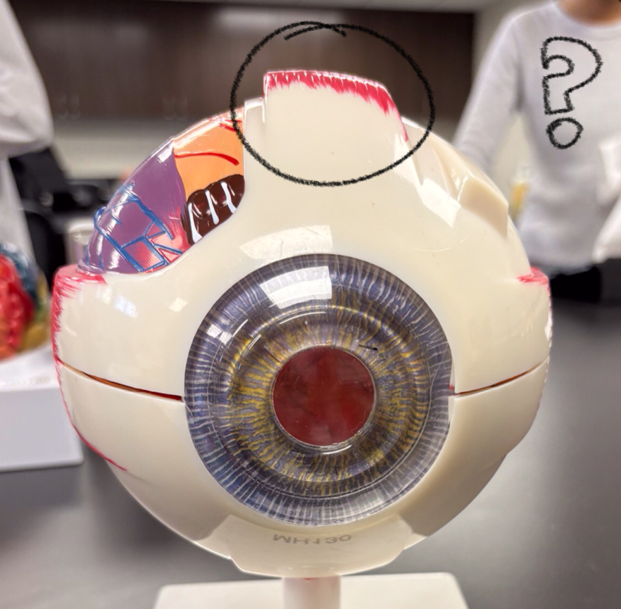
medial rectus model
inferior rectus model
superior oblique model
inferior oblique model
optic nerve model
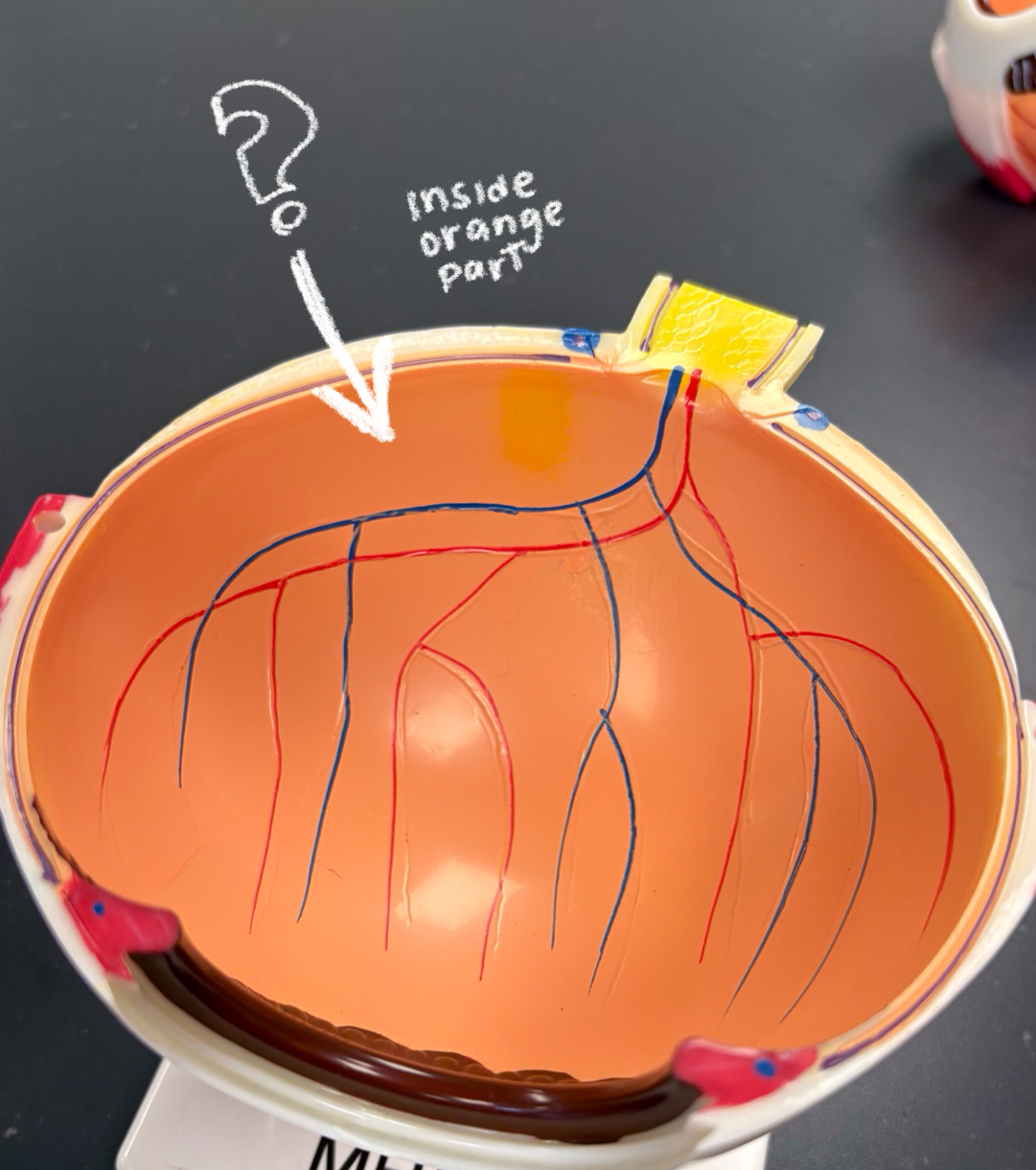
fovea centralis model
hole/ yellow part by retina (might be below white circle)
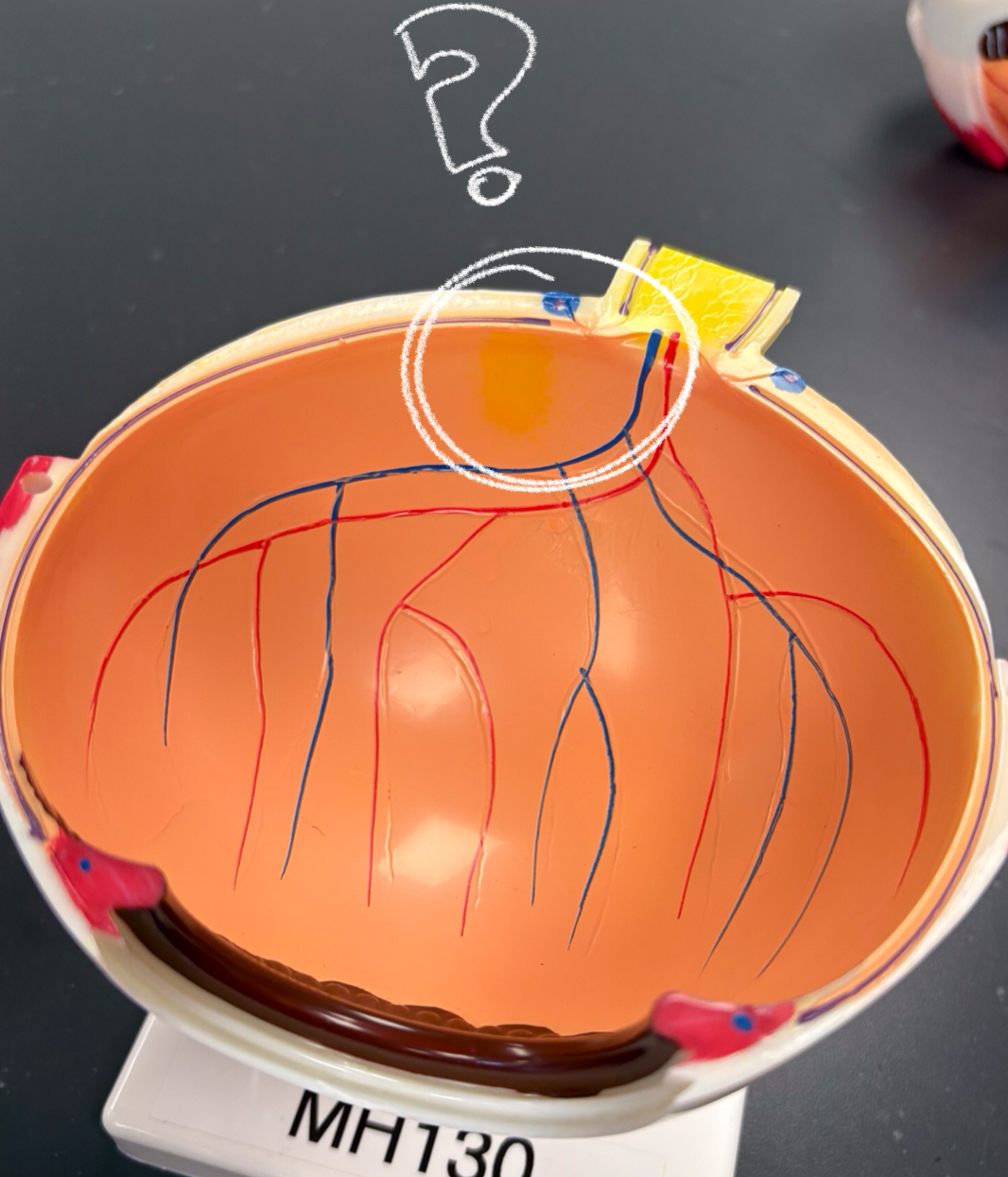
retina model
optic disc model
lens model
vitrous humor model
ciliary body model
light reaches photoreceptors cells → activates rods/cones → receptors send signals forward thru bipolar cells to ganglion cells → gang cells send action potential down axons → axons from gang cells extend towards optic disc → then it exits thru posterior of eye → combines to form optic nerve
signal pathway