External and Middle Ear Disorders
1/67
Earn XP
Description and Tags
Topics included: cerumen impaction, otitis externa, Cholesteatoma, Otitis media, TM perforation, mastoiditis
Name | Mastery | Learn | Test | Matching | Spaced |
|---|
No study sessions yet.
68 Terms
Cerumen impaction
A condition of the ear that is due to over production of ear wax, a tortuous canal, or Q-tip cleaning
keeps canal acidic (inhibits fungal/bacterial growth), repels water (prevent culture growth), traps fine debris
What is the physiological role of cerumen?
external canal (EAC - 2.5 cm long, S shaped), cerumen (skin oils, dead cells), sebaceous glands (lubrication and protection)
The external ear is made up of
cereumen impactation
Patient presents to the clinic with decreased hearing and a feeling of ear “fullness.” While collecting a history, the patient reports frequent Q-tip use. On an otoscopic exam the you note a yellowish-brown mass in the EAC. What are we thinking team?
foreign body, tumor, blood clot (if mass looks red)
Differentials for cerumen impactation
patients has no history of ear infections, perforations, or otologic surgery
In order to give cerumenolytics what must we ensure?
suspected TM damage (ear drainage, ear pain), frequent ear infections as a child
When should we avoid cerumenolytics
H2O2-urea (debrox), Carbamide peroxide (clearcanal), H202-H20 (equal parts), mineral oil, liquid docusate sodium
Cerumenolytics that can be applied at home or in clinic (5-10 drops 2x daily for 4 days)
rule out perforation
Before preforming a lavage for a cerumen impaction, what do we need to do?
otomicroscopic debridement
What is the safest way to remove cerumen and is done by an ENT specialist?
infection from iatrogenic EAC trauma
Complications of otomicroscopic debridement
Eustachian Tube
What drains the middle ear into the nasopharynx?
Adenoid hypertrophy, nasopharyngeal carcinoma
Differentials of Eustachian tube dysfunction (ETD)
ETD
Patient presents to the clinic with a cold. While collecting a history your patient notes that her ears feel “stocked up” and when she swallows she notes crackling/popping when swallowing. While conducting a physical, you note a retracted TM of normal color and decreased mobility with valsalva. What are we thinking team?
No antibiotics, treat URI/allegies (decongestants topical/oral, intranasl corticosteroids), valsalva several times a day, avoid high pressure (air travel, scuba)
How are treating a ETD
conductive hearing loss, acute serous otitis media, chronic serous otitis media, TM perforation, cholesteatoma
Complications due to a negative pressure (vacuum) in ETD
Otitis externa (swimmers ear)
An infection of the EAC do to an infectious, allergenic, or dermatological etiology characterized by the breakdown of skin in cerumen barrier that causes inflammation leading to swelling and obstruction of the ear canal resulting in itching and increases the pH of the ear (more common in the summer)
swimming, trauma (scratching/cleaning), devices that occlude EAC (hearing aids), allergic contact dermatitis, psorasis, atopic dermatitis
Risk factors of otitis externa
acute otitis media (AOM), contact dermatitis, chronic suppurative otitis media, psoriasis, carcinoma of ear canal, TM perforation
Differentials of otitis externa
otitis externa
Patient reports to the clinic for ear pain that he rates 8/10. He also reports slight trouble hearing and pruritus. On a physical exam you note the EAC is red and swollen but nothing that should be an 8/10. While examining the ear the patient notes increased pain when you palpate the tragus and helix. Thoughts?
infection has spread to middle ear/mastoid
If a patient comes in for otitis externa and there’s a fever what do we suspect?
Pseudomonas (most common)
If you see green purulent discharge in otitis externa what infection is most likely?
Staph A
If you see yellow purulent discharge in otitis externa what infection is most likely?
candida (main cause with hearing aid patients)
If you see cheesy purulent discharge in otitis externa what infection is most likely?
Aspergillus (usually after bacterial otitis externa)
If you see fluffy/sporus discharge in otitis externa what infection is most likely?
clotrimazole 1% (2x daily for 10-14 days)
How are we treating our fungal otitis externa?
culture with sensitives, KOH prep
If your patient sees no improvement in their otitis externa symptoms with inital treatment what can we do?
myringitis, conductive hearing loss, cellulitis, osteomyelitis of skull base
Complications of otitis externa
Acetic acid/hydrocortisone ear drops (Acetasol HC - Note: alters pH)
How are we treating the mild otitis externa (minor discomfort, itching, minimal edema)?
Cipro/hydrochloride/hydrocortis ear drops (ciprodex HC), Neomycin/polymyxin B sulfates/hydrocortisone ear drops (cortisporin)
How are we treating moderate otitis externa (intermediate pain/itching, partially occluded ear canal)?
Cipro HC, Corticosporin on a Pope otowick (replac in 48 hours)
How are we treating severe otitis externa (intense pain, fever, canal is swollen shut, periauricular erythema, regional lymphadenopathy)?
NSAIDS, keep water out of ear
Regarding all forms of otitis media, what can we do to improve symptoms?
ENT
If otitis externa does not respond to treatment within 6 days, who we calling?
rapid ascent/descent, overpressure, infection, myringotomy
What causes a TM perforation?
TM perforation
Patient presents to the clinic with extreme ear pain. He also reports tinnitus. While collecting a history you discover that he is a scuba diver. On a physical exam you note bloody otorrhea and a visualize a torn TM. Diagnosis?
cholesteatoma, severely retracted TM
Differentials of TM perforation
refer to ENT, tympanoplasty
How are we treating a large TM perforation?
refer to ENT, usually heal spontanously
How are we treating a linear or small TM perforation?
swim (prevent water contaminants from getting into the middle ear)
What are you not allowed to do with a TM perforation?
Cholesteatoma
What is a abnormal, noncancerous, keratinous skin growth from the middle ear that is a result of chronic negative middle ear pressure or retraction of pars flaccida into the middle ear.
vertigo, facial nerve twitch/paralysis
Rare symptoms of cholesteatoma
cholesteatoma
Patient reports to the clinic with hearing loss in her right ear. She states that over the last few months she has notice pus coming out of her ear and tinnitus but thought nothing of it until her hearing loss got worse. On a physical exam you note a attic retraction so you order a CT which shows unilateral growth in the ear canal, what is this consistent with?
surgery (if untreated it can advance to mastoid sinus and then we’d have to remove the mastoid bone)
How are we treating our patient with cholesteatoma?
acute otitis media (AOM)
What is an ear condition common in childhood that peaks in those less than 2 and then sharply declines after 7.
Most commonly viral, but can be S. pneumonia, H. Influenza, Moraxella catarrhalis
Etiology of acute otitis media
sepsis workup
If a neonate has acute otitis media, what are we doing team?
Otitis externa, referred otalgia, serous otitis media (SOM), barotrauma, bullous myringitis
Differentials for AOM
amoxicillin-clavulanate for 10 days (cephalosporin if allergic) (antipyretics, analgesics)
A 5 y/o patient presents to the ER with flu-like symptoms. The child is pulling at her ears saying, “it hurts, it hurts.” Mother reports that this has been going on for 3 days. Vitals are stable with the exception of a fever. Otoscopy shows a red, bulging TM with decreased mobility and otorrhea is present. What’s our treatment plan?
antipyretics, analgesics, antibiotics
When treating AOM in patients less than 6 months
antipyretics, analgesics, antibiotics or observation (if immune competent, no craniofacial issues, unilateral, mild symptoms, etc)
When treating AOM in patients 6 months to 2 y/o what are we doing?
otalgia for more than 2 days, temp over 102.2 in the past 2 days, bilateral, otorrhea, uncertain follow-up
When treating AOM in patients above the age of 2 when do we give antibiotics?
serous otitis media (SOM)
A condition of the ear the is characterized by middle ear fluid without acute signs of infection that may occur after AOM, URI, or ETD (AKA otitis media with effusion)
nasopharyngeal tumor
If you see chronic or unilateral SOM in adults, this is a red flag for
hearing loss, blocked/plugged feeling in ears
Symptoms of SOM
retracted immobile TM, air fluid levels/bubbles
What are you going to see on an otoscopy in a patient with SOM?
Audiogram, because the TM is compromised with fluid
How and why do you evaluate hearing loss in SOM?
Thug it out (time, most resolve w/o intervention), treat the symptoms (decongestants, intranasal corticosteroids, valsalva)
Treatment plan for SOM
poor school performance, adhesive otitis media
Complications of SOM
Chronic, recurrent otitis media
What is common in young children and infants as a result of lack of symptomatic improvement 48-72 hours after starting antibiotics for AOM (predisposing factors, compliance issues, or wrong meds for organisms)
3+ AOM in 6 months, 4+ AOM in 12 months, persistent/recurrent ear discharge, SOM for longer than 3 months
At what point do we call it chronic/recurrent otitis media?
refer to ENT
If we are thinking chronic, recurrent otitis media → what do we need to do?
tympanostomy tubes, ventilating tubes, PE tubes
Treatment plan for chronic recurrent otitis media (last 6 months to 1 year)
T-tube placement
If a child develops infections after the small tubes for chronic recurrent otitis media what’s the next step?
helps ear drain, buys time until eustachian tubes develop
What is the goal of placing tubes in the TM in chronic, recurrent otitis media?
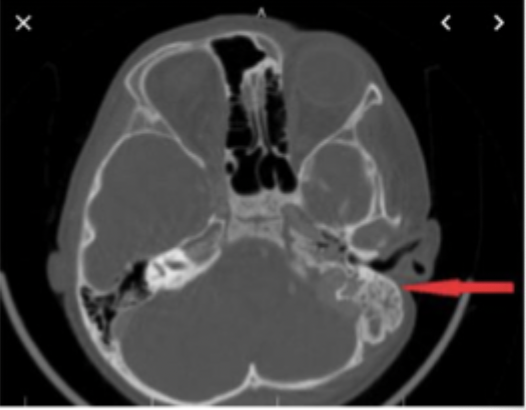
mastoid air cell CT
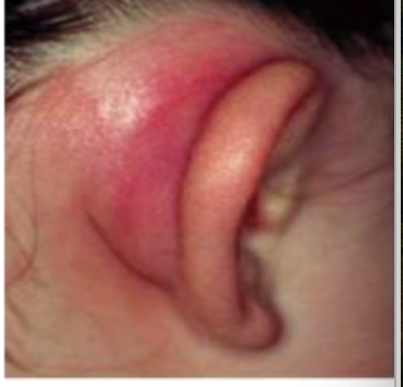
Patient presents to the clinic for ear pain and malaise. Patient reports that they were recently diagnosed with otitis media. Vitals are stable with the exception of a 102.4 temperature. On a physical exam you note, the mastoid is tender and warm on palpation, retroauricular edema, and erythema is present. You also notice a protruding auricle and the loss of the postauricular crease. What do we need to order.
opacification, possible subperiosteal abscess and intracranial extension
In a mastoid air cell CT positive for mastoiditis, what would we find?
IV vanc (hospitalization), ENT referral
What’s our treatment place for mastoiditis?
No improvement after 24 hours, intracranial complications, subperiosteal collection, facial paralysis, worsening of condition
When is a mastoidectomy indicated?