Neurobiology
1/47
There's no tags or description
Looks like no tags are added yet.
Name | Mastery | Learn | Test | Matching | Spaced | Call with Kai |
|---|
No analytics yet
Send a link to your students to track their progress
48 Terms

Neurons
Main signaling units of the nervous system
The neuron is an anatomical unit—the
fundamental structural and functional
unit of the nervous system.The neuron is composed of three parts:
cell body, dendrites, and axons.Neurons are discrete cells that are not
continuous with other cells.The points of connection between
neurons are called synapses.The neuron is a physiological unit.
Electrical activity flows through the
neuron in one direction (from dendrites
to the axon, via the cell body)
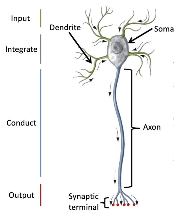
this is an image of a generic multipolar neuron
neurons have distinct neurological domains
neurons are an anatomical unit
fundamental structural unit of brain and broader nervous system
discrete cells, not continuous with other cells, contiguous —> form synapses
are physiological units
dentrites —> cell body —> axon —> axon terminals
this implies that the information is integrated within the soma and then the product of that integration in the form of an action potential is conducted across the axon
neurons are specialized for electrical signaling
Dendrite
neurite that conveys information toward
the cell body (receives information) recieves signals and then pass those signals on toward the cell body
Soma
Cell body (contains nucleus) integrates
information , also the primary metabolic center
integrative region that puts those inputs together
Axon
neurite that conveys signals and information away from
the soma, sometimes over short or long distances
(sending information) interneurons = short distance
projection neurons = long distances
conductor of signal away from neuron
action potential conducted across neuron
Synaptic terminal
site of output from a given neuron output, passed along to target
where neurotransmitters are released at a given synapse, passing it from one neuron to another forming a circuit
neurotransmitter acts as a chemical signal communicating the signal from one neuron to another
Neuronal morphology
*listen back in recording
larger dendritic arbor —> more inputs it can recieved, more synapses can form
large arbors = more convergence and computational power
Convergence
The number of inputs to a single
neuron; reflects the ability to integrate signals.
Divergence
The number of targets innervated by
any one neuron.
Non-neuronal Cells
Many cells in the nervous system are non-neuronal cells
– Glia
• Differences: no active electrical response, small, have symmetrical branches
• Why are glial cells so important to the nervous system?
– Provide physical support
– Regulate extracellular environment to maintain homeostasis
– Provide protection, have defensive roles (immune cells)
– Produce myelin
morphology of neurons
unipolar neuron
multipolar neuron
most common type found in brain
pseudounipolar neuron
Macroglia
Astrocytes and Oligodendrocytes
Astrocytes
• Provide structural support
• Act as glial guide wires for neurons during development
• Maintain ion balance around neurons
• Participate in reuptake of neurotransmitter adjacent to synapse
• Surround blood vessels (BBB)
• Migrate to site of neuronal injury and proliferate to aid in repairing damaged neuronal
tissue (specialized set of astrocytes called Reactive Astrocytes)
- Unfortunately if unsuccessful can lead to gliosis – glial scarring
- glial cells dominate injury site
Protoplasmic astrocytes
Delicate w/ many branched processes
Occur in gray matter
Fibrous astrocytes
More fibrous and robust
Radiate long processes in all directions from soma
Surround blood vessels (Blood Brain Barrier)
Occur in white matter
Cover exterior surface of CNS
Oligodendrocytes
Predominate in white matter
Extend mulitiple arms to myelinate multiple axons within the CNS ONLY
Insulating cells in the CNS
(in PNS – this function is done by the Schwann Cell)
Microglial Cells
Macrophages (scavengers)
Survey the CNS to combat infection
Activate and infiltrate injured zones of CNS to scavenge for infection and damage
Some exist within the CNS and others infiltrate from the blood
The Central (CNS) and Peripheral (PNS) Nervous Systems
A. Structural (anatomical) – 2 systems
1) Central Nervous System (CNS)
– brain (cerebrum, cerebellum, brainstem) and spinal cord
2) Peripheral Nervous System (PNS)
– cranial and spinal nerves
Afferent
towards CNS, sensory
Efferent
away from CNS; motor
Extracellular recording
shows the relative frequency and pattern
of action potentials in neurons that form the neural circuits for
the myotatic reflex
Functional Analyses
Functional Brain Imaging
• Positron Emission Tomography (PET scan)
– E.g., radioactive glucose
• Functional Magnetic Resonance Imagine (fMRI)
– Blood Oxygen Level Dependent (BOLD sign
fMRI is a technique which
allows for a good balance
between spatial and temporal
resolution.
Magnetoencephalography
(MEG) is a technique that
allows for mapping brain
activity at high temporal
resolution, but low spatial
resolution.
Positron emission tomography
(PET) acquires signals more
slowly and approaches the
spatial resolution of fMRI.al)
Anterograde tracer
maps connections from source to their termination
Retrograde tracer
maps connections from termination to origin
Tract Tracing
– Pros
• High(est) spatial resolution, i.e, ultrastructural level
(microscale)
• Produces a wealth of quantifiable data
• Generates testable, functional hypotheses
– Cons
• Invasive
• Time intensive
Brain Mapping: Diffusion Tensor Imaging
Diffusion tensor imaging (DTI), is a technique
that allows bundles of axons that carry
information between brain regions to be
visualized.
DTI works by measuring magnetic fields to
calculate the diffusion of water molecules.
The diffusion of water in tissue varies with
direction (anisotropic) which can indicate the
underlying tissue orientation.
Diffusion Tensor Imaging (DTI)
– Pros
• Non-invasive; can be conducted in/ex-vivo
• Fast
• Vast clinical applicability
– Cons
• Low Resolution (Macroscale)
• Indirect Technique