anatomy unit 6: digestive system
1/95
There's no tags or description
Looks like no tags are added yet.
Name | Mastery | Learn | Test | Matching | Spaced | Call with Kai |
|---|
No analytics yet
Send a link to your students to track their progress
96 Terms
functions of digestive system
digestion and absorption
processes of digestion
Ingestion
Propulsion
Food breakdown- mechanical digestion
Food breakdown- chemical digestion
Absorption
Defecation
Mechanical Digestion
mixes and divides food for further degradation by enzymes.
Examples
Mixing of food in the mouth by the tongue
Churning of food in the stomach
Segmentation in the small intestine
includes peristalsis and segmentation
Chemical Digestion
a sequence of steps in which large food molecules are broken down into their building blocks by enzymes.
mouth, stomach, small intestine
where does mechanical digestion happen
starts in mouth and stomach, small intestine
where does chemical digestion happen
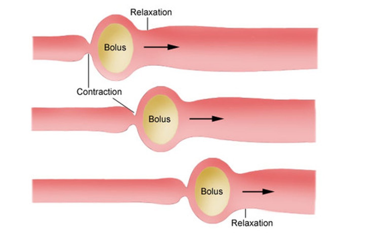
Peristalsis
adjacent segments contract and relax, pushing the food along the tract.
This can be found throughout the system, from the pharynx down to the anus.
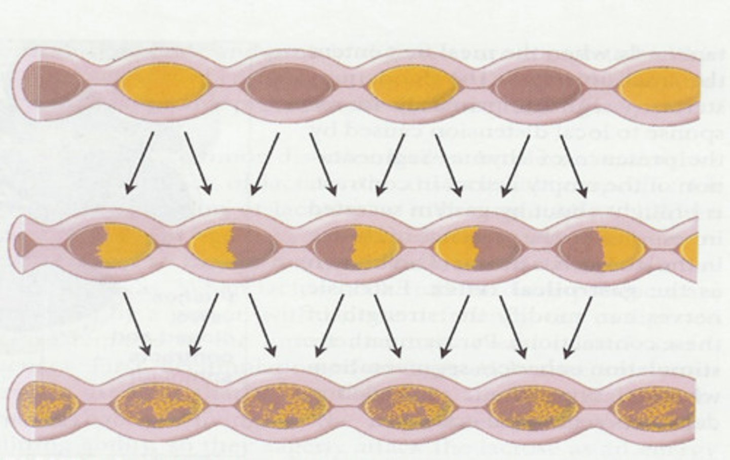
Segmentation
single segments alternatively contract and relax. The food is moved forward and then back again. So, the food becomes mixed rather than just propelled along the tract.
Found only in the small intestine
alimentary canal
The Alimentary Canal (also known as the Gastrointestinal Tract) is a continuous, coiled, hollow, muscular tube that winds through the body from mouth to anus
The canal is open at both ends.
9 meters/30ft in in a cadaver
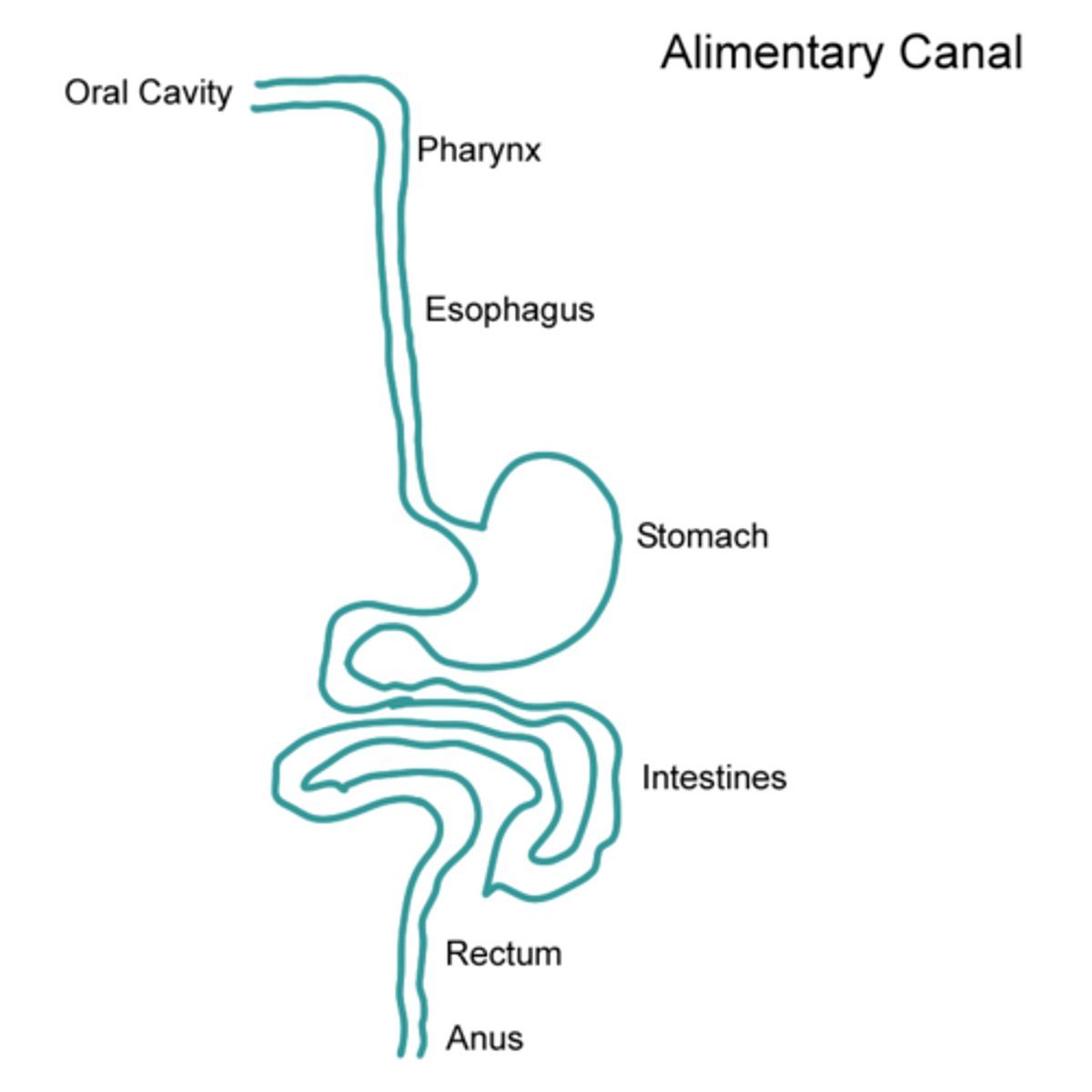
Mouth
Pharynx
Esophagus
Stomach
Small intestine
Large intestine
The Organs of the Alimentary Canal
lumen
The hole/tunnel that the food material moves through
mucosa, submucosa, muscularis externa, serosa
the four basic tissue layers that make up the walls of the alimentary canal organs from the esophagus to the large intestine
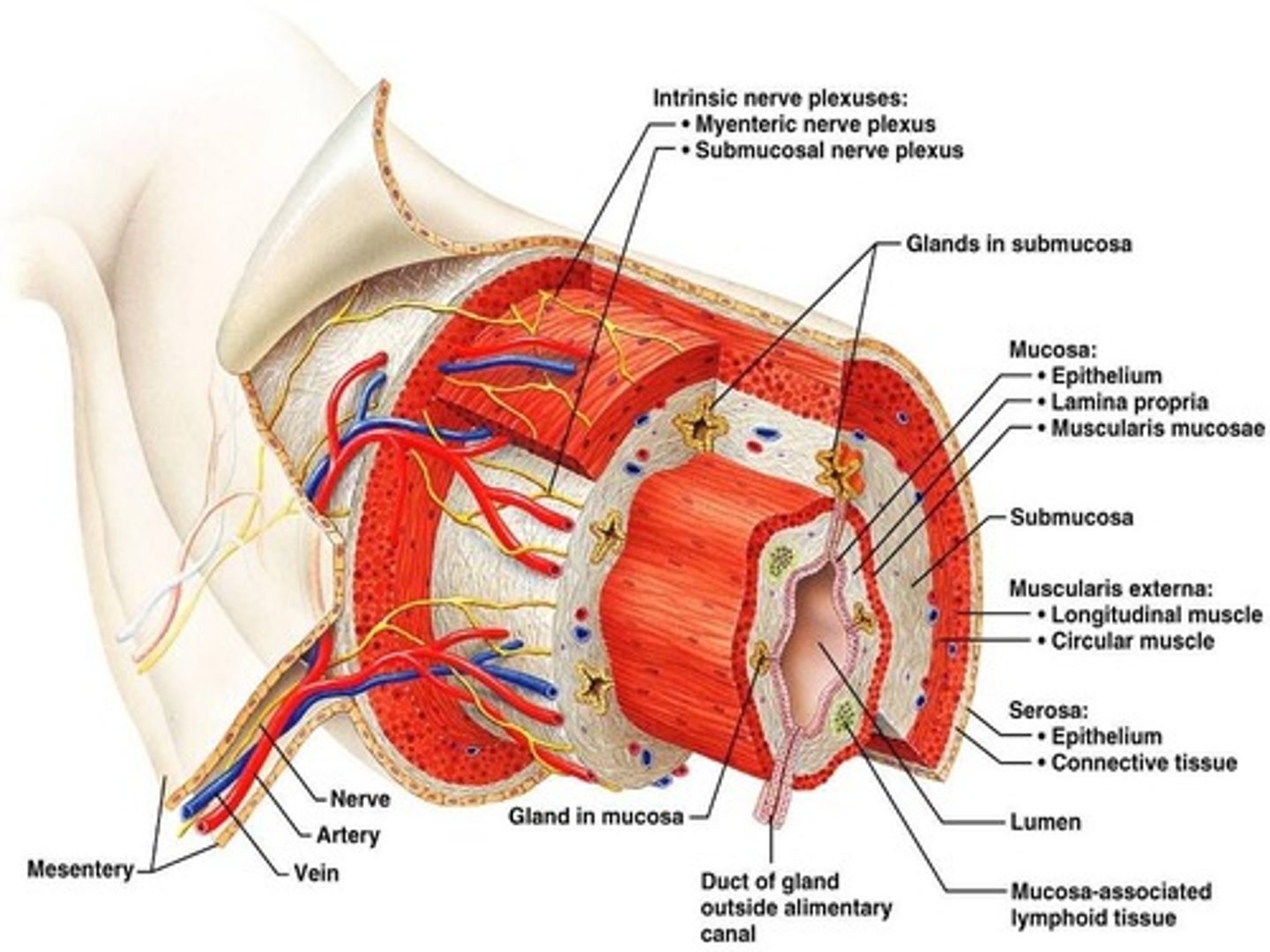
Mucosa
the innermost layer, a membrane that secretes mucus and lines the lumen, of the organ. Consists primarily of epithelium. (Simple columnar epithelial)
Submucosa
found just beneath the mucosa. It is a soft connective tissue layer containing blood vessels, nerve endings, lymph nodules, and lymphatic vessels.
Muscularis externa
is a muscle layer made up of smooth muscle cells.
The serosa is the outermost layer of the wall.
mouth/oral cavity
opening where food enters the body and undergoes the first process of digestion
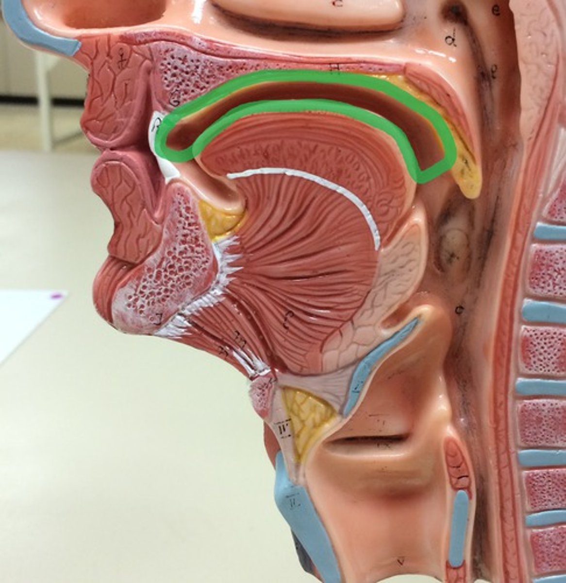
Palate
the roof of the mouth
Hard is in the front
Soft is in the back
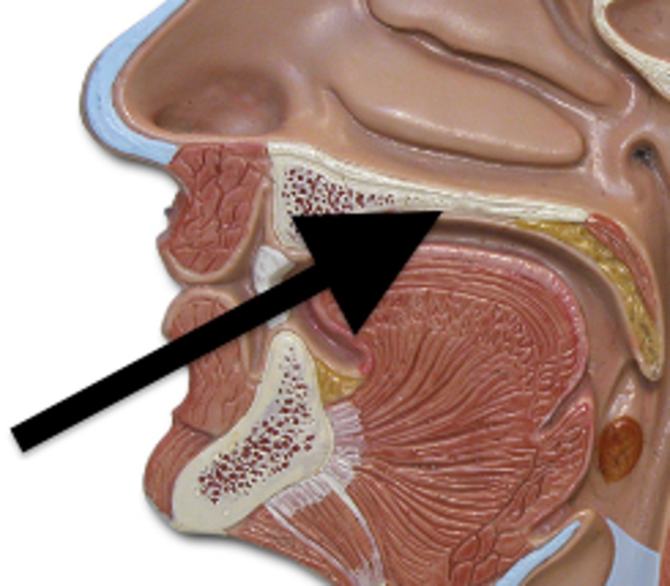
Uvula
a fleshy finger-like projection of the soft palate, which extends downward.
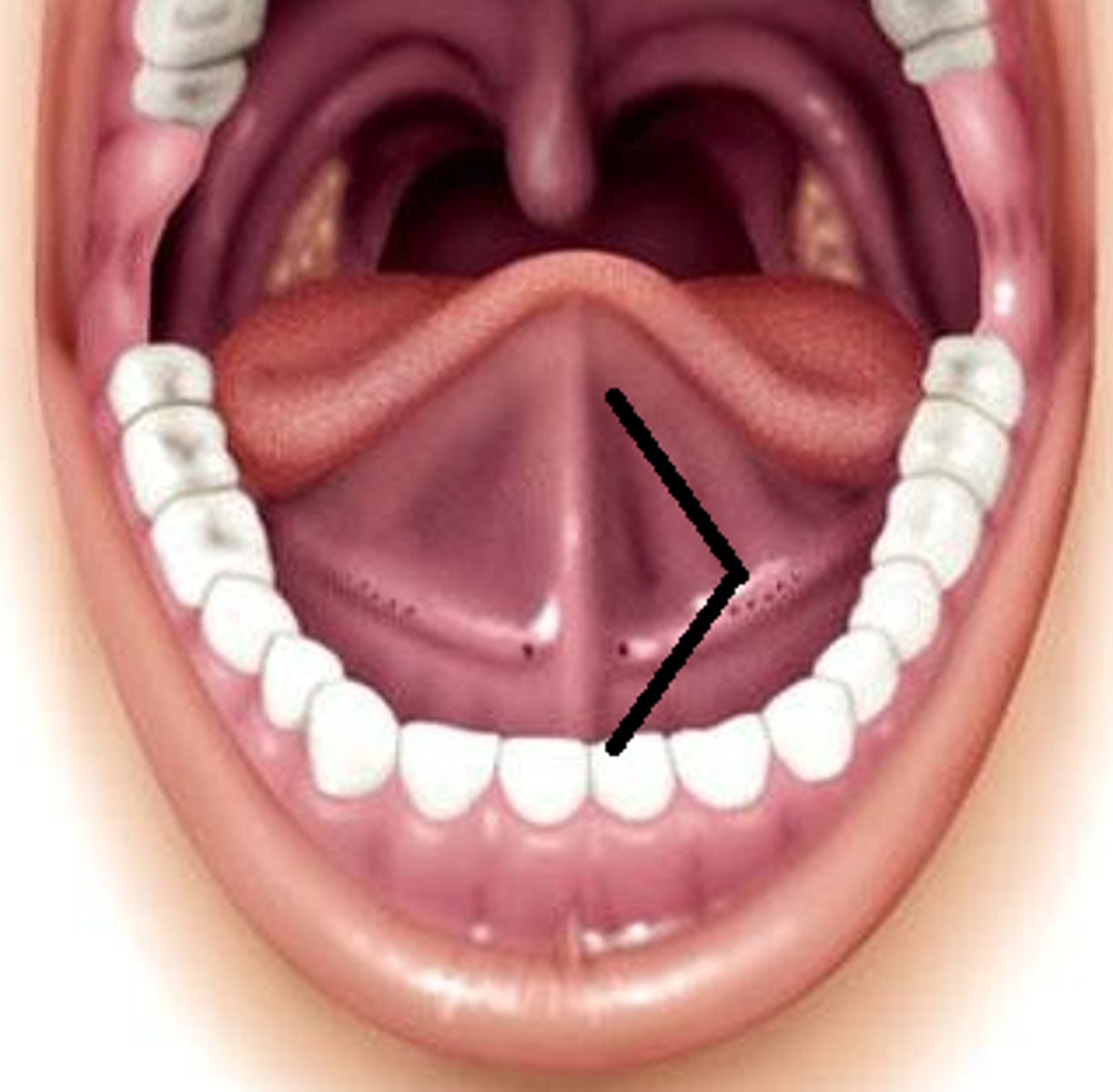
Oral cavity proper
area contained by the teeth
Lingual frenulum
a fold of mucous membrane, it secures the tongue to the floor of the mouth and limits its posterior movements.
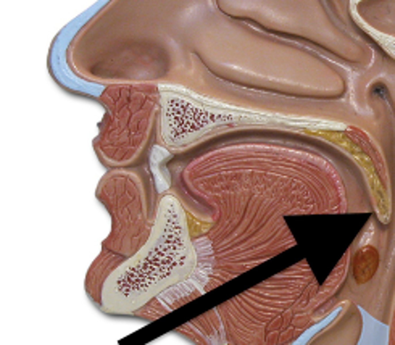
Pharynx
From the mouth, food passes posteriorly into the oropharynx and laryngopharynx, both of which are common passageways for food, fluids, and air.
Oropharynx
posterior to the oral cavity
Laryngopharynx
continuous with the esophagus below.
Epiglottis
flap of elastic cartilage that covers the air passageway whenever food passes through the pharynx and into the esophagus.
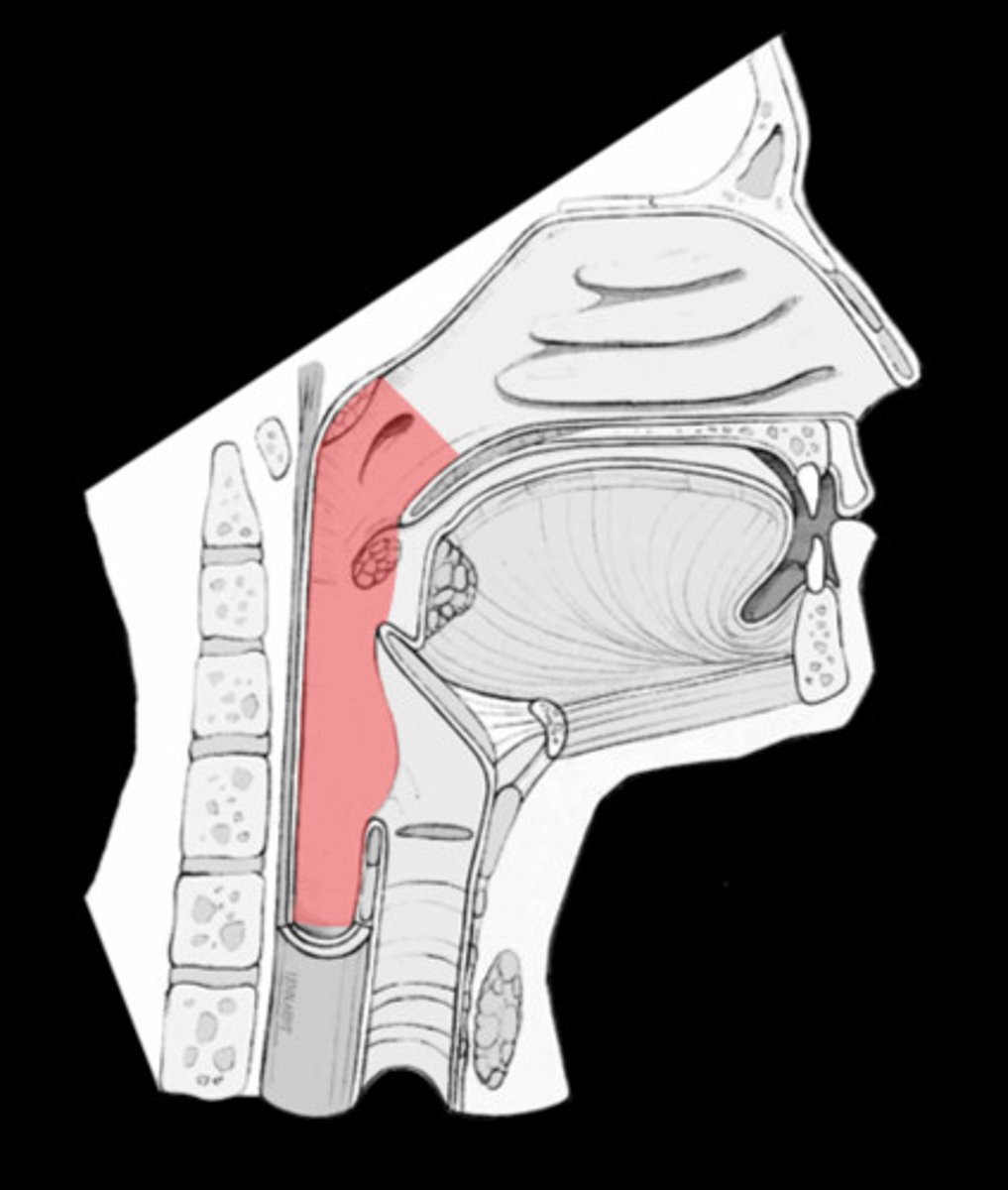
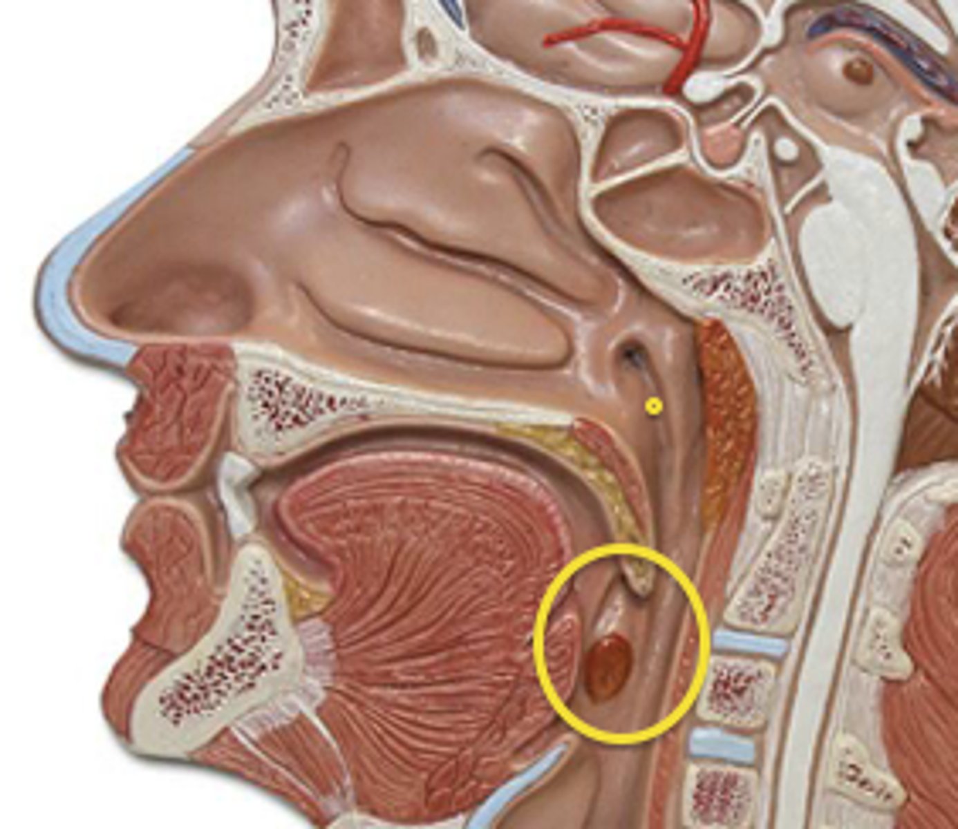
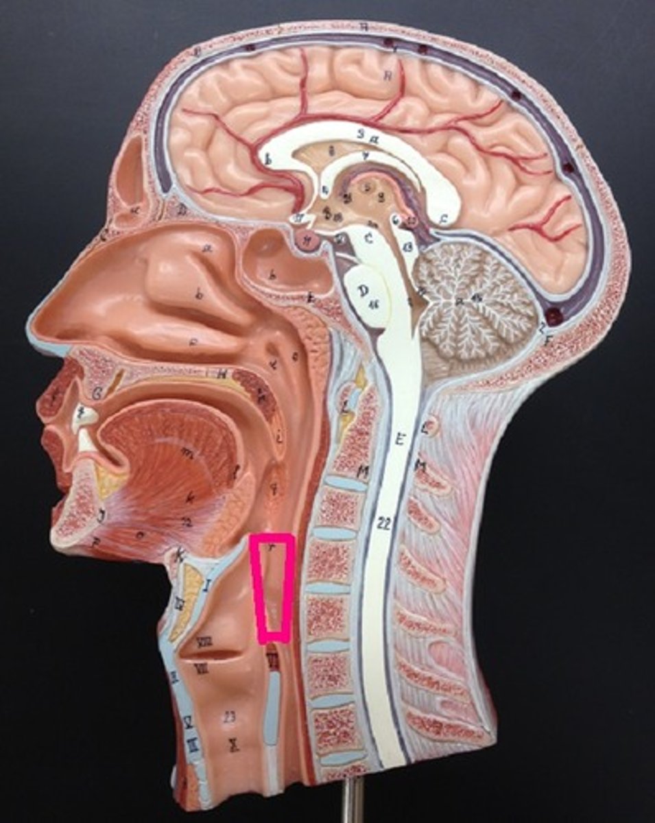
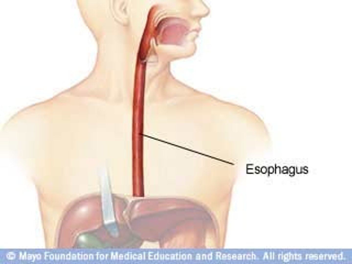
esophagus (gullet)
runs from the pharynx through the diaphragm to the stomach.
Two sphincters
Upper esophageal sphincter- separates the pharynx from the esophagus
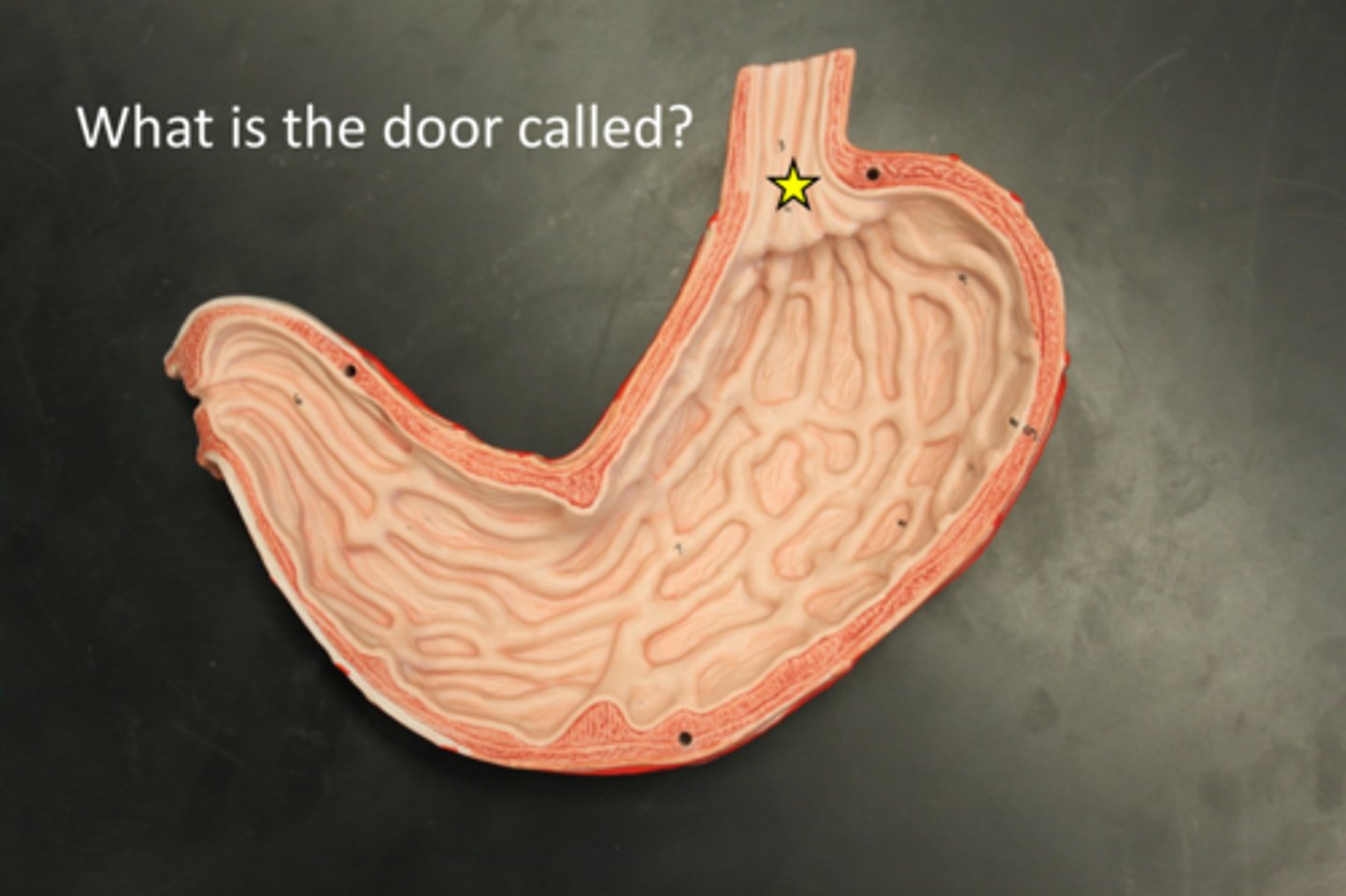
Cardioesophageal sphincter- separates the esophagus from the stomach (aka: gastroesophageal sphincter OR lower esophageal sphincter)
Upper esophageal sphincter
separates the pharynx from the esophagus
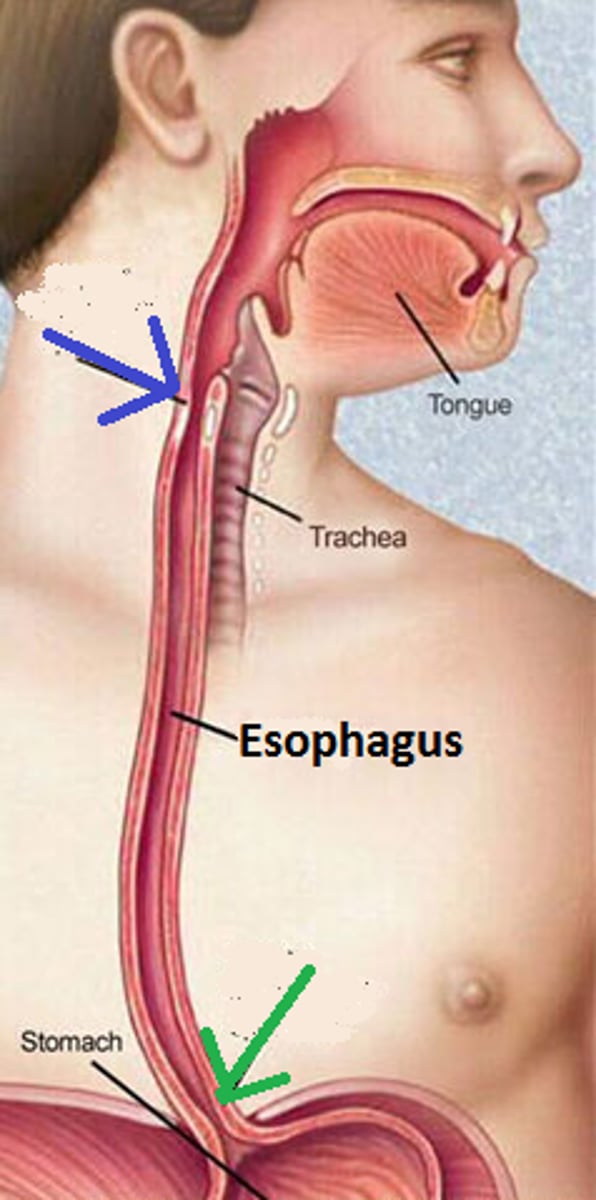
Cardioesophageal sphincter
separates the esophagus from the stomach (aka: gastroesophageal sphincter OR lower esophageal sphincter)
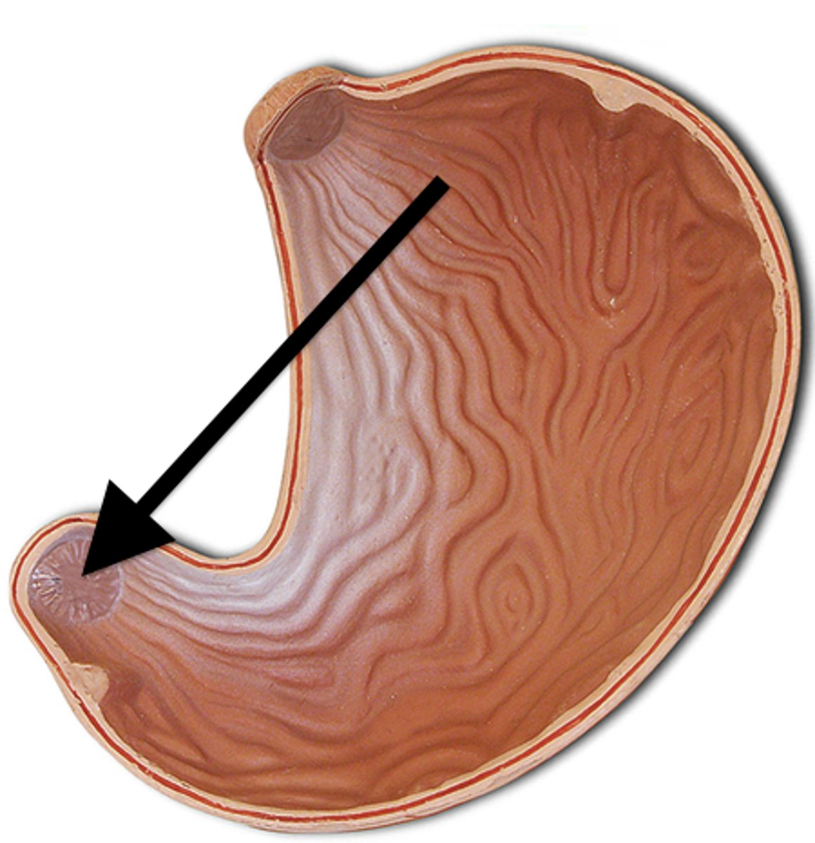
cardiac region, fundus, body, pylorus
regions of stomach
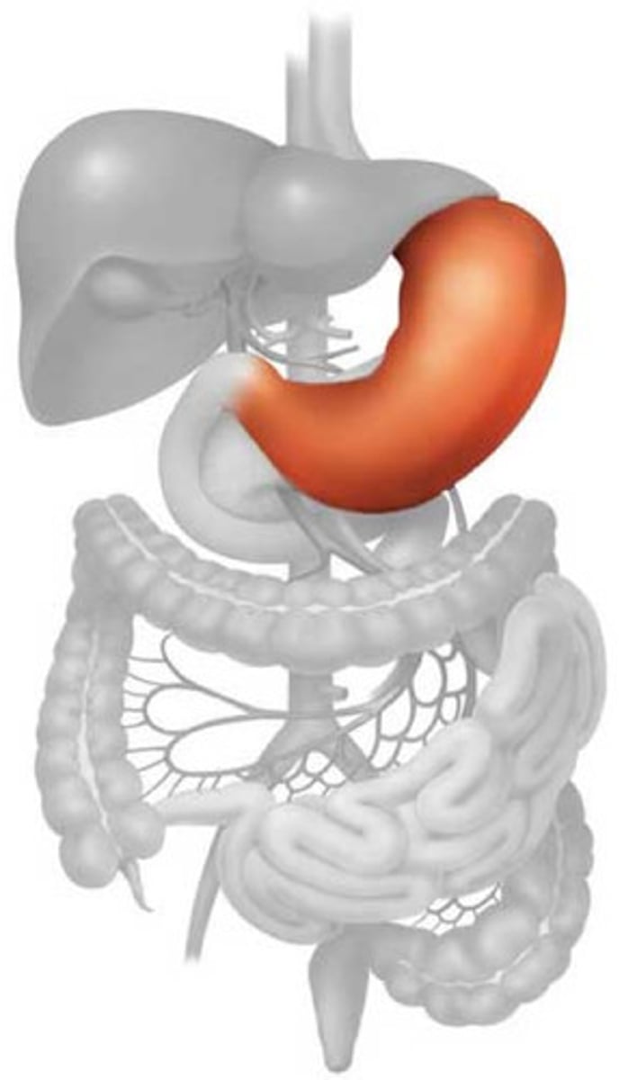
stomach
The stomach is a C-shaped organ on the left side of the abdominal cavity, nearly hidden by the liver and the diaphragm.
cardiac region
(named for its position near the heart) surrounds the cardioesophageal sphincter, where food enters the stomach from the esophagus.
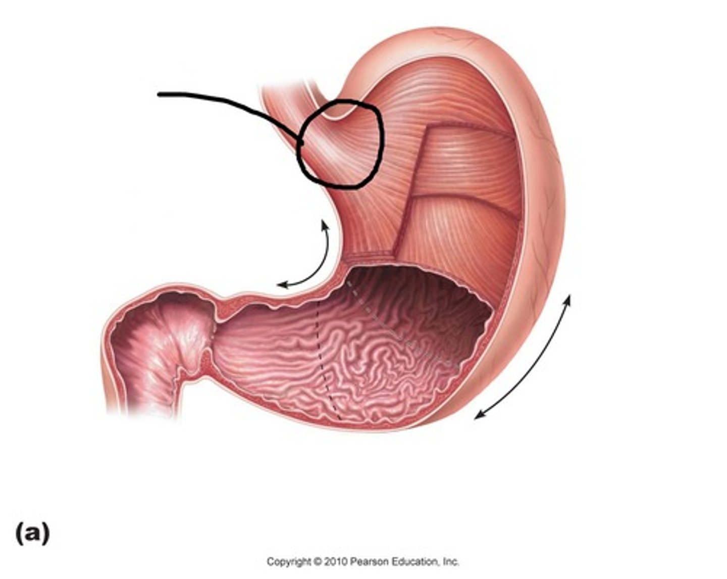
Fundus
the expanded part of the stomach lateral to the cardiac region
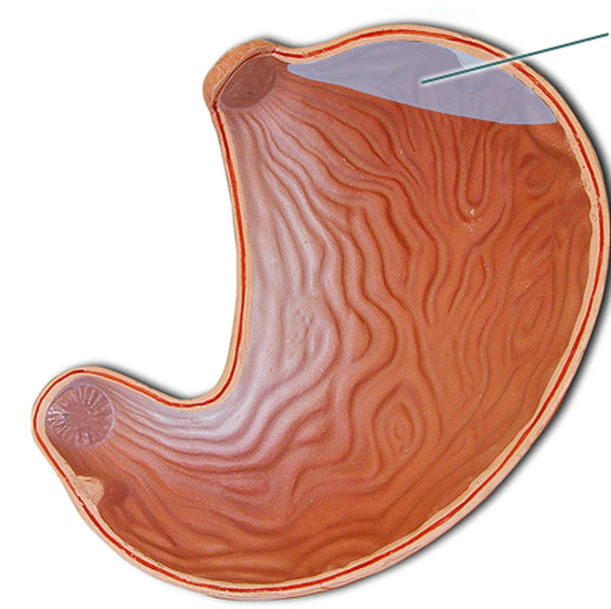
Body
the midportion of the stomach
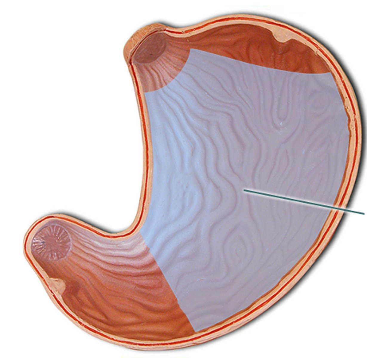
Pylorus
is the terminal part of the stomach
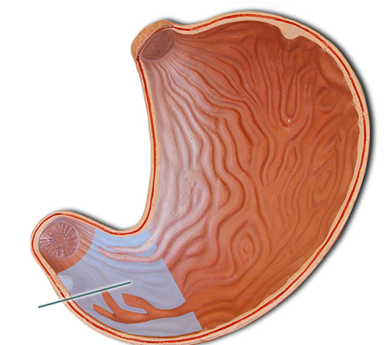
Greater curvature
the convex lateral surface of the stomach
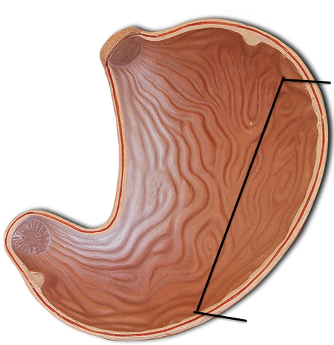
Lesser curvature
the concave medial surface of the stomach
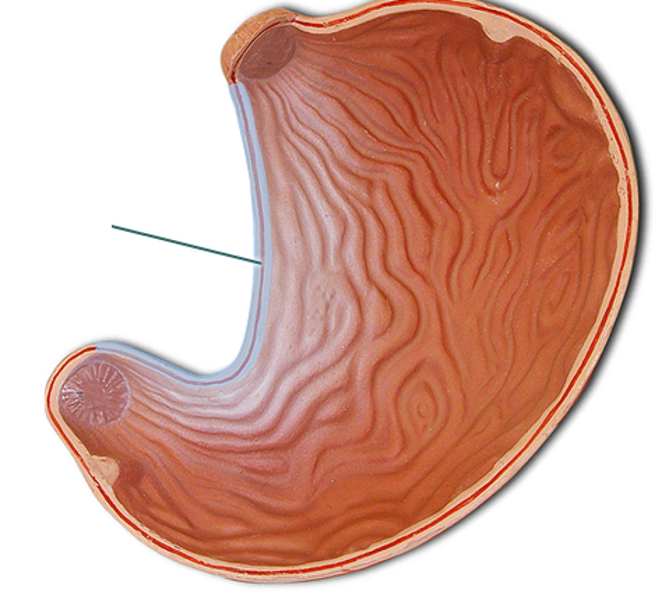
Rugae
wrinkles of mucosa tissue on the stomach wall. (Increases surface area)
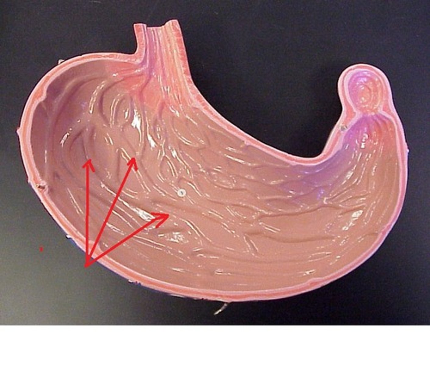
Gastric Pits
found in the walls of the stomach, within the mucosa layer.
Gastric Glands
the interior of the gastric pits. Consists of Mucous neck cells, Parietal Cells, and Chief cells.
Mucous neck cells, Parietal Cells, and Chief cells.
cells of the gastric pits
Chief cells
located in the Gastric Glands, produce pepsinogen.
Parietal cells
secrete Hydrochloric Acid (HCl), which transforms the pepsinogen into pepsin.
Neck cells
produce a sticky alkaline mucus, which clings to the stomach mucosa and protects the stomach wall from being damaged by the acid and digested by the enzymes.
Pepsin
enzyme that digests protein within the stomach.
how is food moved from stomach to intestine
Peristaltic waves act primarily in the inferior portion of the stomach to mix and move chyme through the pyloric valve.
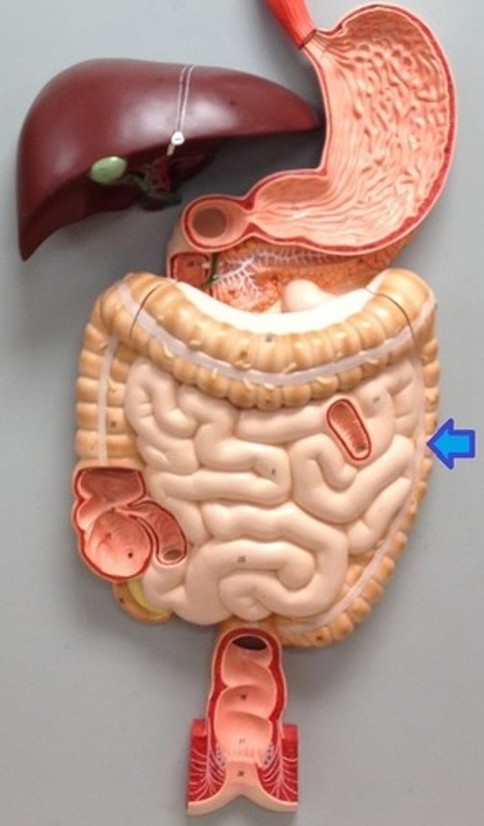
Small intestine
The small intestine is the body's major digestive organ. Here usable food is finally prepared for its journey into the cells of the body.
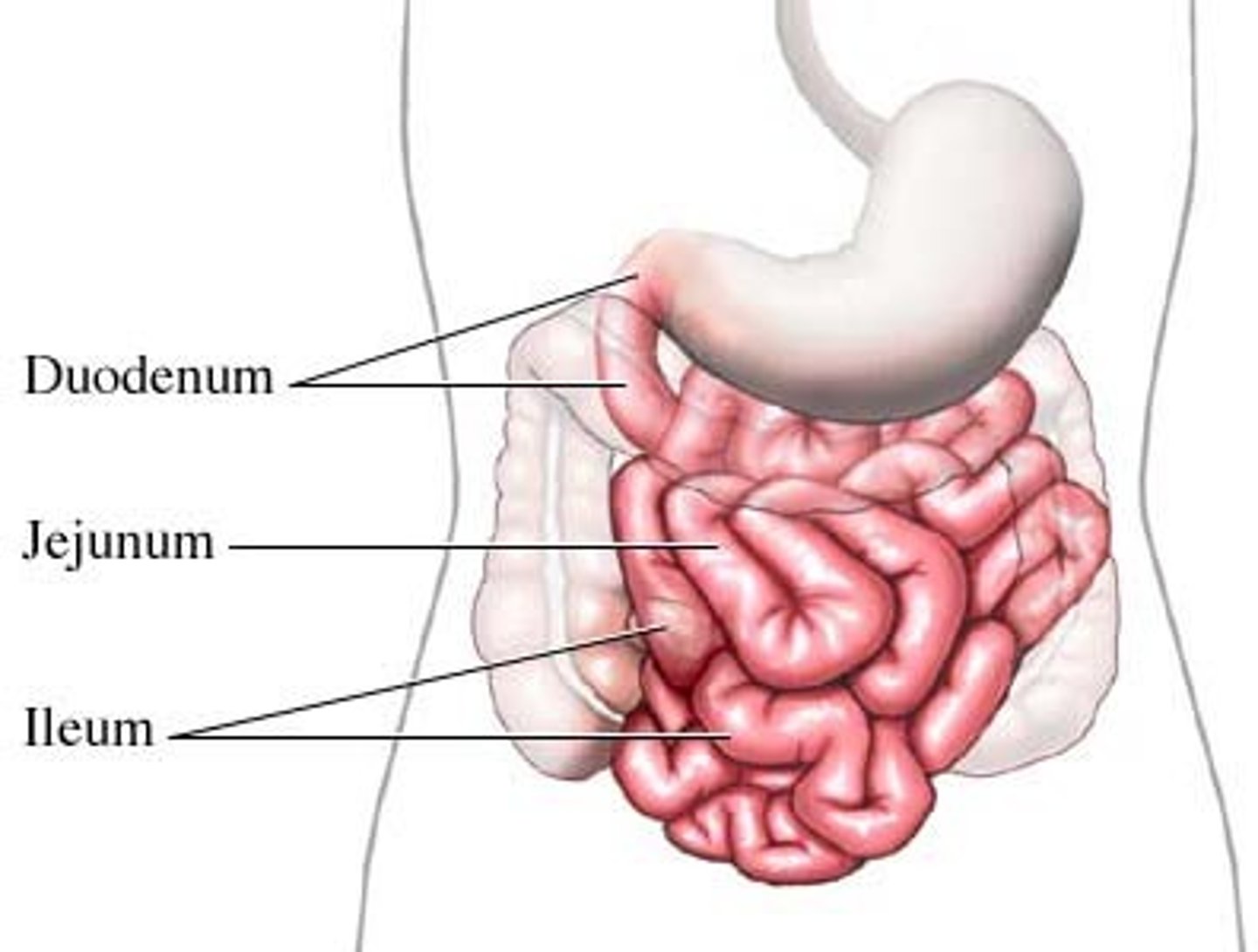
pyloric sphincter
entrance to the small intestine. Controls food movement into the small intestine and prevents the small intestine from being overwhelmed.
Ileocecal valve
end of the small intestine, leads into the large intestine
Duodenum
curves around the head of the pancreas, about 25cm long (10 inches).
Jejunum
extends from the duodenum to the ileum, about 2.5m long (8 feet).
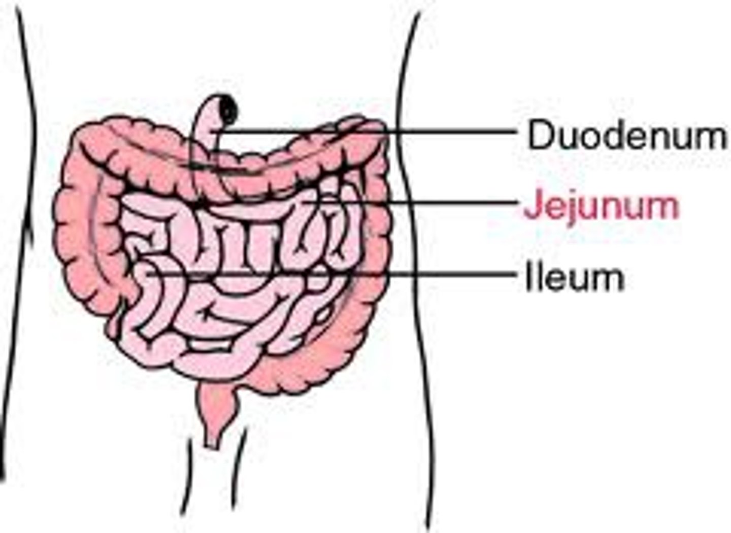
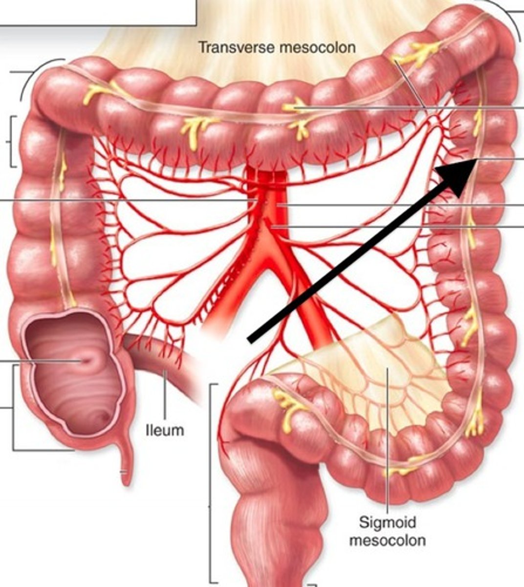
Ileum
the terminal part of the small intestine, about 3.6m long (12 feet).

circular folds, villi, microvilli
structures that increase surface area of small intestine
Circular folds
also called plicae circulares, are deep folds of both mucosa and submucosa layers. (Increases surface area)
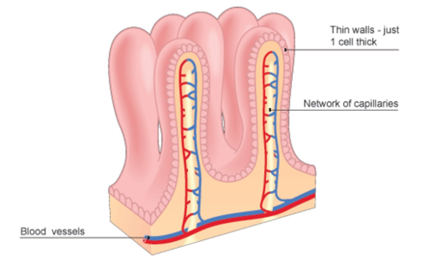
Villi
finger-like projections of the mucosa that give it a velvety appearance and feel, much like the soft texture of a towel. (Increases surface area)
Microvilli
are tiny projections of the plasma membrane of the mucosa cells that give the cell surface a fuzzy appearance, also called the brush border. (Increases surface area)
Each Villus has a rich capillary bed and a modified lymphatic capillary, called a lacteal.
The digested foodstuffs are absorbed through the mucosa cells into both the capillaries and the lacteal.
Lacteals are really good at absorbing fats/ lipids; and the capillaries absorb just about everything else.
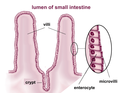
brush border
Surface of a cell covered with microvilli. increases surface area of a cell for absorption
lacteals
modified lymphatic capillary that absorbs fat and lipids
capillaries
absorbs everything else in the small intestine
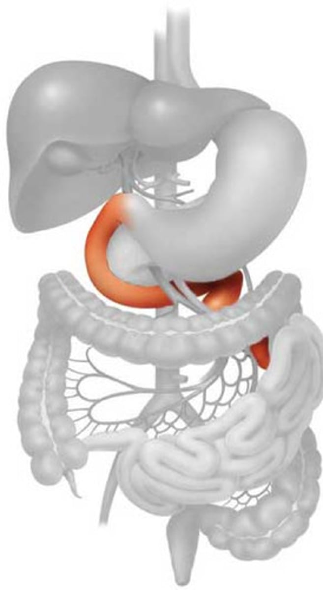
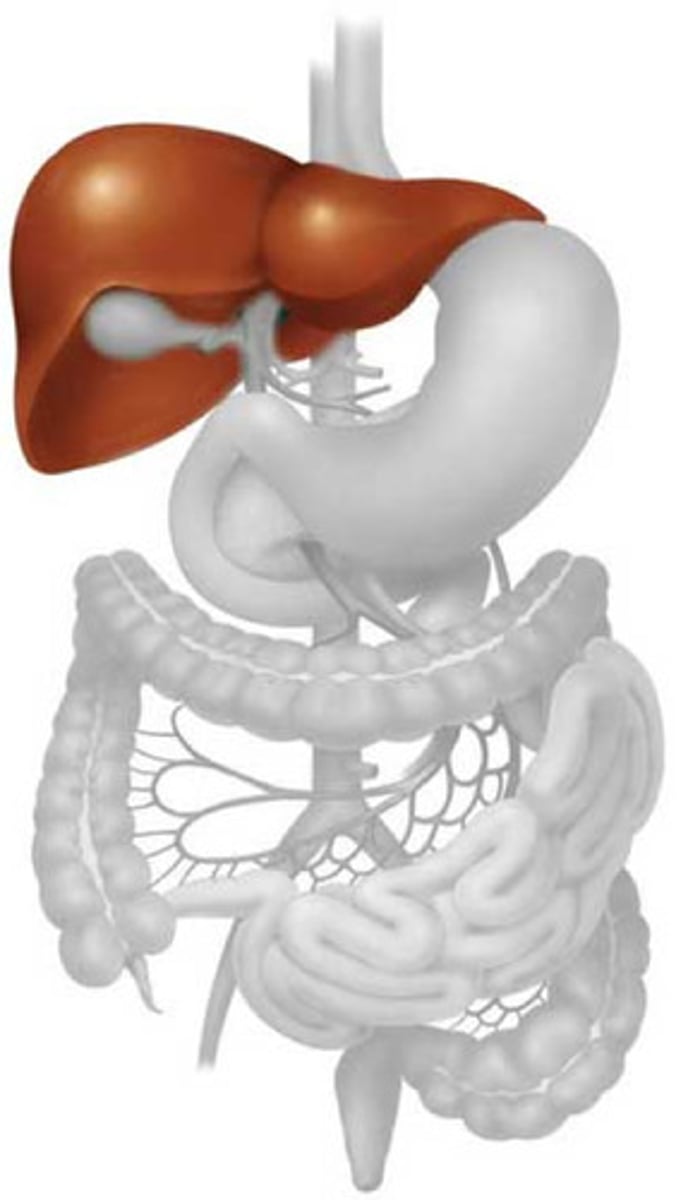
Liver
Triangular shaped.
The liver is the largest gland in the body.
Located under the diaphragm next to the stomach.
Functions:
To detox the blood.
Produce bile to help emulsify fats.
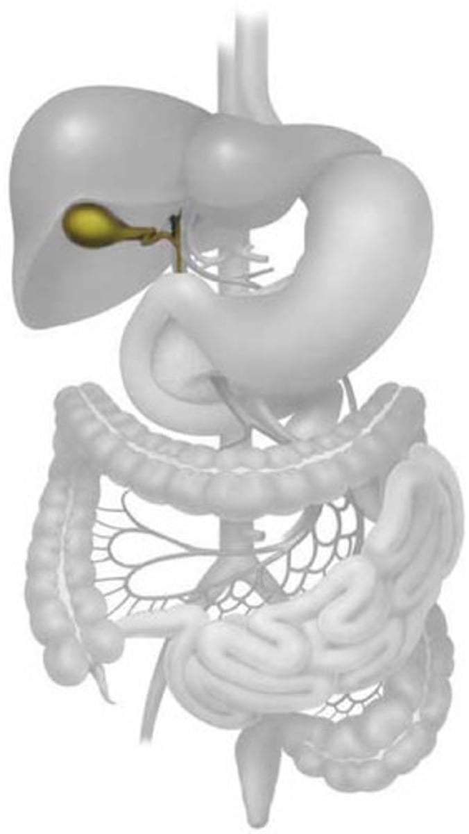
Gallbladder
Bulb shaped organ
Sits beneath the liver
Function:
Stores the bile that the Liver produces.
Problem: if it stores too much cholesterol with the bile, gallstones can form
Pancreas
A weird shaped organ, it kind of looks like an elf's ear.
It is tucked underneath the stomach and liver.
The Pancreas is a dual organ, meaning it works with both the digestive system and the endocrine system.
For the Digestive System: produces pancreatic juice (juice that neutralizes the stomach acid and provides a lot of pancreatic enzymes to break down foods.)
For the Endocrine System: helps regulate blood sugar levels
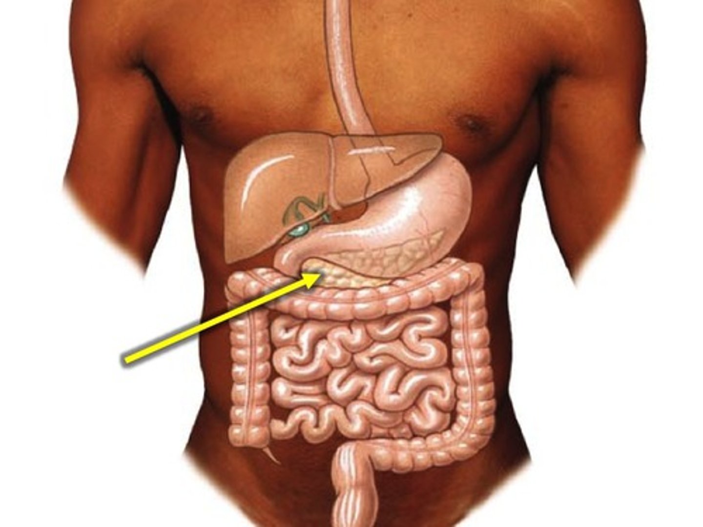
Pancreatic duct
- tube that drains enzymes and pancreatic juice out of the pancreas
Bile
(formed by the liver to emulsify fats) enters the duodenum through the bile duct.
The pancreatic juice, enzymes, and bile join in the hepatopancreatic ampulla, then travel together through the major duodenal papilla into the duodenum.
hepatopancreatic ampulla
where the bile duct and pancreatic duct meet
major duodenal papulla
a rounded projection in the duodenum where the main pancreatic duct and the common bile duct empty,
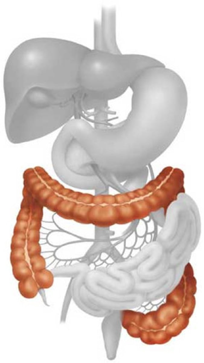
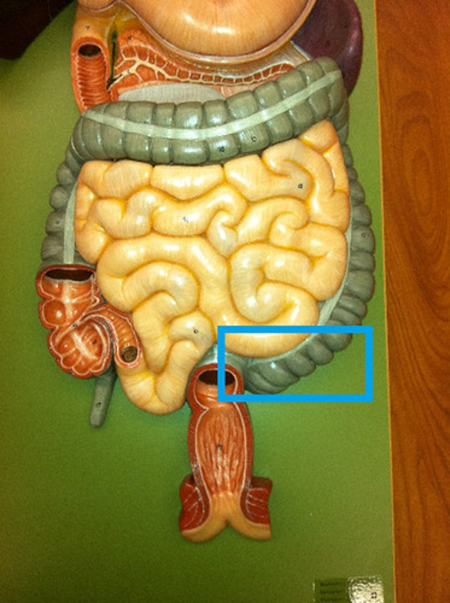
large intestine
extends from the ileocecal valve to the anus.
Functions: absorbs water and electrolytes from the remaining indigestible food matter, converts the liquid waste into solid feces, and stores it until it can be eliminated from the body through defecation
- two sphincter

ileocecal valve and anus
Two sphincters are found in the large intestine.
Anus
the exit of the intestine and the entire digestive system. Controlled by both smooth muscle and skeletal muscle.
cecum, appendix, colon, rectum, anal canal
parts of large intestine
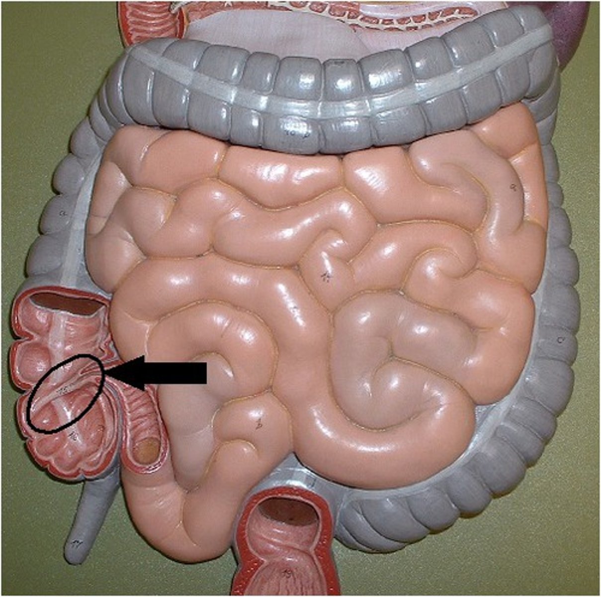
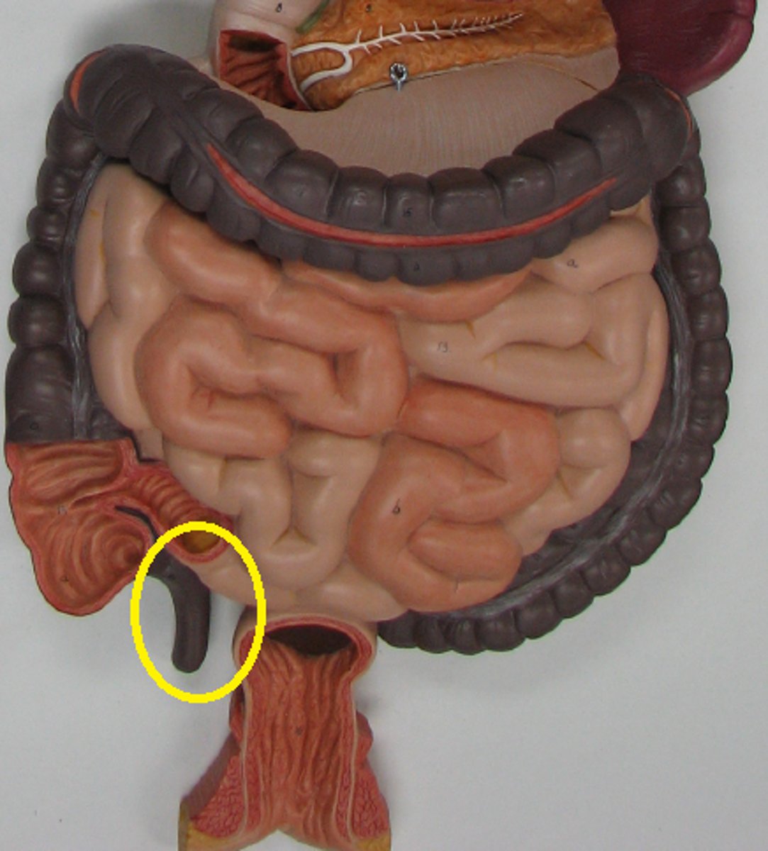
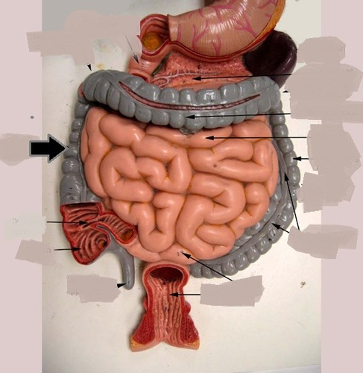
Cecum
sac-like and is the first part of the large intestine.
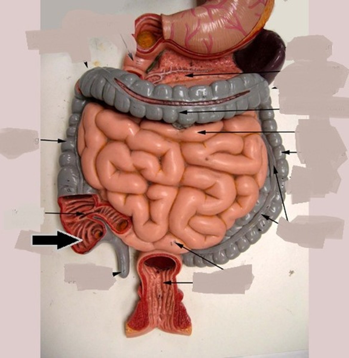
Appendix
hangs from the cecum, a potential trouble spot because it is usually twisted and it is an ideal location for bacteria to accumulate.
Colon
divided into several distinct regions: ascending, transverse, descending, and sigmoid
Rectum
lies in the pelvis comes after the sigmoid colon.
Anal canal
end of the large intestine and end of the digestive system.
Ascending colon
travels up the right side of the abdominal cavity and makes a turn at the right colic
Transverse colon
travels across the abdominal cavity and makes a turn at the left colic
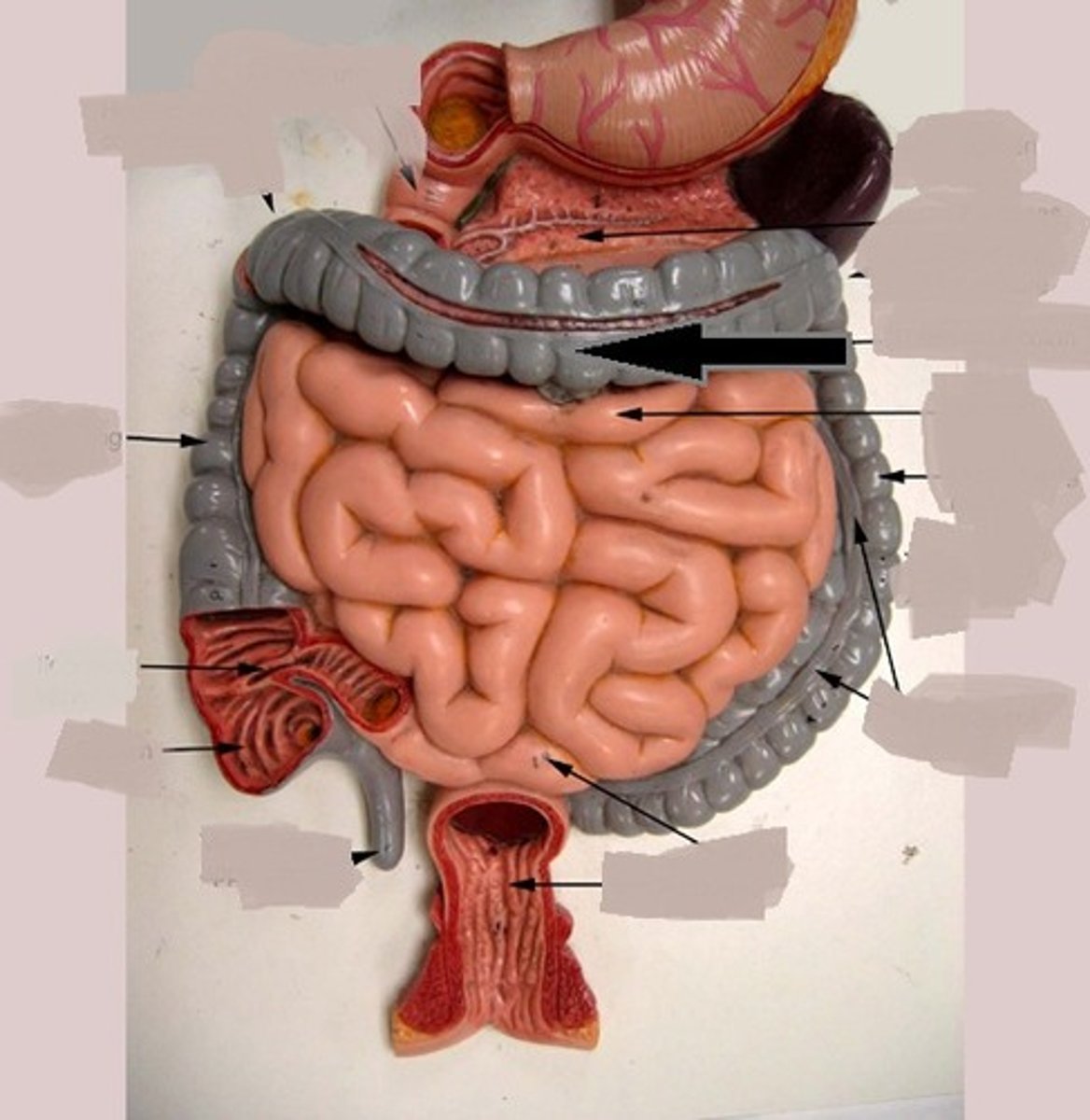
Descending colon
travels down the left side to enter the pelvis, where it becomes the S-shaped sigmoid colon.
Sigmoid colon
the end of the colon, lies within the pelvis
Haustra
Sac-like pouches that line the inner surface of the large intestine (colon). They are formed by the contraction of three longitudinal bands of muscle called teniae coli.
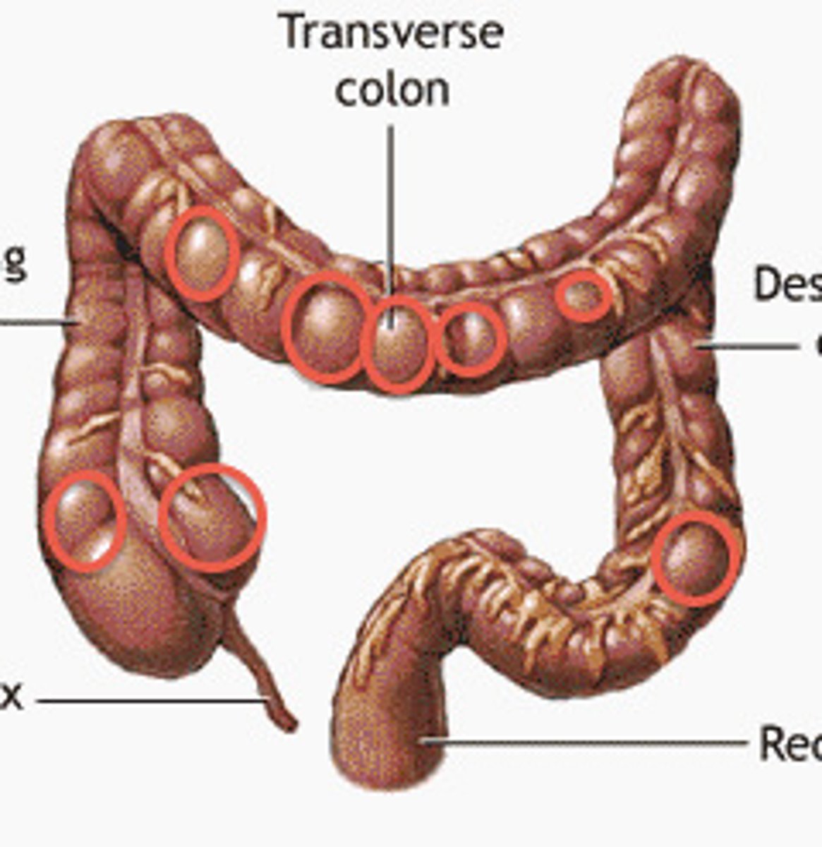
teniae coli
Smooth muscle in the wall of large intestine that pulls on the wall causing haustra
Salivary amylase and pancreatic amylase
Enzymes that break down carbohydrates.
Pepsin
Trypsin
Chymotrypsin
Carboxypeptidase
enzymes that break down protein
Bile salts
Pancreatic Lipase
Brush border enzymes
enzymes that break down fat/lipids
Pancreatic Nucleases
Brush border enzymes
enzymes with nucleic acids as nutrients
enzymes in small intestine
Pancreatic amylase
Trypsin
Chymotrypsin
Carboxypeptidase
Bile salts
Pancreatic Lipase
Brush border enzymes
Pancreatic Nucleases
Brush border enzymes
Gastroesophageal reflux disease (acid reflux)
-occurs when stomach acid frequently flows back into the esophagus.
Gallstones
Gallstones are hardened deposits of bile that can form in your gallbladder.
Celiac Disease
is an immune reaction to eating gluten, a protein found in wheat, barley and rye. Over time, this reaction damages your small intestine's lining and prevents absorption of some nutrients.
Crohn's Disease
is an inflammatory bowel disease (IBD). It causes inflammation of your digestive tract, which can lead to abdominal pain, severe diarrhea, fatigue, weight loss and malnutrition.
Hemorrhoids
-are swollen veins in your anus and lower rectum, similar to varicose veins.
Irritable bowel syndrome
a common disorder that affects the large intestine. Signs and symptoms include cramping, abdominal pain, bloating, gas, and diarrhea or constipation, or both.
Ulcerative colitis
is an inflammatory bowel disease (IBD) that causes long-lasting inflammation and ulcers (sores) in your digestive tract. Ulcerative colitis affects the innermost lining of your large intestine (colon) and rectum.
Pyloric stenosis
Pyloric muscles thicken and become abnormally large, blocking food from leaving the stomach.
vestibule and oral cavity proper
mouth regions
vestibule
the space between the lips and cheeks (externally) and the teeth and gums (internally).
bolus
a small rounded mass of chewed food at the moment of swallowing.
chyme
the pulpy acidic fluid which passes from the stomach to the small intestine, consisting of gastric juices and partly digested food.