Unit 4: Cell Communication and Cell Cycle
1/81
Earn XP
Description and Tags
Ch 11, 43, 40.2-40.3, 12
Name | Mastery | Learn | Test | Matching | Spaced | Call with Kai |
|---|
No analytics yet
Send a link to your students to track their progress
82 Terms
Signal Transduction Pathway
pathways involved in cell communication
Reception
The target cell’s detection of a signaling molecule (ligand) coming from outside the cell.
A chemical signal is “detected” when the signaling molecule (ligand) binds to a receptor protein located at the cell’s surface or inside the cell.
Transduction
converts signal reception into cellular response.
Accomplished via activation of proteins through phosphorylation, or a change in intracellular conditions (a change in ion concentration).
Response
changes in gene expression, activation of preexisting inactive proteins
Ligand
small, nonpolar (hormones)
can easily diffuse through membrane
uses intracellular receptors
large, polar, water soluble
extracellular
other: nitroglycerin -> nitric oxide
Nitroglycerin (travels through bloodstream) uses nitric oxide to travel to cells (nitric oxide has short half life, cannot travel far)
Think of: Nitroglycerin: bus, nitric oxide: elderly folks who can’t walk far
signaling molecules
(properties of) Receptor protein
1. An area of the protein that interacts with the signaling molecule (ligand).
2. An area of the protein that transmits (transduces) the signal to another protein.
3. Signal transmission is accomplished through conformational change in receptor, which activates transduction pathway in cell.
protein that exists embedded in membrane or on surface of nucleus that has receptors that ligands can bind to (reception and transduction happen here)
G-Protein Coupled Receptors (GPCRs)
Ligand attaches to G protein coupled receptor
Causes conformational shape change, causing phosphorylation of G protein
G protein phosphorylates next protein in pathway
membrane receptor
Ligand Gated Ion Channel
Ligand binds to the channel, conformational change opens channel, and ions move freely in to cell.
This change in ion concentration will then trigger cellular responses by changing the shape of various proteins.
channel that can be opened once ligand binds to receptor on surface
Tyrosine Kinase Receptors
A type of membrane receptor involved in cell signaling that has enzymatic activity. When a ligand (e.g., a growth factor) binds to the receptor, it activates the receptor's tyrosine kinase domain, which adds phosphate groups to specific tyrosine residues on itself or other proteins. This phosphorylation triggers a signaling cascade that can influence cell growth, division, or differentiation.
Key Points:
Involved in pathways controlling cell proliferation and survival.
Often forms dimers (two receptors come together) upon activation.
Phosphorylation Cascade
number of proteins involved in transduction increases EXPONENTIALLY as more and more proteins are phosphorylated
2nd Messengers
Ex. Cyclic AMP: when present in a cell, activates various catabolic metabolic pathways.
Internal signaling molecules, often activated by multiple external signals.
They do not have enzymatic activity—rather they act to regulate target enzymes by binding to them noncovalently.
Pathogen
Any organism or virus that causes disease (e.g., bacteria, viruses).
Antigen
A molecule (often on the pathogen) that triggers an immune response.
Antibody
A Y-shaped protein produced by B cells that binds specifically to antigens to neutralize them.
Macrophage
A large white blood cell that engulfs pathogens and debris and presents antigens to T cells.
T cell
A type of white blood cell that plays a role in cell-mediated immunity.
Helper T Cells: Activate B cells and cytotoxic T cells by releasing cytokines.
Cytotoxic T Cells: Directly kill infected or cancerous cells by releasing toxic chemicals.
Memory Cells
Long-lived B and T cells that "remember" pathogens for a faster response next time.
Innate Immunity
The non-specific, first line of defense against pathogens (e.g., skin, macrophages).
Antigen Presenting Cell (APC)
B cells, macrophages that ‘eat’ pathogens, process and display pathogen particles (antigens) on surface to trigger immune response (recognition & destruction)
Humoral Response
Part of the adaptive immune response involving B cells and antibodies to neutralize pathogens.
Positive Feedback
end product continue loop/cycle, amplifying it
Negative feedback
end product ends/deescalates loop/cycle
Homeostasis
balance in body
Neuron
A specialized cell that transmits electrical and chemical signals in the nervous system.
B cell
A type of white blood cell that produces antibodies and differentiates into plasma and memory cells.
Plasma Cells: Secrete large amounts of antibodies.
Memory B Cells: Long-lived cells that "remember" antigens for faster future responses.
Dendritic Cell
A type of antigen-presenting cell that activates T cells and initiates adaptive immunity.
Major Histocompatibility Complex (MHC)
Proteins on APCs that present antigens to T cells
Adaptive Immunity
A specific immune response involving B cells and T cells tailored to specific pathogens.
Cell Mediated Response
Part of the adaptive immune response involving T cells that target infected or abnormal cells.
Cytokines
Signaling molecules released by immune cells to coordinate an immune response.
Clonal Selection
When lymphocytes mature, those that have receptors that react with “self” molecules are eliminated.
When lymphocytes are presented with an antigen, only those with reactive receptors are allowed to reproduce.
Immunological Memory
The ability of memory B and T cells to mount a faster and stronger response to previously encountered pathogens.
Dendrite
Branch-like extensions of a neuron that receive signals from other neurons or sensory inputs.
Axon
The long extension of a neuron that transmits signals to other neurons or effectors (muscles, glands).
Myelin Sheath
A fatty layer that insulates axons, increasing the speed of signal transmission.
Synapse
The junction between two neurons or a neuron and its target cell where signals are transmitted.
Neurotransmitter
A chemical released at synapses to transmit signals between neurons.
Resting Potential
The electrical charge across a neuron's membrane when it is not transmitting a signal.
Synaptic Vesicles
Membrane-bound sacs in the axon terminal that store neurotransmitters.
Signal Transmission
The process of sending signals through the neuron and across synapses to other neurons or cells.
Cell Cycle
The series of events a cell goes through as it grows and divides.
Interphase
The phase of the cell cycle when the cell grows and prepares for division (G1, S, G2 phases).
G1 Phase: Cell growth and preparation for DNA replication.
S Phase: DNA replication occurs.
G2 Phase: Preparation for mitosis; organelles replicate.
Mitosis
The process of nuclear division, producing two genetically identical nuclei.
Prophase: Chromosomes condense; spindle fibers form.
Metaphase: Chromosomes align at the cell's equator.
Anaphase: Sister chromatids separate and move to opposite poles.
Telophase: Nuclear envelopes reform around separated chromosomes.
Cytokinesis
Division of the cytoplasm, resulting in two daughter cells.
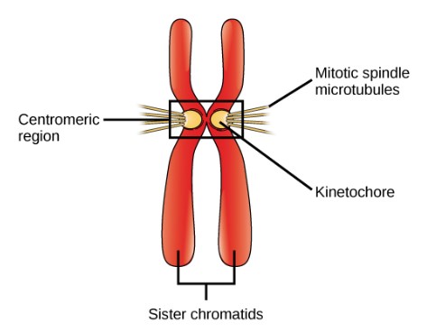
Chromatin
The uncondensed form of DNA and protein in the nucleus.
Chromosome
The condensed, organized form of DNA during cell division.
Centromere
The region of a chromosome where sister chromatids are joined.
Spindle Fibers
Microtubules that help separate chromosomes during mitosis.
Centrosome
The structure that organizes spindle fibers during mitosis.
Cell Cycle Checkpoints
Control points that regulate the cycle (e.g., G1, G2, M checkpoints).
Cyclins
Proteins that regulate the progression of the cell cycle.
Cyclin-Dependent Kinases (CDKs)
Enzymes that activate cyclins to control the cell cycle.
Apoptosis
Programmed cell death, essential for normal development and health.
Cancer
Uncontrolled cell division due to the failure of cell cycle regulation.
Intracellular Receptors
Receptors located inside the cell, either in the cytoplasm or nucleus, that bind to small, hydrophobic molecules (like steroid hormones) that can cross the cell membrane. Once activated, these receptors often function as transcription factors, directly affecting gene expression.
Key Points:
Examples: Steroid hormone receptors, thyroid hormone receptors.
Work by influencing DNA transcription.
Protein Kinase
An enzyme that transfers phosphate groups from ATP to specific amino acids (like serine, threonine, or tyrosine) on target proteins, a process called phosphorylation. This modification can activate or deactivate the target protein, altering its function and triggering signaling pathways.
Key Points:
Central to signal transduction cascades.
Phosphorylation often amplifies the signal in a pathway.
Protein Phosphate
An enzyme that removes phosphate groups from proteins, reversing the action of protein kinases. Dephosphorylation helps to turn off signaling pathways or reset proteins for future activation.
Key Points:
Balances the activity of protein kinases to regulate cell signaling.
Critical for stopping or fine-tuning signals.
Feedback Regulation in Signaling
The process where the output or end product of a signaling pathway influences the activity of the pathway itself.
Positive Feedback: Enhances or amplifies the original signal, leading to an increase in the pathway’s activity. Example: Platelet activation during blood clotting.
Negative Feedback: Reduces or inhibits the original signal, helping maintain homeostasis. Example: Insulin signaling lowering blood sugar levels.
Key Points:
Keeps signaling pathways under control.
Maintains balance and prevents overactivation.
Specificity of Signal Response
The ability of a cell signaling pathway to produce a precise and appropriate response depending on the type of cell, the receptors expressed, and the downstream proteins involved.
Key Points:
Different cell types can interpret the same signal differently.
Achieved through distinct combinations of receptors, transduction proteins, and target molecules.
Ensures that the correct cellular response is elicited in the right context.
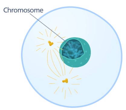
Prophase
Chromatin condenses into chromosomes. Nucleolus disappears.
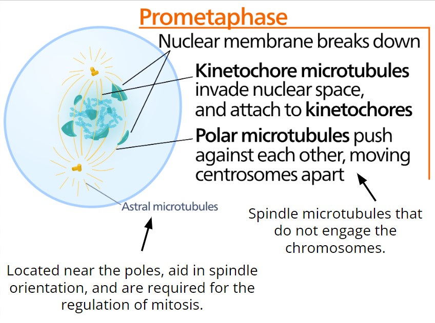
Prometaphase
Nuclear membrane breaks down.
Kinetochore microtubules invade nuclear space, and attach to kinetochores.
Polar microtubules push against each other, moving centrosomes apart.
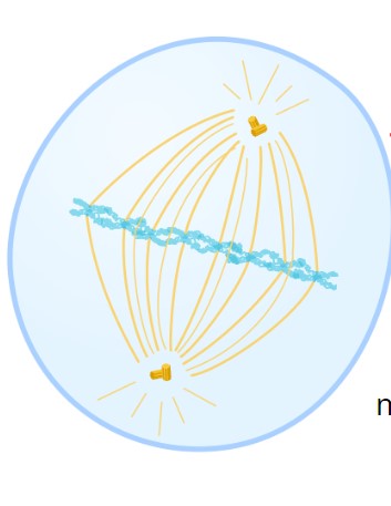
Metaphase
Chromosomes line up against metaphase plate (imaginary plane).
Each chromosome has a microtubule attached to opposite sides of its kinetochore.
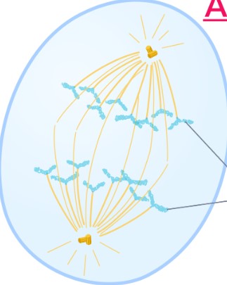
Anaphase
Chromosomes break at centromeres
Sister chromatids move to opposite sides of the cell
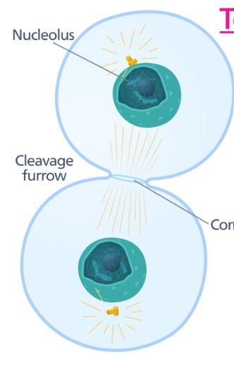
Telophase & Cytokinesis
Nuclear membrane reforms
nucleoli reappear
chromosomes unwind into chromatin
Myosin II and actin filaments ring contract to cleave cell in two
Mitosis Promoting Factor
Controls specifically the “Mitosis checkpoint” of the cell. If MPF is not present, a cell will not enter mitosis.
MPF is the result of the combination of two other proteins:
Cdk (cyclin-dependent kinase): always present in the cell. (inactive until bound to cyclin)
Cyclin: only made once a cell has met the criteria needed to enter mitosis.
Other checkpoints are also regulated with other versions of both Cdk and Cyclin.
G1 Checkpoint
Cell is large enough, sufficient nutrients available. Regulated by the presence of S-cyclin-Cdk complexes. Cell cycle continues if it receives the go-ahead.
G2 Checkpoint
Cell is large enough & replication of chromosomes has been successfully completed. When sufficient MPF accumulates, the G2 checkpoint is passed, and mitosis is promoted.
M Checkpoint
The kinetochores must all be attached to spindle fibers during metaphase. This will activate an enzyme (separase), which allows the sister chromatids to separate, and anaphase will proceed.
Autocrine
Cell targets itself
Cell secretes signaling hormone to outside of cell
Signaling molecule attaches to extracellular receptors on outside of the cell to communicate to cell to begin response
Gap junctions
Cell targets another cell through gap junctions
Paracrine
Cell targets nearby cell
Secretes ligands that flow and bind to nearby cell’s receptors
Ligands are produced by cells and diffuse to local target cell populations.
Endocrine
A cell targets a distant cell through the bloodstream
Interferons
chemicals released from damaged, invaded cells used to warn other cells of incoming danger—other cells increase their defenses to interfere with the ability to be attacked.
SAVE YOURSELF
or my fav: KILL YOURSELF
MHC II
All antigen presenting WBC’s possess these. _____ holding an antigen shows other WBC’s what to look for and kill.
MHC I
All cells OTHER THAN WBC’s possess these. ______ placed on the cell surface holding an antigen tells WBC’s that the cell itself is infected.
What happens to chromosomes during prophase?
Chromosomes condense and become visible.
Mitotic spindle begins forming.
Nuclear envelope starts breaking down.
What happens to the nuclear envelope in prometaphase?
It breaks apart completely.
Spindle fibers attach to kinetochores (protein structures at centromeres
Where are chromosomes lined up during metaphase?
At the metaphase plate (equator of the cell).
Each chromosome is attached to spindle fibers from opposite poles.
What key event happens during anaphase?
Sister chromatids are pulled apart to opposite poles.
(Now each side has identical sets of chromosomes.)
What reforms during telophase?
Nuclear envelopes re-form around each set of chromosomes.
Chromosomes begin decondensing.
Spindle fibers break down.
What happens in cytokinesis?
The cytoplasm divides.
Two separate daughter cells are formed.
In animal cells: cleavage furrow.
In plant cells: cell plate.
What is the correct order of mitosis?
Prophase → Prometaphase → Metaphase → Anaphase → Telophase → Cytokinesis