cell theory states that
both plants and animal tissue is composed of cells
cells are the basic unit of all life
cells only develop from existing cells
why is light microscopy important?
easily available
relatively cheap
can be used out in the field
can be used to observe living organisms as well as dead, prepared specimens
development of cell theory
cell first observed - robert hook
first living cells observed - anton von leeuwenhoek
evidence for the origin of new plant cells - barthélemy dumortier
nucleus first observed - robert brown
birth of a universal cell theory - matthias schleiden, jan purkynê, theodor schwann
evidence for the origin of new animal cells - robert remake, rudolf virchow
spontaneous generation disproved - louis pasteur
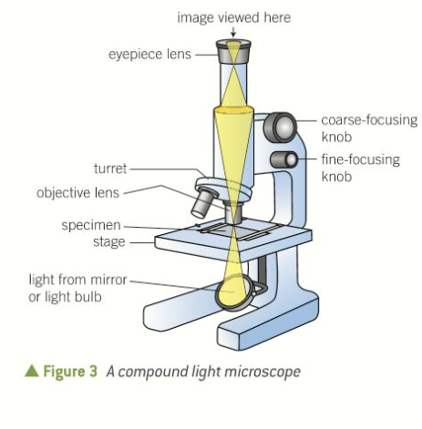
how many lenses does a compound light microscope have?
what does the objective/eyepiece lens configuration allow for?
2 (objective - placed near to specimen, eyepiece - through which specimen is viewed). objective produced magnified image, which is magnified again by the eyepiece
allows for a much higher mag + reduced chromatic aberration than that in simple microscope
what usually provides illumination?
a light underneath the sample
opaque specimens can be illuminated from above w some microscopes
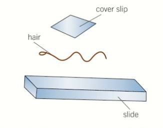
DRY MOUNT sample prep
solid specimens, whole or cut into v thin slices w sharp blade (called sectioning)
placed on centre of slide + cover slip over sample
eg hair, pollen, dust, insect parts can be viewed whole in this way
eg muscle tissue, plants can be sectioned + viewed in this way
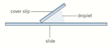
WET MOUNT sample prep
specimens suspended in a liquid (eg water or an immersion oil)
cover slip placed on from an angle
eg aquatic samples or other living organisms can be viewed in this way
SQUASH SLIDES sample prep
wet mount first prepared
lens tissue used to gently press down the cover slip
depending on material, potential damage to cover slip can be avoided by squashing sample between 2 microscope slides
good technique for soft samples
care needs to be taken that cover slip is not broken when being pressed
eg. root tip squashes used to look at cell division
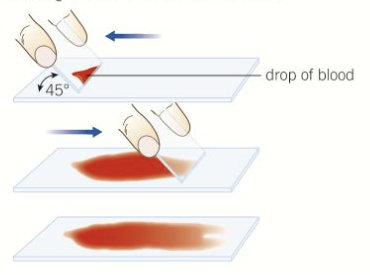
SMEAR SLIDES sample prep
edge of a slide used to smear sample, creating thin, even coating on another slide
cover slip then placed over sample
example of a smear slide - sample of blood
good way to view cells in blood
how is the sample illuminated in basic light microscopy?
what are the properties of the images and why?
how is the resolution?
what is diffraction?
from below with white light + observed from above (brightfield microscopy)
whole sample illuminated at once (wide-field microscopy)
images tend to have low contrast as most cells dont absorb a lot of light
resolution limited by wavelength of light and diffraction of light as it passes through the sample
the bending of light as it passes close to the edge of an object
what is the cytosol of a cell?
what parts of the cell are often transparent?
what do stains do?
how do u prepare a sample for staining?
aqueous interior
the cytosol and other cell structures
increase the contrast as diff components within a cell take up stains to diff degrees. this allows components to become visible so can be identified
place sample on slide + allow to air dry, then its heat-fixed by passing through flame, specimen will adhere to microscope slide n will take up stains
crystal violet / methylene blue
dyes such as nigrosin / congo red
positively charged dyes, r attracted to negatively charged materials in cytoplasm leading to staining of cell components
negatively charged, repelled by negatively charged cytosol. dyes stay outside cells, leaving cells unstained, which then stand out against stained background. negative stain technique
what can differential staining do?
distinguish between 2 types of organisms that would otherwise be hard to identify
can also differentiate between diff organelles of a single organism within a tissue sample
what is gram stain technique used for?
steps
to separate bacteria into 2 groups: gram-positive bacteria, gram-negative bacteria
crystal violet first applied to a bacterial specimen on slide
then iodine which fixes due
slide washed w alcohol
GP retain crystal violet stain → blue / purple under microscope
GN thinner cell walls so lose stain
then stained w safranin dye (counterstain) → appear red
GP susceptible to penicillin (antibiotic) → inhibits formation of cell walls
GN much thinner cell wall so not susceptible to pencillin
what is acid-fast technique used for?
what is a lipid solvent used for?
steps
to differentiate species of mycobacterium from other bacteria
to carry carbolfuchsin dye into the cells being studied
cells then washed w dilute acid-alcohol solution
mycobacterium X affected by the acid-alcohol + retain carbolfuchsin stain (bright red)
other bacteria lose stain + exposed to methylene blue stain (blue)
the 4 stages in the production of slides (pre-prepared)
fixing - chemicals like formaldehyde used 2 preserve specimens in as near-natural a state as possible
sectioning - specimens dehydrated w alcohols n placed in mould w wax or resin 2 form hard block. then can be sliced thinly w knife called a microtome
staining - specimens often treated w multiple stains to show diff structures
mounting - specimens then secured 2 a microscope slide n cover slip placed on top
magnification
how many times larger the image is than the actual size of the object being viewed
(interchangeable objective lens on compound light microscope allow user to adjust)
resolution definition
what is it limited by?
the ability to see individual objects as separate entities
the diffraction of light as it passes through the samples (n lenses)
diffraction
tendency of light waves to spread as they pass close to physical structures such as those present in the specimens being studied
(structures present in specimens v close to eachother n light reflected from individual structures can overlap due to diffraction → no longer seen as separate entities and detail lost)
what structures cannot be seen separately (resolved) in optical microscopy?
structures that are closer than 1/2 the wavelength of light
how can resolution be increased?
why does this work?
by using beams of electrons which have a wavelength 1000s of times shorter than light
electron beams are still diffracted but shorter wavelength means that indiv beams can be much closer b4 they overlap → objects which r much smaller n closer tg can be seen separately without diffraction blurring image
what do you have to use to measure the size of a sample?
why do you have to use this?
an eyepiece graticule
true mag of the diff lenses of a microscope can vary slightly from the mag stated, so every microscope + every lens has to be calibrated individually using eyepiece graticule and slide micrometer
what is an eyepiece graticule?
what increases w each increase in mag?
glass disc marked w a fine scale of 1 to 100
no units on scale
remains unchanged whichever obj.lens is in place
relative size of the divisions
what is a stage micrometer + what is it used for?
to calibrate the scale on the graticule
microscope slide w a very accurate scale in micrometres engraved on it
usually 100 divisions = 1mm so 1 division = 10micrometres
what happens if you measure the same cell once all 3 lenses are calibrated?
you should get the same acc measurement each time
what is the limiting factor in light microscopy?
resolution
increase mag can be achieved easily using appropriate lens, but if image blurred, no more detail will be seen
what is used to illuminate the specimen in electron microscopy?
why can more detail of cell ultrastructure be seen?
what types of images can be produced?
a beam of electrons w wavelength <1nm
electrons have a much smaller wavelength than light waves
images w mags up to x500,000 + still have clear resolution
disadvantages to electron microscopes
expensive pieces of equiptment
can only be used inside carefully controlled environment in dedicated space
specimens can be damaged by electron beam
bc prep process is v complex, is a problem w artefacts (structures that are produced due to the prep process)
what does TEM stand for?
how does it work?
what is its resolving power?
transmission electron microscope
beam of electrons transmitted through a specimen + focused to produce an image
similar to light microscopy
best resolution, resolving power of 0.5nm
what does SEM stand for?
how does it work?
what is its resolving power?
scanning electron microscope
beam of electrons is sent across the surface of a specimen + reflected electrons collected
resolving power 3-10nm, so resolution not as good as TEM
stunning 3 dimensional images of surfaces produced → valuable info about appearance of diff orgamisms
what is the inside of an electron microscope like? why?
what does specimen prep involve bc of this?
vacuum - ensure electron beams travel in straight lines
fixation using chemicals or freezing
staining w heavy metals
dehydration w solvents
samples for a TEM then set in resin + may be stained again
samples for a SEM may be fractured to expose inside + will need to be coated w heavy metals
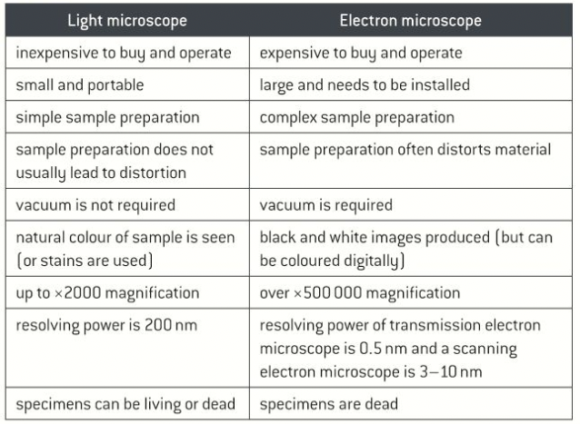
light vs electron microscope
cost, size, sample prep, distortion, vacuum, colour, mag, resolving power, specimens
what is an artefact?
what type of microscopy do they appear in?
example of artefact: the bubbles that get trapped under…
a visible structural detail caused by processing the specimen + not a feature of the specimen
both light + electron
… the cover slip as u prep a slide 4 light microscopy
what is inevitable when preparing specimens 4 electron microscopy?
examples
changes in the ultrastructure of cells during the processing that samples must undergo
loss of continuity in membranes, distortion of organelles, empty spaces in cytoplasm of cells
what is mesosome?
name given to invaginations (inward foldings) of cell membranes that were observed using electron microscopes after bacterial specimens had been chemically fixed
what do conventional optical microscopes use to illuminate specimens?
what do they use to produce a magnified image?
visible light
a lens
what is used in fluorescent microscopes to illuminate a specimen?
what is fluorescence?
a higher LI that has been treated w a fluorescent chemical (fluorescent ‘dye’)
absorption + re-radiation of light
light of longer wavelength + lower E emitted n used to produce magnified image
what does a laser scanning confocal microscope do?
what does this cause?
moves a single spot of focused light across a specimen (point illumination) → 2D image
3D can be produced by creating images @ diff focal planes
causes fluorescence from the components labeled w a ‘dye’
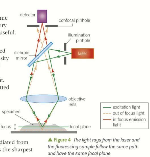
in a laser scanning confocal microscope, through what is the emitted light from the specimen filtered?
which light is detected?
what would reduce the resolution and cause blurring? how is this prevented?
what is used to get higher intensities, improving illumination?
a pinhole aperture
only light radiated from very close to the focal plane (the distance that gives the sharpest focus)
light emitted from other parts of the specimen. does not pass through pinhole → not detected
a laser instead of light
why can very high resolution images be obtained from LSCM?
thin sections of specimen is examined n light from elsewhere is removed
4 facts about LSCM
non-invasive
currently used in diagnosis of diseases in eye
being developed for use in endoscopic procedures
also used in development of new drugs (bc can be used to see distribution of molecules within cells)
what do the future uses 4 advanced optical microscopy include?
virtual biopsies, particularly in case of suspected skin cancer
what is the beamsplitter?
dichroic mirror
only reflects one wavelength (from laser) but allows other wavelengths (produced by sample) to pass through
what do the positions of the 2 pinholes mean? (LSCM)
what does this mean?
light waves from laser (illuminating sample) follow same path as light waves radiated when sample fluoresces
will both have the same focal plane (hence term confocal)
why do we use antibodies w fluorescent ‘tags’?
target specific features → studied by confocal microscopy w much omre precision than when using staining + light microscopy
what is GFP?
what does it do?
what have GFP molecules been engineered to do?
green fluorescent protein
emits bright green light when illuminated by UV light
fluoresce diff colours, meaning diff components of specimen can be studied at same time
what happens to the gene for GFP?
what does the fluoresce indicate?
examples of things that have been modified to express this gene + fluoresce
what does the use of these fluorescing proteins provide?
gene 4 this protein isolated + can be attached, by genetic engineering, to genes coding 4 proteins under investigation
a protein is being made + is used to see where it goes within cell or organism
bacteria, fungal, plant + human cells
a non-invasive technique to study the production + distribution of proteins in cells + organisms