Week 4 - Muscle excitation & contraction coupling
1/10
There's no tags or description
Looks like no tags are added yet.
Name | Mastery | Learn | Test | Matching | Spaced | Call with Kai |
|---|
No analytics yet
Send a link to your students to track their progress
11 Terms
Skeletal muscles overview
Function: Maintain body temp (heat production), movement & metabolism
All skeletal muscles are connected to bones via tendons.
under voluntary control
Can only be stimulated by an action potential arriving from a somatic motor neuron
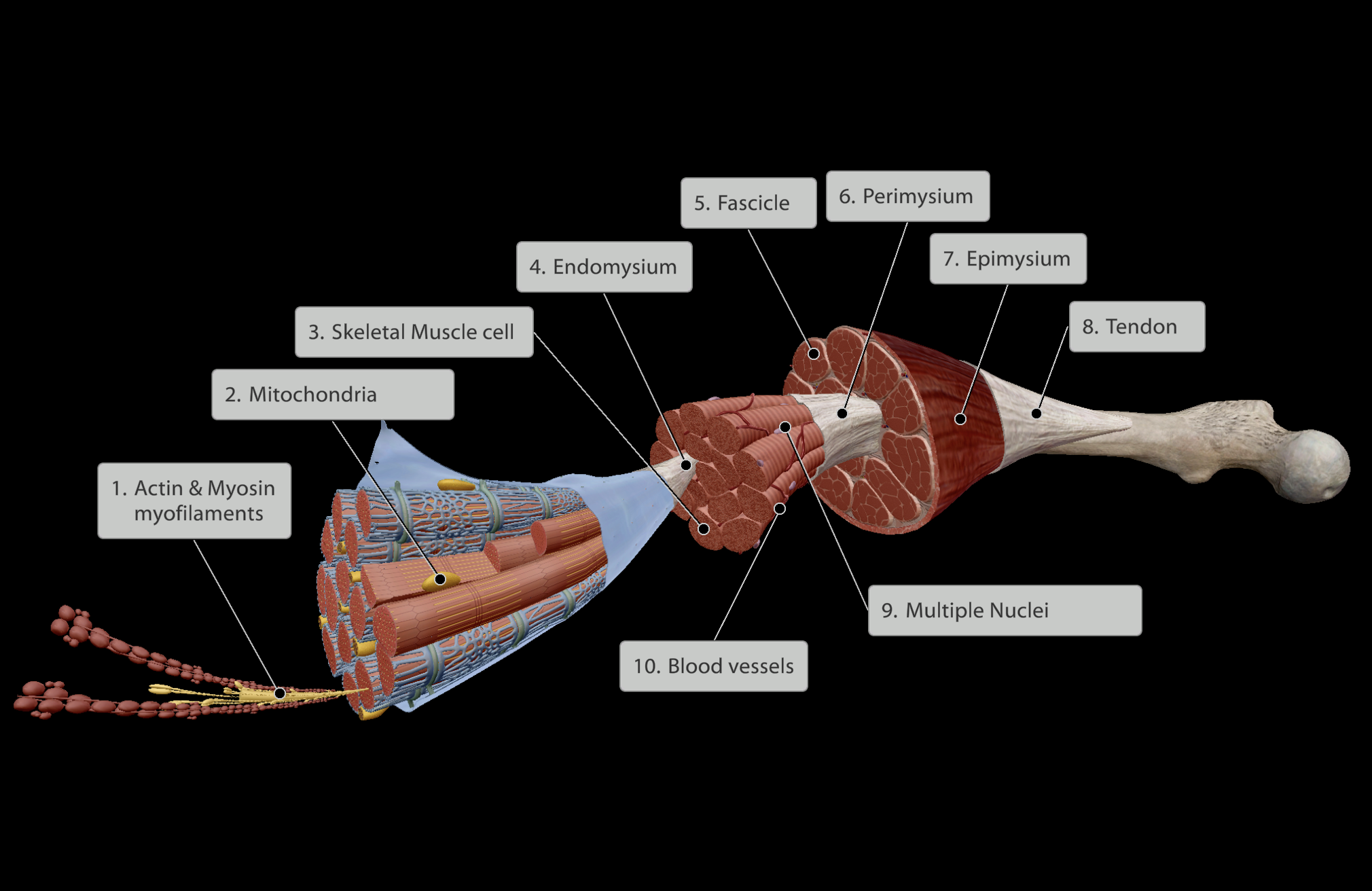
Skeletal muscles, subdivisions
Levels of the Muscle Belly (Inner to Outer):
Actin & Myosin myofilaments within skeletal muscle cell
Skeletal Muscle cell/skeletal muscle fibre
Endomysium - Connective tissue surrounding skeletal muscle cell
Fascicle - Groups of skeletal muscle cells each wrapped in endomysium
Perimysium - Connective tissue surrounding each fascicle
Epimysium - Connective tissue surrounding all fascicles in muscle belly
Tendon - Joining point of all inner connective tissue (endomysium, perimysium, epimysium) that links muscle to bone.
a. Dense fibrous regular connective tissue.
b. Links muscle directly into bone
All muscle is highly vascularised, however the tendons at the end of muscles have less blood supply.
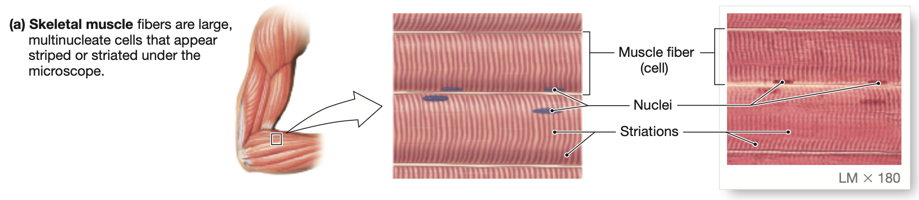
Why are skeletal muscles striated?
All skeletal muscles are striated. Each skeletal muscle cell has alternating dark and light bands.
Each muscle cell is multinucleated, and spans the length of the ENTIRE muscle.
Hence, can be the largest cells in the body
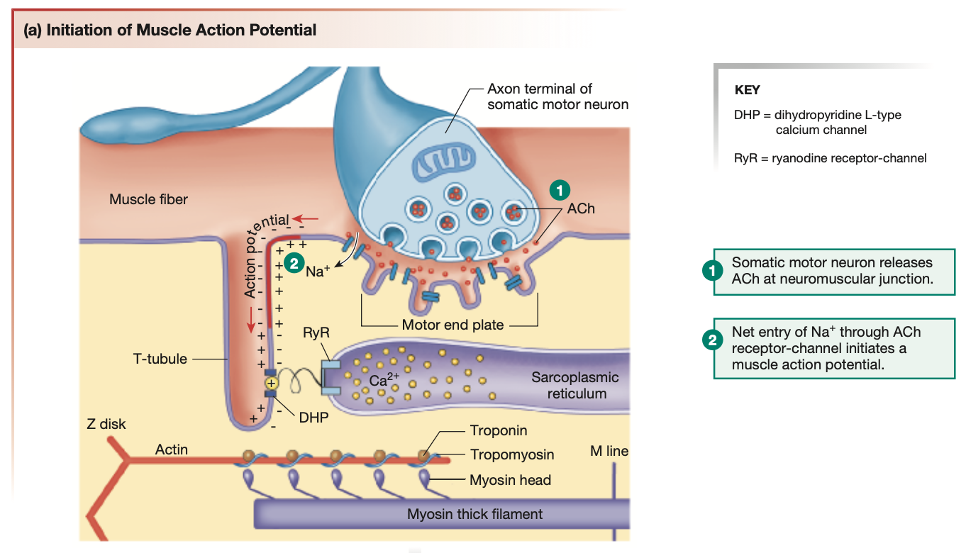
Skeletal muscle CONTRACTION process: Somatic motor neuron
Action potential travels down to axon terminal of somatic motor neuron
Due to processed (refer to previous notes), Ca2+ voltage-gated ion channels open, allowing Ca2+ to rush into the neuron’s axon terminal
Ca2+ sensing protein “synaptotagmin” found on the surface of vesicles within the axon terminal activate
These vesicles containing neurotransmitters (in muscle contractions, Ach), fuse with the axon terminal’s cell membrane
Ach is released into the neuromuscular junction via exocytosis.
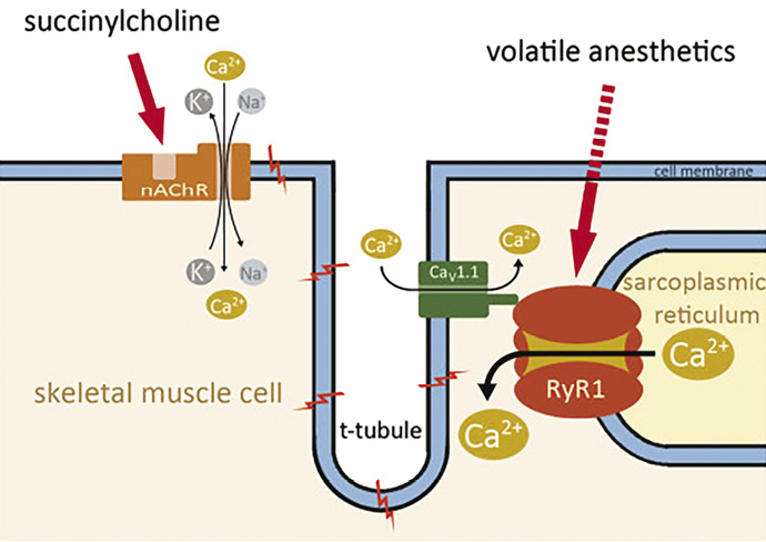
Ach binds to receptors on the chemically-gated/ligand-gated Na+ channels on skeletal muscle cells.
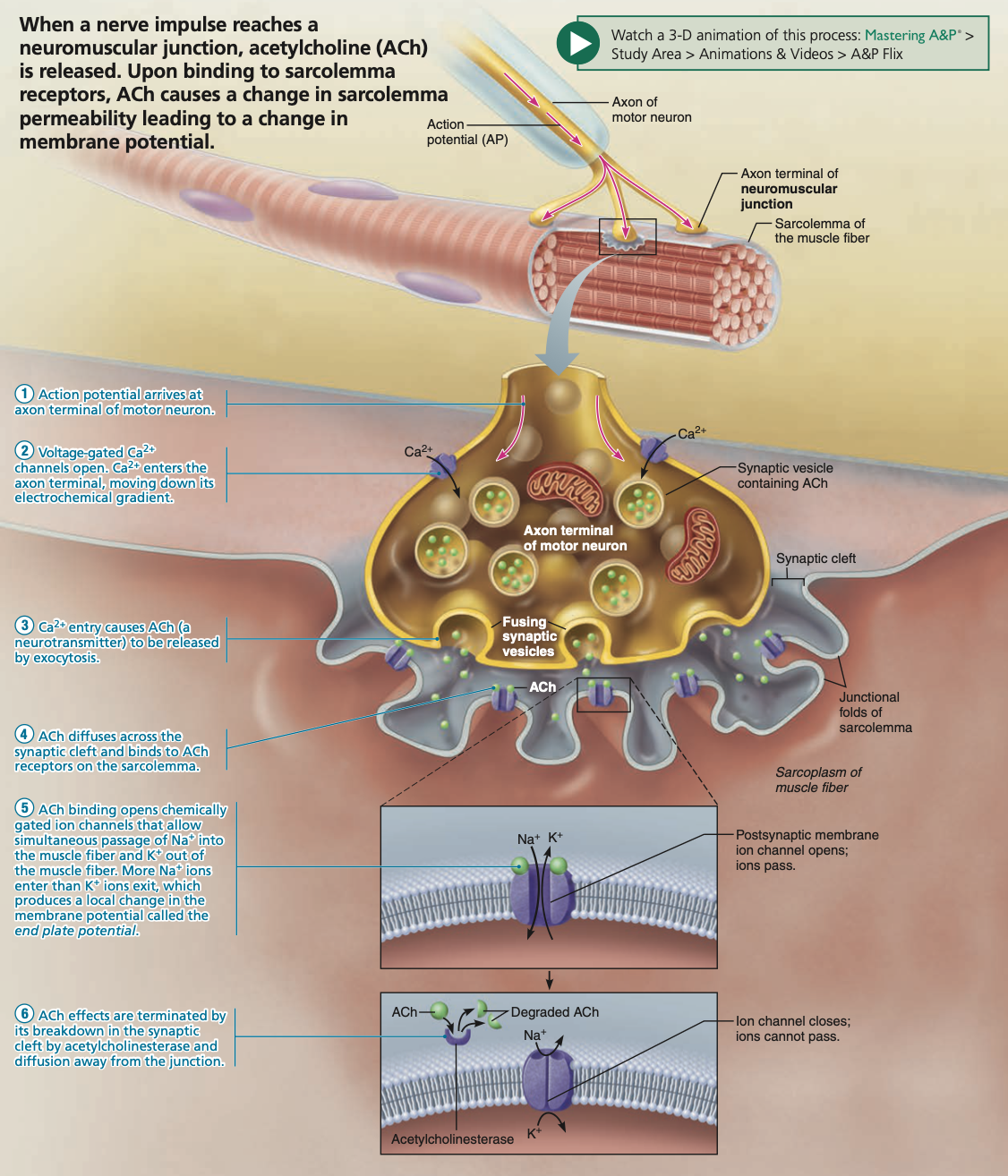
Skeletal muscle CONTRACTION process: Sarcolemma
Opening these chemically-gated/ligand Na+ channels (activated by Ach) allows Na+ to flow down the concentration gradient, into the muscle cell.
Depolarisation of the skeletal muscle cell membrane/surface (called ‘sarcolemma’) occurs.
Moving away from the motor end-plate/neuromuscular junction, chemically-gated Na+ channels, activated by Ach, become voltage-gated Na+ channels, enabling depolarisation to occur.
Action potentials propagate in all directions from the neuromuscular junction
Action potentials travel down into ridges/tunnels on the sarcolemma, called T-tubules.
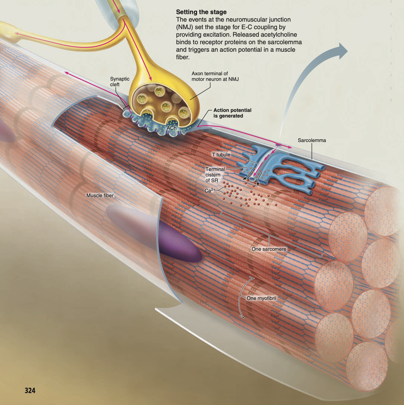
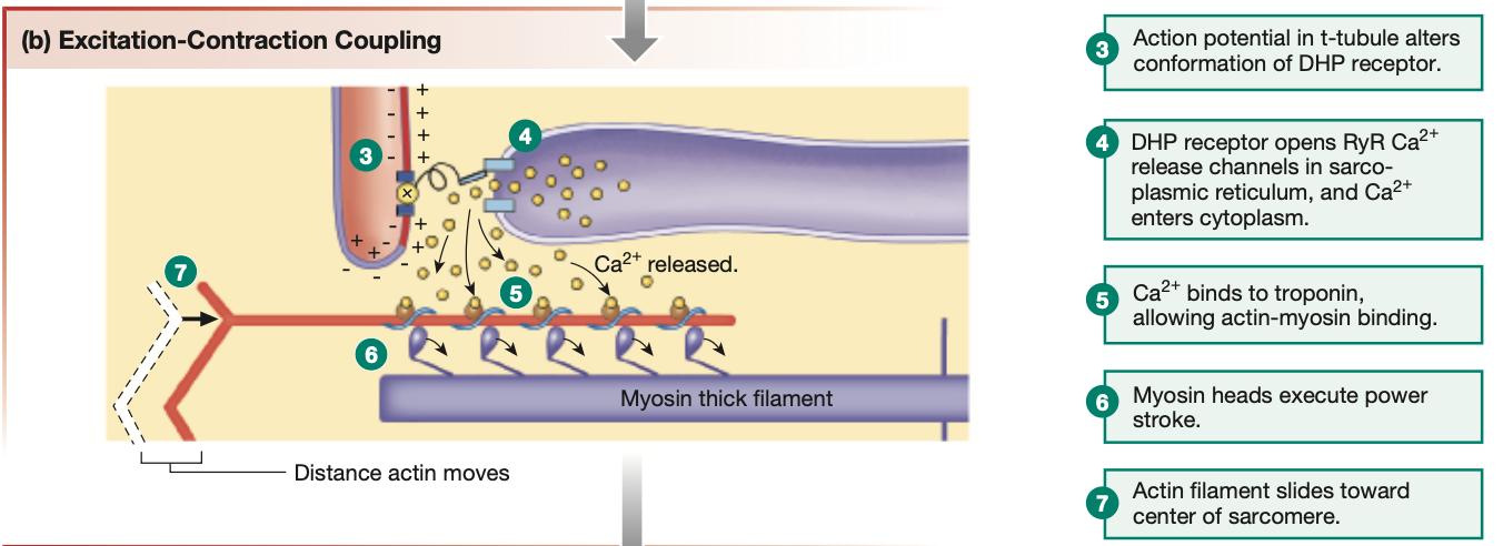
Skeletal muscle CONTRACTION process: T-tubules
At the base of T-tubules, are complexes called DHP (dihydropyridine L-type
calcium channel). DHPs are voltage receptors, hence respond to the depolarisation of the sarcolemma.
DHPs are physically connected to RyR channels (ryanodine receptor-channel). Hence, when DHPs are activated, RyR channels follow suit.
RyR channels are found gating the sarcoplasmic reticulum found within the muscle cell.
Skeletal muscle CONTRACTION process: Sarcoplasmic reticulum
RyR channels are physically pryed open, letting the contents of the sarcoplasmic reticulum flow into the skeletal muscle cell. (RyR channels are mechanically-gated ion channels)
The sarcoplasmic reticulum contains Ca2+ stores.
Hence, when RyR channels open, Ca2+ rushes into the skeletal muscle cell.
The more Ca2+ released by the sarcoplasmic reticulum, the more force produced in a movement
Ca2+ that floods the skeletal muscle cell binds to troponin on actin myofilaments
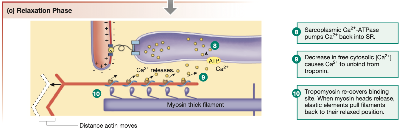
Skeletal muscle RELAXATION process
Action potentials stop being send down the somatic motor neuron
Ach is no longer released as a neurotransmitter, and Ach is removed from chemically-gated/ligand-gated Na+ channels on the sarcolemma.
Sarcolemma repolarises, voltage-gated Na+ channels close.
T-tubules repolarises
DHP no longer senses a voltage change in the T-tubule, hence DHP inactivated & RyR channel physically closes.
Sarcoplasmic reticulum is closed, Ca2+ stops flooding the skeletal muscle cell
Ca2+ already bound to troponin on actin myofilaments is released
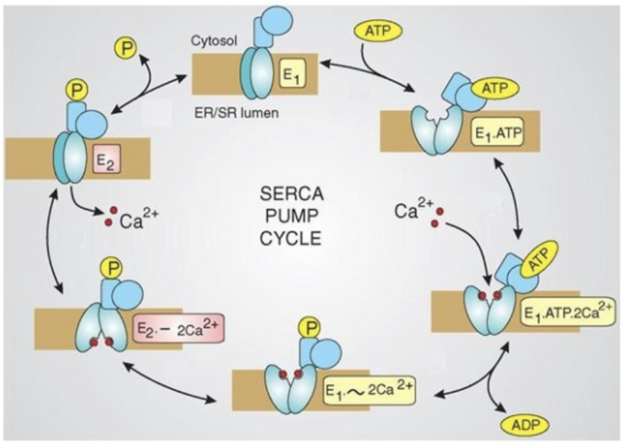
SERCA pumps (Sarcoplasmic/Endoplasmic Reticulum—Calcium ATP pump) reintakes Ca2+ into the sarcoplasmic reticulum at the cost of ATP.
Muscle cramps
Occurs when somatic motor neurons continuously send action potentials, leading to continuous propagation of Ach neurotransmitters, leading to muscles continuously staying contracted.
Can lead to permenant muscle damage
EXTRA KNOWLEDGE: SERCA pumps & Enzyme function
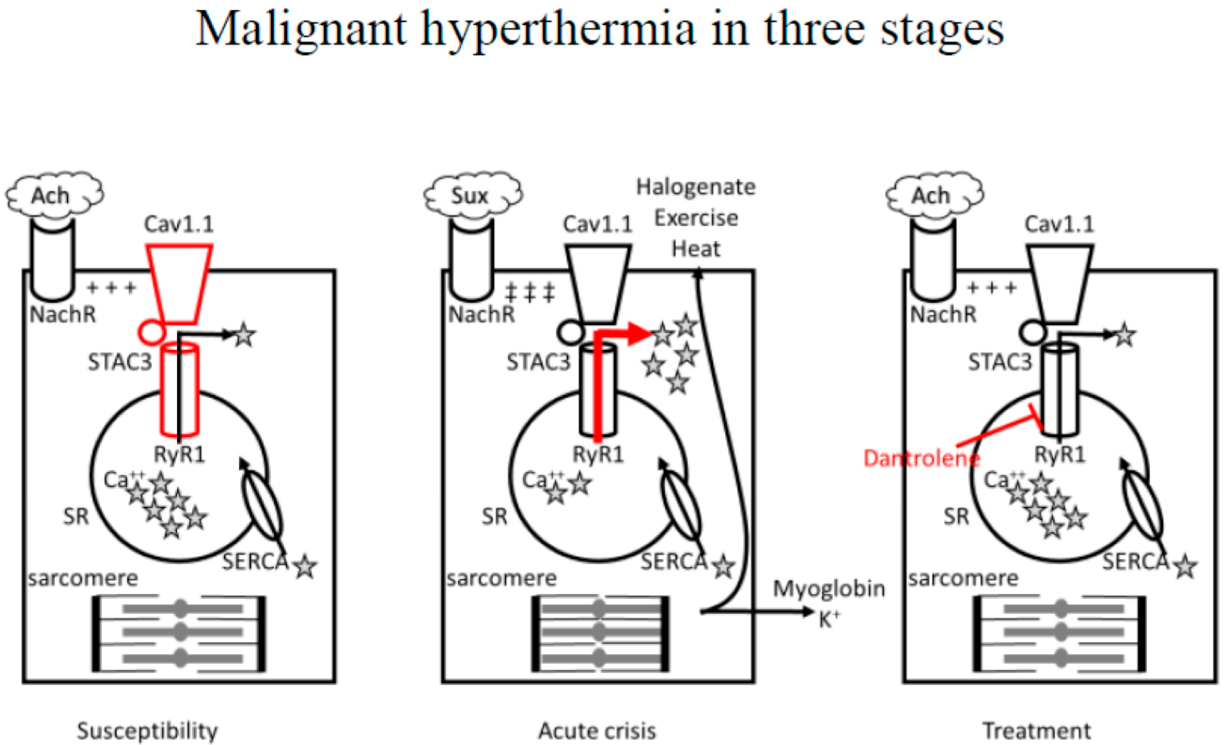
EXTRA KNOWLEDGE: Malignant Hyperthermia
Malignant Hyperthermia - Genetic condition where abnormal RyR receptors remain open longer than normal, allowing excess Ca2+ to be released from the sarcoplasmic reticulum.
Excess Ca2+ in the cytoplasm, turnover by SERCA pumps cannot catch up, leading to Ca2+ remaining bound to troponin for longer than necessary.
Thus, myosin heads remain clamped onto actin filaments for long periods of time
Muscle remains contracted for an extended period of time - ensuing muscle cramps
SERCA pumps work overtime (consuming lots of ATP → ADP + Pi), releasing heat in the catabolic reaction
Muscle cells/fibres can melt from the heat
Also excess Ca2+ triggers enzymes that cause muscle-breakdown (rhabdomyolysis) - resulting in dark/red urine
If left untreated, can lead to kidney failure or even death
Often found during surgery, as certain anaesthetics can trigger the RyR channels directly
Muscle relaxant Succinylcholine can also trigger MH