Intraoral Maxillary Anatomy
1/78
There's no tags or description
Looks like no tags are added yet.
Name | Mastery | Learn | Test | Matching | Spaced |
|---|
No study sessions yet.
79 Terms
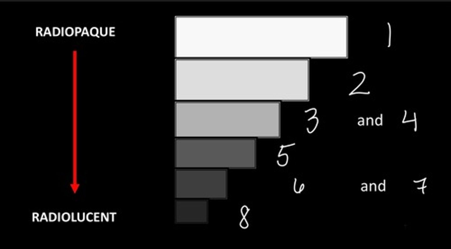
radiolucent structures
that interact (absorbed or scattered by the material.)‘ less with x-rays, meaning they let more X-rays pass through them
example of radiolucent structures
soft tissues and air lke
radiopaque structures
structures that interact more with x-rays
example of radiopaque structures
dental tissues, bone
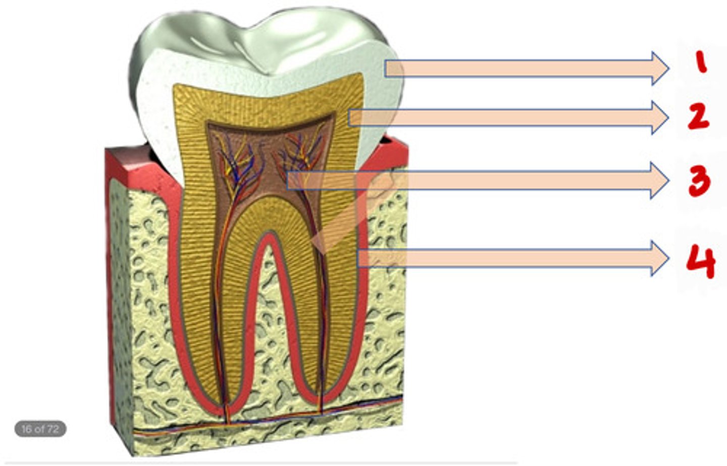
enamel
1

dentin
2

pulp cavity (camber and canals)
3

cementum
4
alveolar crest
1

periodontal ligament space
2

lamina dura
3
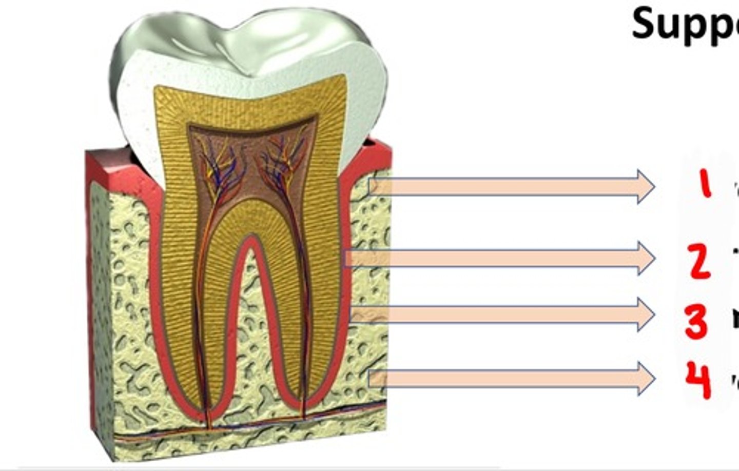
alveolar bone
4

enamel
1

cortical bone
2

dentin
3

cement
4

alveolar bone
5

pulp chamber
6

canals
7

periodontal ligament space
8
enamel description
densest structure found in the human body
enamel radiographic
outermost radiopaque layer of the crown of a tooth on maxiallary
dentin description
-is found beneath the enamel layer of a tooth and surrounds the pulp cavity
-makes up the majority of the tooth structure
dentin radiographic informaion
appears radiopaque, but not as radiopaque as enamel
cementum description informaion
is not usually apparent radiographically because the contrast between it and dentin is so low and the cementum layer is so thin
hypercementosis
pulp chamber and root canals description
contains blood vessels, nerves, and lymphatics
pulp chamber and root canals radiographic informaion
appears relatively radiolucent
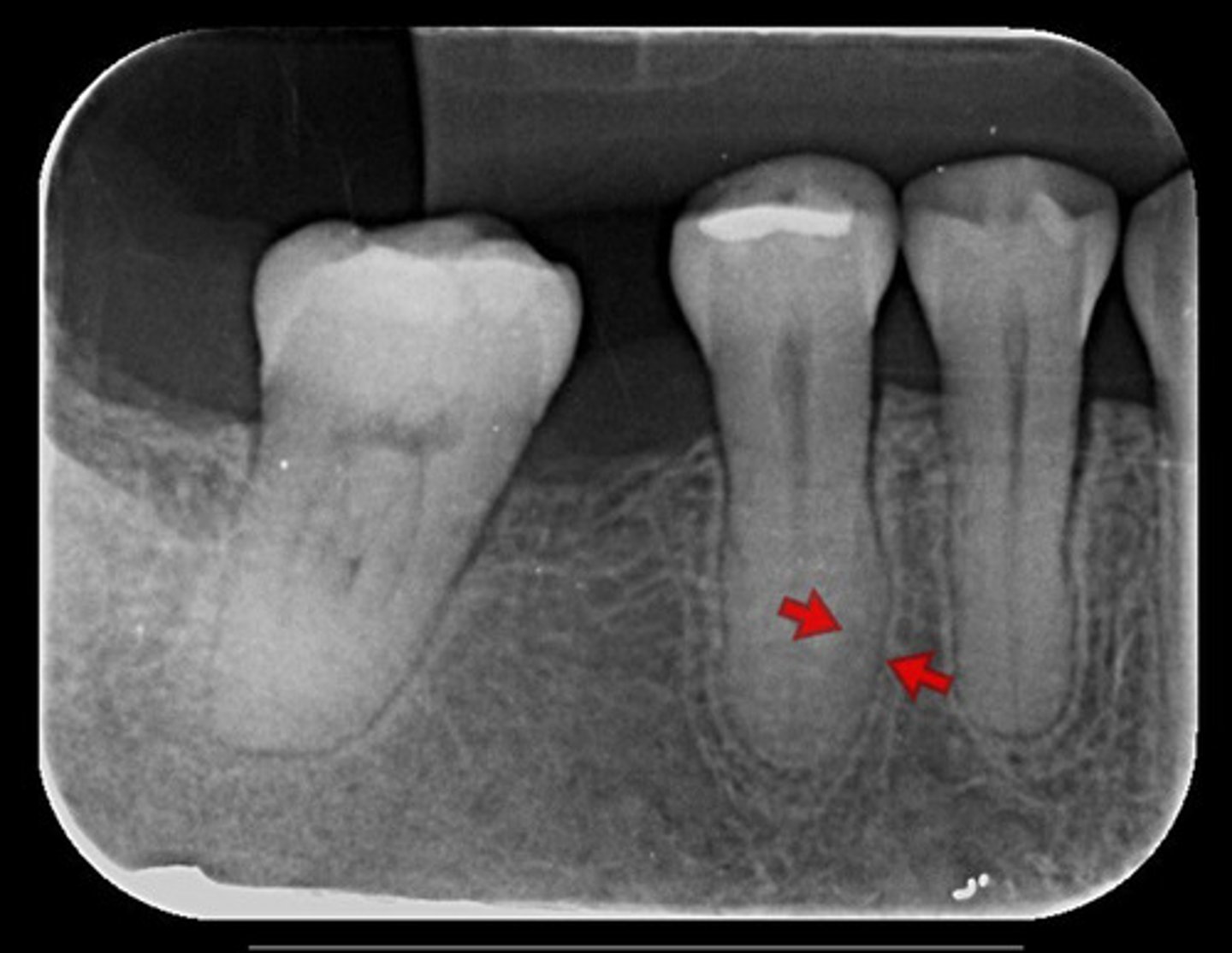
periodontal ligament space description
-space between the root of the tooth and the lamina dura
-contains connective tissue fibers, blood vessels, and lymphatic ducts
periodontal ligament radiographic informaion
-a thin radiolucent line around the root of a tooth
-in the healthy periodontium: continuous radiolucent line of uniform thickness
lamina dura description
-wall of the tooth socket that surrounds the root of a tooth
-made up of dense cortical bone
lamina dura radiographic informatipn
dense radiopaque line that surrounds the root of a tooth
alveolar crest description
-most coronal portion of alveolar bone found between teeth
-made up of dense cortical bone and is continuous with the lamina dura
alveolar crest radiographic infomation
appears radiopaque and is typically located 1.5 to 2mm below the junction of the crown and the root surfaces (the cemento-enamel junction)
alveolar bone description
supports and encases the roots of teeth
alveolar bone radiographic informaion
trabeculae and medullary spaces
trabeculae informaion
radiographic line
medullary spaces informaion
radiolucent areas
dense cortical bone
1
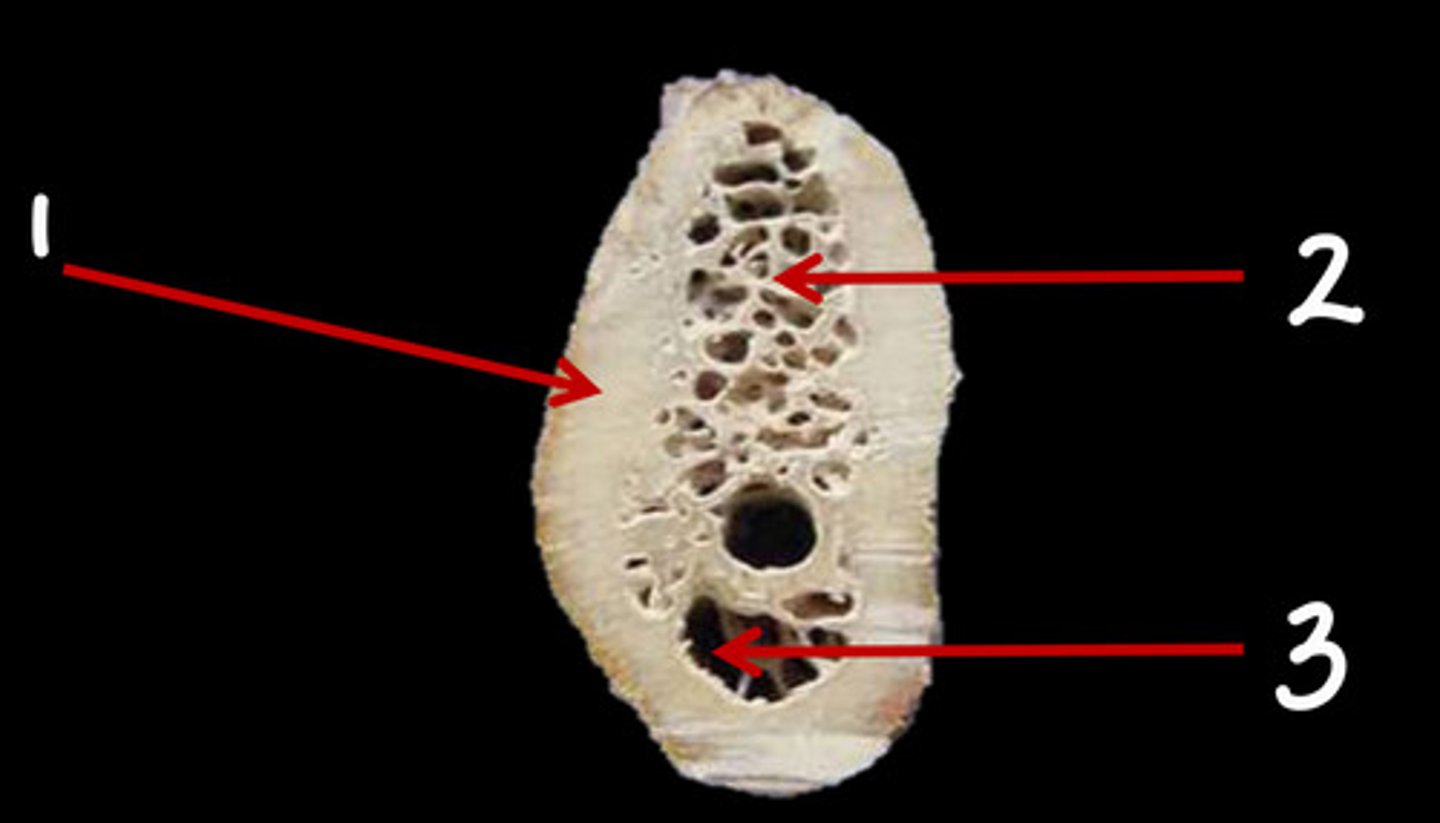
trabeculae
2

medullary spaces
3

dental papilla space radiogrpahic informaion
radiolucent region at the apex of teeth in formation
dental follicle space radiographic informaion
radiolucent area between the crown of an unerupted tooth and the surrounding bone
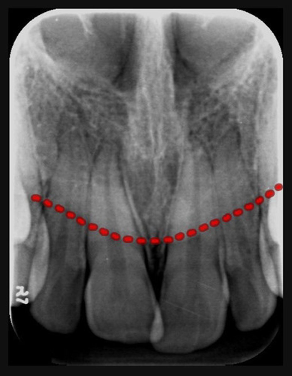
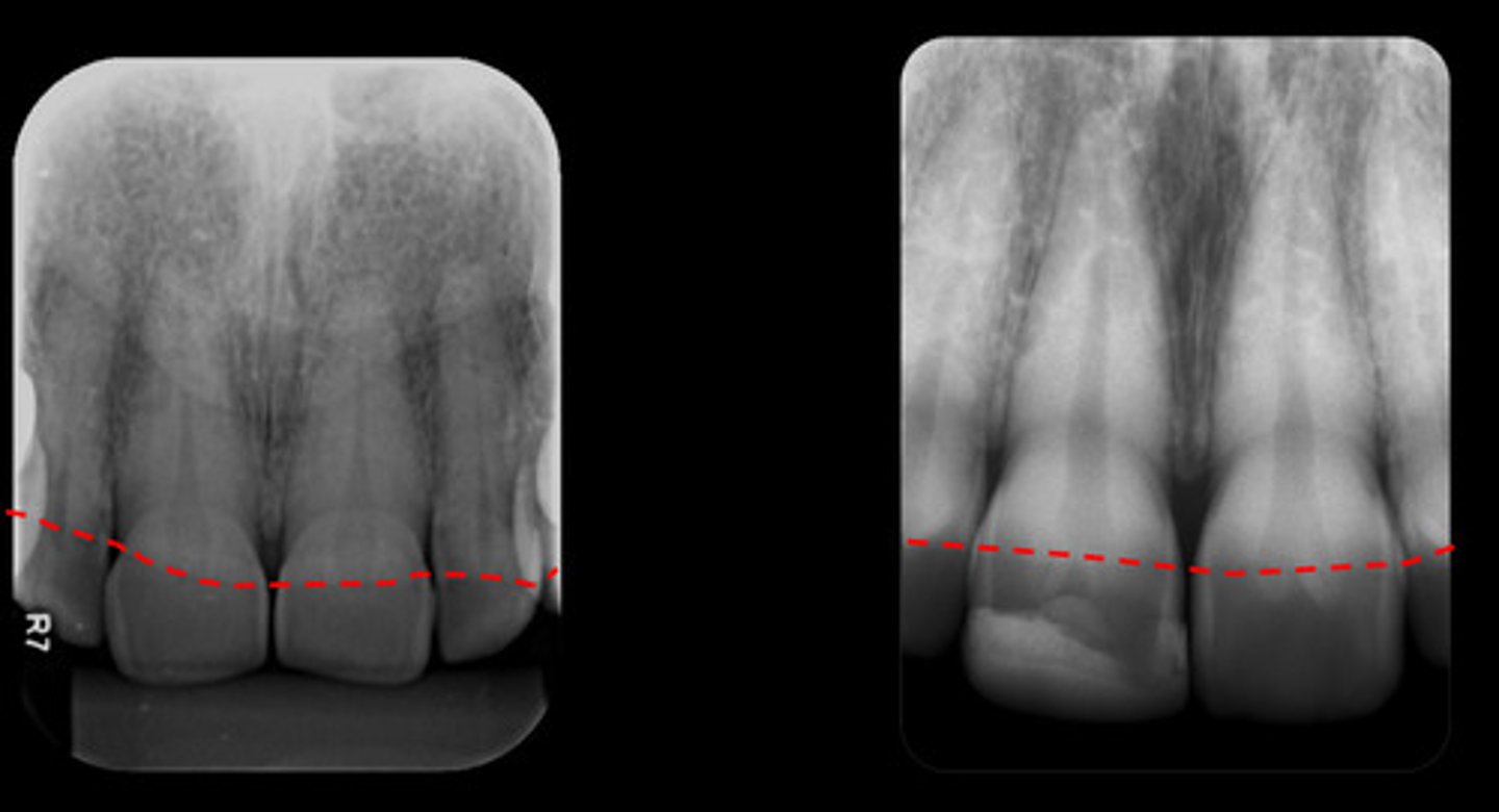
intermaxillary suture description
suture present between the palatine processes and the anterior portion of the maxilla
intermaxillary suture radiographic appearance informaion
radiolucent line, in the midline, between the central incisors
incisive foramen description
exit of the nasopalatine canal
incisive foramen radiographic informaion
ovoid radiolucency, often with diffuse borders
nasal cavity-nasal fossae description
bone cavities filled with air divided by the nasal septum
nasal cavity-nasal fossae radiographic informaion
radiolucent area
inferior nasal conchae description
wafer-thin, curved plates of bone that extend from the lateral walls of the nasal cavity
inferior nasal conchae radiographic informaion
radiopaque structure extending from the lateral walls of the nasal cavity
nasal septum description
divides the nasal cavity into two fossae
nasal septum radiographic informaion
vertical radiopaque structure
floor of the nasal cavity radiographic
radiopaque line located in the lower portion of the nasal cavity (in the projection of incisors and canines)
anterior nasal spine description
bony projection formed by the fusion of the two maxillary bones at the intermaxillary suture
anterior nasal spine radiographic
radiopaque V-shaped line
tip of the nose
upper lips
naso-labial fold
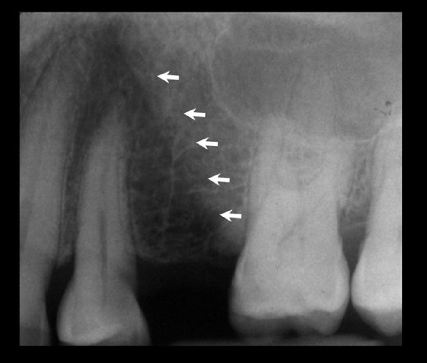
lateral fossa radiographic
radiolucent area located between the canine and the lateral incisor
maxillary tuberosity description
the most posterior region of the alveolar process of the maxilla
maxillary tuberosity radiographic
bone radiopacity
pterygoid hamulus description
hook-like process at the lower part of the medial pterygoid plate of the sphenoid bone
pterygoid hamulus radiographic
radiopaque hook-like image
maxillary sinus radiographic
wide radiolucent area bounded by a radiopaque line
floor of the maxillary sinus radiographic informaion
thin radiopaque line located in the lower portion of the maxillary sinus
alveolar extension of the maxillary sinus pneumatization radiographic information
radiolucent areas that extend towards the alveolar ridge
tuberosity extension of the maxillary sinus radiographic informaion
radiolucent areas that extend towards the maxillary tuberosity
anterior extension of the maxillary sinus radiographic informaion
radiolucent areas that extend anteriorly
Y line of Ennis (inverted Y) description
superimposition of the floor of the nasal fossa and the anterior border of the maxillary sinus
Y line of Ennis radiographic informaion
radiopaque line in inverted "Y" format
more x-rays of Y line of Ennis

nutrient canals
anatomic structures of the alveolar bone through which neurovascular elements transit
zygomatic process of maxilla description
bony projection of the maxilla that articulates with the zygomatic bone
zygomatic process of maxilla radiographic information
J-shaped or U-shaped radiopacity located superior to the maxillary first molar region
zygomatic bone description
-articulates with the zygomatic process of the maxilla
-is composed of dense cortical bone
zygomatic bone radiographic informaion
appears as a diffuse radiopaque band extending posteriorly from the zygomatic process of the maxilla
coronoid process of the mandible description
marked prominence of bone on the anterior ramus of the mandible
coronoid process of the mandible radiographic informaion
triangular radiopacity superimposed over, or inferior to, the maxillary tuberosity region