SPINAL CORD AND REFLEXES- JONES
1/28
There's no tags or description
Looks like no tags are added yet.
Name | Mastery | Learn | Test | Matching | Spaced | Call with Kai |
|---|
No analytics yet
Send a link to your students to track their progress
29 Terms
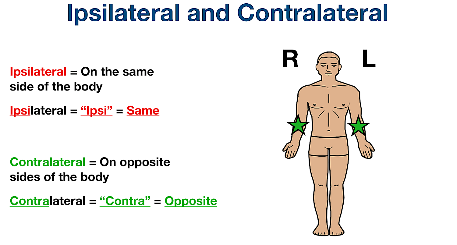
What does Ipsilateral mean?
What does Contralateral mean?
Ipsi= same side
Contra= opposite side
What does “tract” mean?
bundle of nerves
What does decussation mean?
crossing/where nerves cross
What’s the difference between cranial and caudal?
cranial- head
caudal- away from head
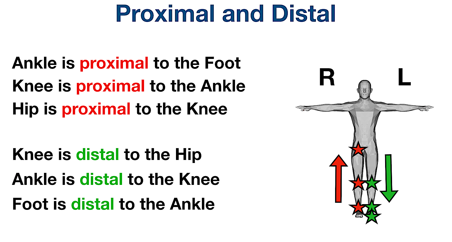
What’s the difference between distal and proximal?
distal- away from torso
proximal- close to torso
What’s the difference between medial and lateral?
medial- close to midline
lateral- away from midline
What’s the difference between anterior and posterior?
anterior- front
posterior- back
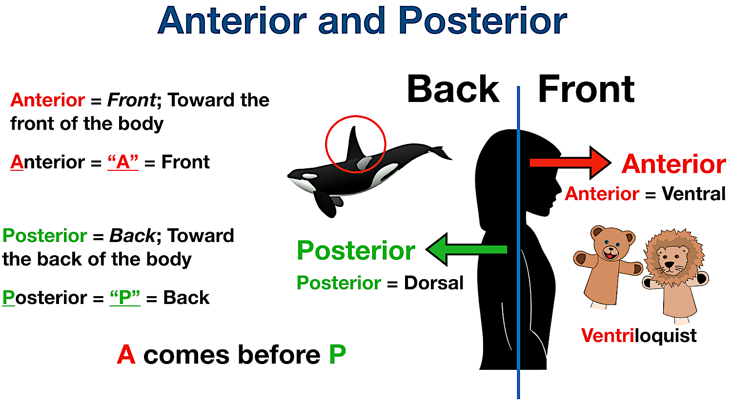
What’s the difference between ventral and dorsal?
ventral- front
dorsal- back

What are the functions of the spinal cord?
conduction (action potentials)
neural integration (respond to stimuli)
locomotion (what signals allow us to move)
reflexes
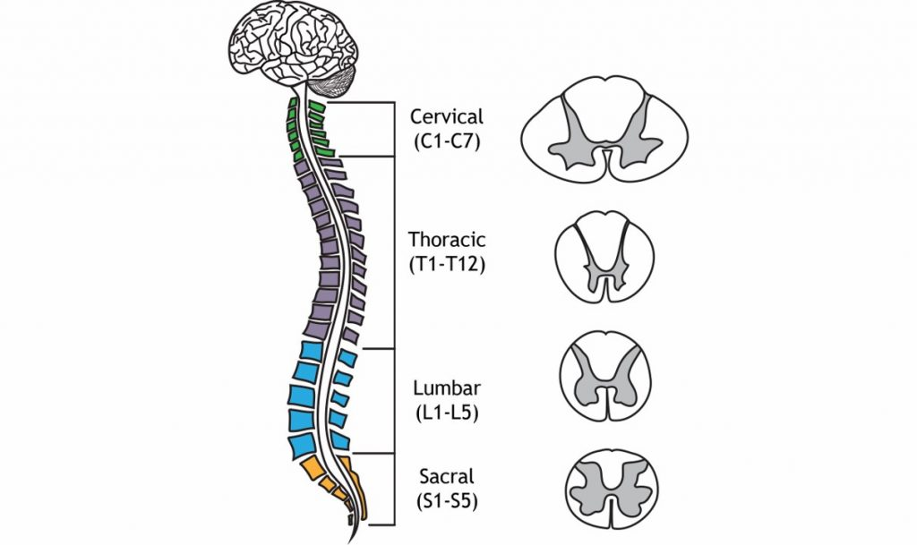
What are the anatomical regions of the spinal cord?
(jones- “know general progression”)
cervical
thoracic
lumbar
sacral
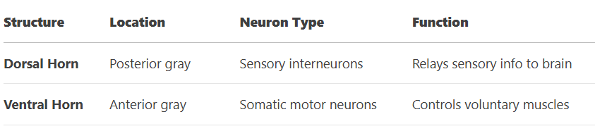
The grey matter of spinal cord is made up of the dorsal and ventral horn.
What’s their function? What do they send/receive?
dorsal- RECEIVES sensory input (from dorsal root ganglion)
ventral- CARRYS motor signals
PRACTICE:
Damage to the dorsal root would cause what deficit?
sensory loss (in that region where the ganglion was damaged)
Describe the white matter of the spinal cord:
axons carrying information to and from the brain
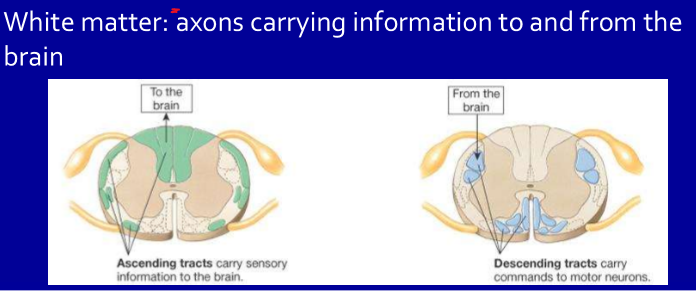
The spinal cord carries sensory information from the body to the brain via _______________________.
a. ascending tracts
b. descending tracts
a.
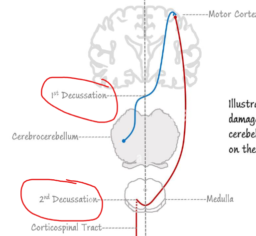
The corticospinal tract is the major pathway for voluntary motor control, carrying signals from the brain to muscles or via _______________________.
a. ascending tracts
b. descending tracts
b.
There are 2 major ascending spinal tracts involved with sensory perception.
The dorsal column-medial lemniscal carries what sensory information?
The spinothalamic system carries what sensory information?
dorsal column-medial lemniscal: touch, pressure, movement
spinothalamic system: pain, heat/cold
Where do each of the following cross to the opposite site or decussate?
dorsal column-medial lemniscal
spinothalamic system
dorsal column-medial lemniscal- medulla
spinothalamic system- spinal cord
In general how many neurons does it take to reach the brain for the dorsal column-medial lemniscal and spinothalamic system?
a. 1
b. 2
c. 3
d. 4
c.
For each of the following does the signal travel Ipsilateral or contralateral on the spinal cord?
dorsal column-medial lemniscal
spinothalamic system
dorsal column-medial lemniscal- Ipsilateral (same side)
ex: Right foot touch → travels up right dorsal column → crosses in medulla → left somatosensory cortex.
spinothalamic system- Contralateral (opposite side)
ex: Left hand pain → crosses in spinal cord → ascends right spinothalamic tract → right thalamus → right somatosensory cortex.
Where does the corticospinal tract cross/ decussate?
medulla
What is a “ganglion”?
cluster of cell bodies outside the CNS ganglion
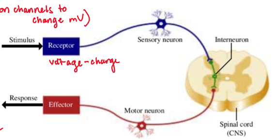
What are the components to a basic/normal reflex?
stimulus
—-
—-
—-
change in skeletal muscle contraction
(not that imp test wise—> good for understanding)
stimulus—> receptor (ion channels to change mV)
sensory neurons have AP’s
SC synapses
motor neurons ±
change in skeletal muscle contraction
What do muscle spindles detect, and what reflex do they trigger?
detect mechanical stretch
trigger stretch reflex
The muscle spindle is made up of modified fibers called what?
nuclear chain + nuclear bag also known as INTRAFUSAL muscle fibers
Explain how the stretch reflex works:
muscle spindle (receptor) detects mechanical stretch
what afferent neurons are then activated?
where is information processed? synapse?
what is then activated?
final result?
muscle spindle (receptor) detects mechanical stretch
1a sensory neurons activated
integration/ in spinal cord synapses with motor neuron
activate ALPHA motor neuron to the same muscle where receptor was
contraction of the muscle
What is the role of gamma motor neurons in the stretch reflex/ muscle spindle reflex?
active when a muscle is contracting and allows the INTRAFUSAL muscle to sense stretch
What is the stimulus for the inverse stretch reflex or golgi tendon reflex?
muscle contraction
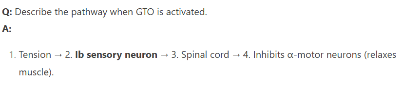
Explain how the inverse stretch reflex or golgi tendon reflex works:
stimulus—> muscle contraction
receptor senses ___________
what afferent neurons are then activated?
where is information processed? synapse?
what is then inhibited?
final result?
stimulus—> muscle contraction
receptor (mechanical) senses pulling in tendon
1b sensory neurons activated
integration/ in spinal cord synapses with motor neuron
inhibits ALPHA motor neuron to the same muscle where receptor was
relaxation of the muscle
Compare and contrast the Golgi tendon reflex with the muscle spindle reflex:
Stretch Reflex (muscle spindle) | Inverse Stretch Reflex (Golgi tendon) | |
receptor | ||
stimulus | ||
afferent neuron | ||
what happens in SC? | ||
result? |
Stretch Reflex (muscle spindle) | Inverse Stretch Reflex (Golgi tendon) | |
receptor | muscle spindle (intrafusal fibers) | golgi tendon organ |
stimulus | muscle STRETCH | muscle CONTRACTION |
afferent neuron | group 1a | group 1b |
what happens in SC? | activates a-motor neurons | inhibits a-motor neurons |
result? | muscle contraction | muscle relaxation |