Telencephalon
1/66
There's no tags or description
Looks like no tags are added yet.
Name | Mastery | Learn | Test | Matching | Spaced |
|---|
No study sessions yet.
67 Terms
Prosencephalon
Primary vesicle during development
Becomes: telencephalon and diencephalon
Cerebral cortex
collection of cell bodies, axons, and dendrites
2 hemispheres, right and left
divided in the middle by the longitudinal fissure
contains layers differentiated by the size + connectivity of the cells contained within the layer —> generalization
The cerebral cortex is the outermost layer of the
brain containing gray matter. Responsible for many “higher-order” functions like language and information processing
layers of the cerebral cortex
generally differentiated by size and connectivity of cells in the layer
layers are organized by function/anatomical feature
layers are called lamina
humans have 3-6 laminae depending on the area of the cortex
layers are typically numbered using roman numerals
Multiform layer
innermost layer of the cerebral cortex
type of cell: primarily fusiform cells
connection: output to thalamus
ex: what is the deepest layer?
Layers of the cerebral cortex
each cortical layer has a primary source of
cells with similar functions tend to be aligned within
long ______ extend throughout each layer
input and primary target for output
the same layer
interneurons
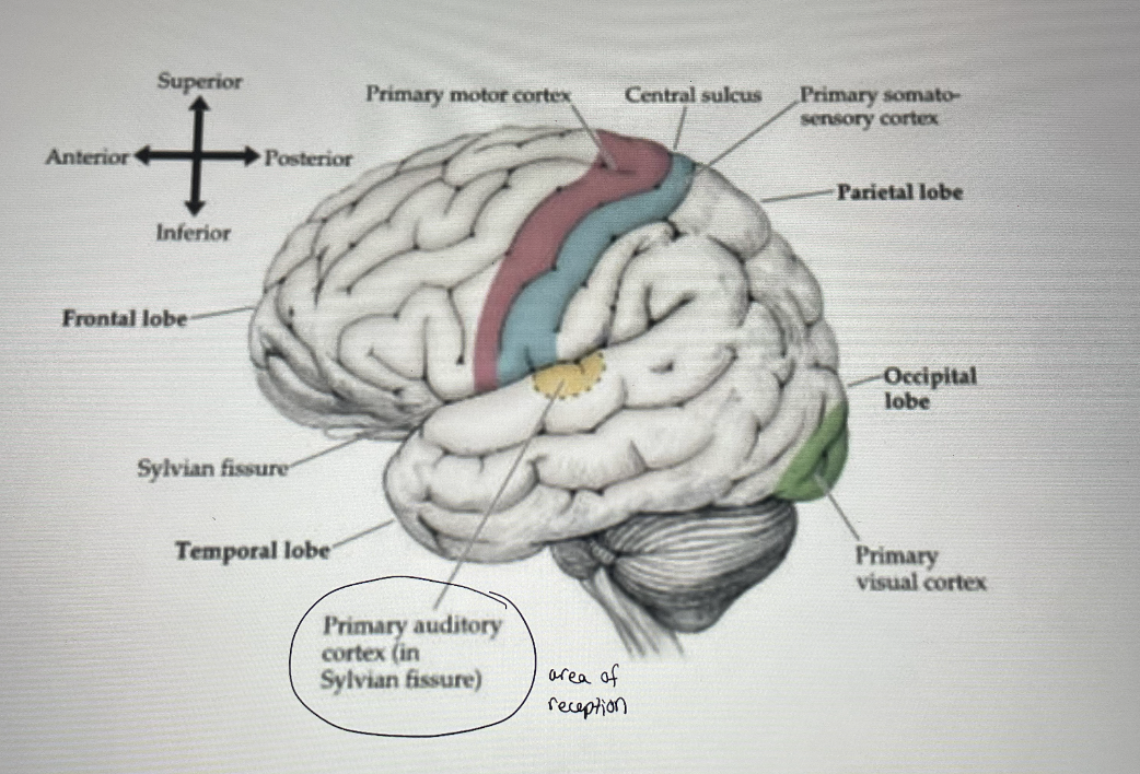
Lobes of the cortex
frontal, parietal, temporal, occipital
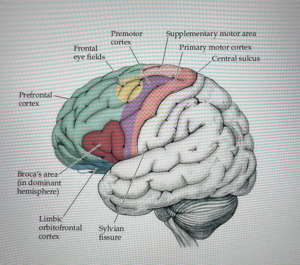
frontal lobe
movement, higher order cognition, decision making and planning
parietal lobe
processing and integration of sensory input
temporal lobe
hearing, learning and memory
occipital lobe
vision
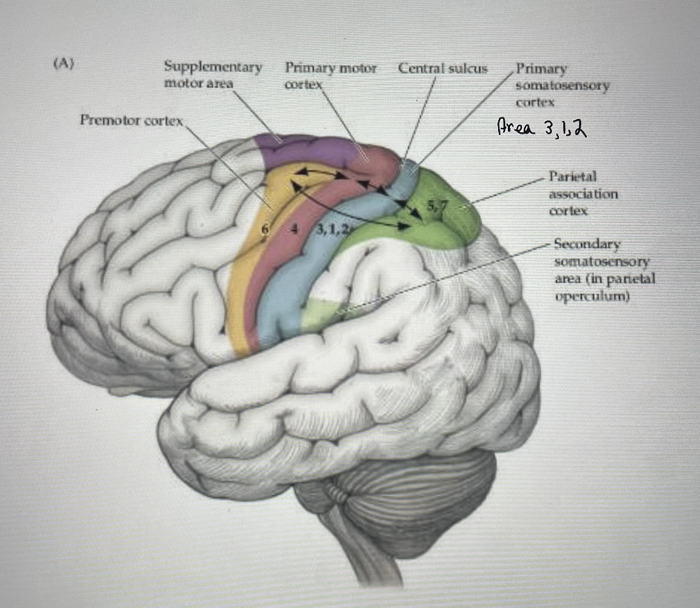
Primary motor cortex: Area 4
Location:
Contains
Controls
Source of
Pre-central gyrus, immediately anterior to central sulcus
complex map of the body
voluntary movement of the opposite side
most neurons in the CST
Other areas of the brain are involved in motor planning:
supplementary motor area
premotor area
Primary motor cortex
input
output
gives rise to the largest
input
basal ganglia
cerebellum
sensory areas (via the thalamus) from muscles AND skin above the muscles
output
efferent (motor) tracts
corticospinal tracts
corticobulbar tracts
Primary motor cortex: function
controls what kind of movement?
controls ____
voluntary movement
contralateral b/c the tract crosses the midline
skilled, precise control of distal limb muscles, especially the flexors of the hand
lower portion of face
more neurons devoted to hand/face than the rest of the body
force and speed of movement
Area 6: Pre-Motor and Supplemental Motor
wrap around towards inside of brain
Location:
anterior to the pre-central gyrus (frontal lobe)
(anterior to primary motor area)
Pre-motor: inferior portion
Supplemental motor: superior portion
Area 6 output
primary motor cortex
area 6 function
generation of
use of _____ and other
movement that relies on
movement that is more
sequences of movement
vision and other sensory inputs to generate movement
various function of the hand
context
planned than automatic
area 6: function part 2
choice of
uses information from the
associates visual input with
orientation of
uses input from
course of action, based on sensory input (especially visual input) and context
PFC and TH to determine movement
movement
orientation of a limb —→ especially hand, to a target
planning bimanual and sequential movements
eyes/head
CB tp provide background postural stability for function
Area 6: Some differences
Pre-motor
NEEDS
activates _____ muscles
30% of the
Supplemental motor
mental rehearsal takes place here —→
activates ____ muscles
NO
Pre-motor
sensory input to function
proximal
CST arises from here
Supplemental motor
learned sequences live here
distal
descending pathway arises from here, not motor tract
Frontal Eye fields: Area 8
Located:
receives input from the
involved in
activated by
connects with the
*notice the overlap of the
anterior to supplemental motor cortex
lateral geniculate, superior colliculus and areas 4,6
conjugate eye movement (esp saccades) and constriction of the pupils
visual stimuli
brainstem and cerebellum
frontal eye fields —→ pre-motor cortex and PFC
Primary sensory cortex: Area 3, 1, 2
location:
receives info from
identifies the location
post central gyrus (parietal lobe)
immediately posterior to central sulcus
receives info from tactile and proprioceptive receptors via the thalamus
of stimuli and discriminates among various shapes, sizes and textures of objects
homunculus main idea
hands and face have lots of real estate
other association areas
an association area is an area of the cortex that receives input from
multiple areas and integrates
Primary visual cortex: Area 17, 18, 19
Location:
receives
info from the ___ side
input is received from the eyes via
outputs from the visual cortex circle back to the
occipital lobe
immediately posterior to parieto-occipital sulcus
and interprets all visual input
left side of the world goes to the right side of the brain
the retina —→ optic nerve —→ thalamus
thalamus and other areas of the brain stem
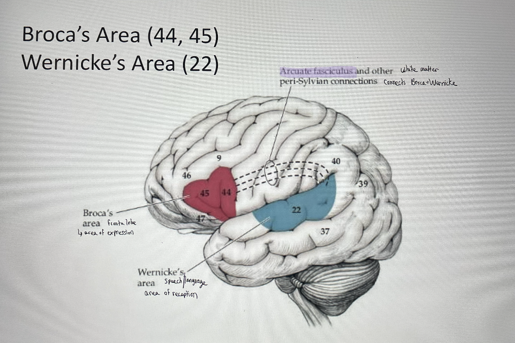
Primary auditory cortex: Area 41, 42
located in the
closely associated with the
the ____ hemisphere is considered the dominant hemisphere for language
temporal lobe
auditory association cortex (Wernicke’s Area): Area 22 and Broca’s area (Area 44, 45)
left
so, although there is an auditory cortex on both sides, the left is “more active”
lesions (and strokes) to the left hemisphere produce more severe language disorders
Broca’s area (44, 45)
location
area of
Wernicke’s area (22)
location
area of
what connects them?
broca’s
frontal lobe
expression
wernicke’s
temporal
reception
Arcuate fasciculus and other peri-Sylvian connects
posterior parietal association area: Area 5, 7
AKA:
Location
Function: integrates
basically helps you make meaning out of the things
so, touch and movement go to
somatosensory association area
superior parietal lobule; just posterior to primary sensory cortex
sensory info and gives meaning; stereognosis; visual-motor perception
you’re touching
3, 1, 2 and get shared to 5, 7 for integration
Pre-Frontal Cortex (PFC): Areas 9 - 12
largest
involved in
has bi-directional
association area in the Frontal Lobe
higher order mental functions- cognitive skills, judgment, abstract thought, personality, communication
connections all over the cortex and sub-cortical structures
“talks to everybody” needs sensory data and info
all association areas, limbic structures, medial nucleus of the thalamus, supplemental motor cortex, reticular system
Possible deficit for frontal lobe
higher order thinking
personality disorder (they don’t smile, they don’t cry)
decision making (abstract thought)
poor judgment
memory
frontal lobe can be damaged by stroke or head trauma (car accident)
Possible deficit for parietal lobe
unable to make meaning out of sensory input/touch
ex: babies putting things in their mouths
you see this in people who have strokes in the right side
Possible deficit for temporal lobe
receptive/Wernicke’s aphasia (not able to receive)
difficulty forming words
memory, trouble remembering what was said
Possible deficit for occipital lobe
vision loss/dysfunction
can’t make meaning based on what you see
very severe and unusual: blindness
Basal Ganglia
planning of
how is the basal ganglia structured?
movement, drives motor plan
it consists of pairs with one on each side of the brain
Basal Ganglia
develops from
basal ganglia means
pairs of
influences movement but no
influences movement by interacting with
telencephalon
large and diverse
gray matter nuclei located deep within the cerebral cortex
direct descending pathways from the BG
many parts of the cortex
What is the most common pathology that occurs when the BG is dysfunctional?
Parkinson’s Disease
Basal Ganglia
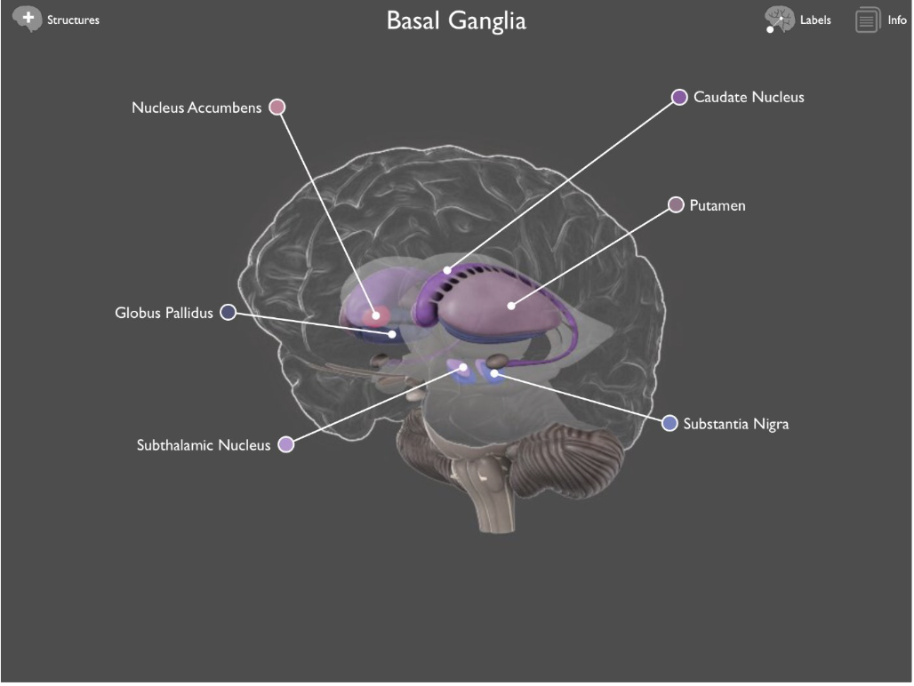
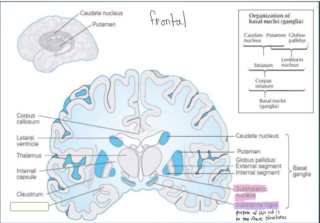
5 main components:
other structure:
Caudate nucleus
putamen
globus pallidus
subthalamic nucleus
substantia nigra
Nucleus accumbens
Caudate/Putamen
Taken together often referred to as the striatum (striatum means “striped”)
Putamen/Globus Pallidus
Taken together, can be referred to as the lentiform (Meaning “lentil” or “lens” shaped)
historically, the ______ was also included as part of the BG
amygdala
BG: caudate
Head/Body/Tail (no specific boundaries)
Amygdala: Anterior top of the tail
BG: putamen
Lateral portion of BG
Ventral Striatum: Anterior/ventral portion, fuses with head of caudate
Nucleus Accumbens —> Forms most of the ventral striatum
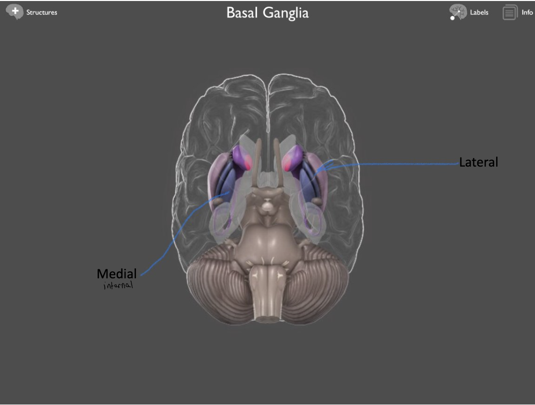
BG: globus pallidus
Medial (internal)
Lateral (external)
Structures that are not attached to the BG
Subthalamic Nucleus
Substantia Nigra
Substantia Nigra consists of
Pars Compacta ** → dysfunctional when someone has Parkinson’s
inner layer (blue)
Pars Reticulata
BG input
Input: From nearly all regions of the cortex Striatum
Data are transferred along a pathway known as “corticostriatal” pathway
BG: output
Output: Via GP and S. Nigra PR to the Thalamus then cortex where it can descend (No direct descending pathways)
Synapses use NT dopamine
BG functions
regulates muscle contraction, tone and force
sequencing of movements
motor learning
motor planning via connections —> through thalamus → into cortex (and eventually into primary motor cortex)
Caudate function
appears to be primarily cognitive
Intrinsic Basal Ganglia Connections
2 intrinsic Basal Ganglia pathways (Fig 16.7)
Direct and indirect
Intrinsic Basal Ganglia Connections: Direct
Striatum → internal segment of globus pallidus or SN pars reticulata
Net effect: excitation of the thalamus
Intrinsic Basal Ganglia Connections: Indirect
Striatum → external segment of globus pallidus and to the subthalamic nuclei
net effect: inhibition of the thalamus
Dysfunction of the BG
movement disorders
Parkinson’s Disease (PD)
Hypokinetic Movement disorder: TOO LITTLE movement
Huntington’s Disease
Hyperkinetic movement disorder: TOO MUCH movement
Physical Therapy and PD
Degenerative disease of the the BG
Etiology: Unknown
Pathology: Degeneration of dopaminergic neurons in the Basal Ganglia, specifically the pars compactus of the Substantia Nigra
Clinical Manifestations → Movement dysfunction
“TRAP” – Tremor/Cogwheel Rigidity/Akinesia/Posture
Internal Capsule
DEF: A collection of projection fibers (white matter) from all parts of the Cortex → Converging towards the Bstem
CST makes up much of the Internal Capsule
A continuous sheet of fiber that forms the medial boundary of the Lentiform N.
Superiorly, the fibers fan out to form a structure knows an the Corona Radiatia
internal capsule can be divided into 3 parts:
anterior limb
posterior limb
genu
Neural Tube also forms cavities:
ventricles
Ventricles contain
Choroid Plexus, located within all ventricles, does what?
Ventricles contain Cerebral spinal Fluid (CSF)
Choroid Plexus, located within all ventricles, synthesizes CSF
ventricles
2 Lateral Ventricles: One in each hemisphere; Extends from frontal lobe to parietal and occipital lobes and reaches laterally into the temporal lobe
3rd Ventricle: Within Diencephalon (Within the thalamus and hypothalamus)
4th Ventricle: Surrounded by the Pons, Medulla and Forms the floor of the Cerebellum
anatomy of the ventricles: lateral ventricles
Frontal (anterior) horn
Body
Occipital (posterior) horn
Temporal (Inferior) Horn
anatomy of the ventricles: third ventricle
cerebral aqueduct
anatomy of the ventricles: fourth ventricle
central canal
C-shaped structures
the lateral ventricles form a C shape
Many structures wrap around the ventricles and thus follow this C-shaped Anatomy
Caudate Nucleus
Corpus Callosum
Fornix
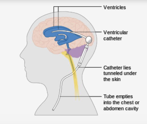
Pathology in the Ventricles: Hydrocephalus
Causes
Excess CSF production → Rare
Obstruction of CSF Flow → Tumor; hemorrhage, congenital malformation
Pathology in the Ventricles: Hydrocephalus
Common Sx:
Treatment
HA, Nausea, Vomiting, decreased/altered vision, altered level of consciousness, altered cognition
may require a drain or shunt → Not done by PT
Meninges defined
Protective layer of tissue that covers the brain and Spinal Cord
Cushions the brain from trauma and protects the brain from infection
Meninges- 3 layers (from outer to inner)
Dura Mater
Hard outer layer
Arachnoid Mater
Creates a space
CSF travels in the space made between the Arachnoid and Pia
Pia Mater
Closely adheres to the brain
Blood Brain Barrier
Specialized permeability barrier between the
Crucial for
Acts as a
Excludes
Specialized permeability barrier between the capillary endothelium and the extracellular space
Crucial for protection and to maintain the homeostasis of the brain
Acts as a physical-chemical-metabolic barrier
Excludes large, water soluble molecules from diffusing into the CNS and restricts drug permeability to the brain
Making many drugs unable to permeate the brain