anatomy exam 2
1/61
There's no tags or description
Looks like no tags are added yet.
Name | Mastery | Learn | Test | Matching | Spaced | Call with Kai |
|---|
No analytics yet
Send a link to your students to track their progress
62 Terms
superior thoracic Aperture (thoracic inlet)
bounded anteriorly by the sternal manubrium and costal cartilages of 1st ribs, T1 posteriorly, and the 1st ribs laterally
transmits trachea, esophagus, aortic arch and branches, superior vena cava and several nerves and blood vessels to and from neck
inferior thoracic aperture (thoracic outlet)
bounded anteriorly by the xiphisternal joint and costal margins (7-10), T12 posteriorly, and 12th ribs laterally
contains diaphragm and transmits esophagus, aorta and inferior vena cava, nerves and blood vessels to and from abdomen
sternal angle (angle of Louis)
formed by articulation of manubrium with body of sternum
at level of 2nd costal cartilage
opposite the intervertebral disc btwn T4 and T5
used to identify the 2nd rib and base of heart
transthoracic plane
RATPLAT
R: rib 2
A: aorta arch start and end
T: trachea (bifurcating)
P: pulmonary artery (bifurcating)
LA: ligamentum arteriosum
L: left recurrent laryngeal nerve
A: azygous vein
T: thoracic duct (part of lymphatic system)
sternum
manubrium:
suprasternal notch (juglar notch)
sternal angle (angle of Louis)
clavicular notch
Body:
costal (hyaline) cartilage
intercostal space
Xiphoid process
xiphisternal joint
ribs
3 types of ribs
true ribs (1-7): direct attach to the body of the sternum
false (8-12)
floating (11-12)
ribs 1, 10, 11, 12 articulate with 1 vertebra
all other ribs articulate with 2 vertebrae
breast anatomy
adipose tissue
mammary glands
lactiferous ducts
suspensory ligaments
nipple
areola
retromammary space
tail of spence
extension of breast tisse into the axilla, in upper outer quadrant
can detect cancer cells in this area through lymph node
upper outer: axillary tail, majority of cancers
respiration musculature
external intercostals
fibers run inferomedially
TP to mammary line
internal intercostals
fibers run superomedially
sternum to scapular line
innermost intercostals
fibers run superomedially
mammary line to scapular line
neurovascular bundle
V: intercostal VEIN
A: intercostal ARTERY
N: intercostal NERVE
4 parts of parietal pleura
costal
mediastinal
diaphragmatic
cervical (pleural or ‘cupula’)
right lung
3 lobes
2 fissures
oblique
horizontal
left lung
2 lobes
1 fissure
oblique
right lung impressions
azygous vein
SVC
IVC
esophagus
liver
left lung impressions
aorta (arch and descending)
left subclavian artery
stomach and spleen
mediastinum
central compartment of thoracic cavity: space btwn the 2 pleural sacs
contains:
heart
great blood vessels:
aorta
SVC
pulmonary trunk & veins
azygous system of veins
trachea
esophagus
thymus gland
thoracic duct & lymph nodes
vagus & phrenic nerves
sympathetic trunks
boundaries & divisions
anterior: sternum
posterior: thoracic vertebrae (T1-T12)
lateral: pleural cavities (lungs)
superior: thoracic inlet (superior thoracic aperture)
inferior: diaphragm
superior mediastinum: above the level of T4-T5 (sternal angle)
contains thymus, aortic arch, brachiocephalic veins, SVC, trachea, esophagus, vagus & phrenic nerves, thoracic duct
inferior mediastinum: below T4-T5 to diaphragm, divided into 3 parts
anterior: small, btwn sternum & pericardium, contains thymus (in children)
middle: contains heart, pericardium & roots of great vessels
posterior: btwn pericardium & vertebral bodies, contains descending aorta, esophagus, thoracic duct, & azygous system
thoracic aorta
continuation of the arch of aorta
begins at inferior border of T4 and descends left of T5-t12
passes through the aortic hiatus at T12
branches: bronchial (1 right, 2 left), esophageal, pericardial, mediastinal
gives posterior intercostal arteries (9 pairs, 3rd-11th spaces), subcostal, and superior phrenic branches
azygos venous system
azygos vein ascends on the right from T12 to T4
connects the superior and inferior venae cavae
drains posterior thoracic and abdominal walls
hemiazygous ascends on the left from T12 to T9 and joins azygos near T9
accessory hemiazygos descends from T5 to T8 and joins azygos at T7/T8
trachea
trachea: fibrocartilaginous tube with C-shaped rings; lies anterior to esophagus
trachea extends from C6 to T4 and bifurcates at the carina near the sternal angle (T4-T5)
esophagus
esophagus extends from C6 to T9 in the thorax and passes through the esophageal hiatus at T10
esophageal constrictions: arch of aorta, left primary bronchus, and esophageal hiatus
thoracic duct
major lymphatic trunk; ascends btwn the thoracic aorta and azygos vein from T5-T12
in the superior medastinum it lies on the left side of the esophagus, deep to the arch of aorta
receives lymph from both lower extermities, lower abdomen, left thorax, left head/neck, and left upper limb
drains into the left brachiocephalic vein
begins inferiorly as the cisterna chyli anterior to T12
phrenic nerve
arise from C3-C5
provide the sole motor supply to the diaphragm and about 1/3 of its sensory supply
right phrenic descends along the right brachiocephalic vein and SVC
left phrenic descends btwn the left subclavian and left common carotid arteries and crosses the arch of the aorta
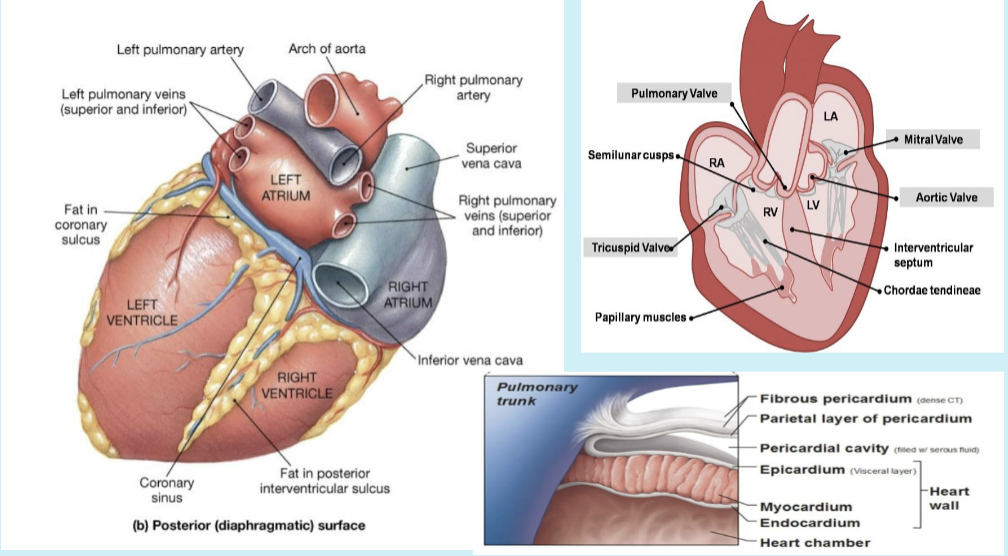
heart
located in the middle mediastinum, btwn lungs
enclosed by pericardium: fibrous (outer) & serous (inner) layers
serous pericardium has parietal and visceral layers (epicardium)
located obliquely in the middle mediastinum
roughly spans from the 2nd-5th intercostal spcaes
2/3 of heart lies left of midline
base (posterior surface): faces posteriorly toward vertebral column (mainly left atrium)
apex: points left & downward to 5th intercostal space, midclavicular line (formed by left ventricle)
resting on: the diaphragm, with pericardium fused
superior border: from 2nd to 3rd costal cartilages, formed by both the atria & great vessels
right border: from 3rd to 6th costal cartilages, mainly right atrium
inferior border: from right border to apex, mostly right ventricle & a little of left ventricle
left border: from apex up to 2nd costal cartilage, mostly left ventricle & part of left atrium
layers of heart wall
epicardium: outer layer (visceral pericardium)
myocardium: thick muscular layer for contraction
endocardium: smooth inner lining of chambers
cardiac surfaces & grooves
sternocostal (anterior) surface:
mostly right ventricle
lies just behind the sternum & costal cartilages
diaphragmatic (inferior) surface:
formed by both ventricles, mainly left ventricle
rests on diaphragm
pulmonary (left) surface:
formed by mainly left ventricle, which makes the cardiac impression on the left lung
base (posterior) surface):
formed mainly by left atrium (receives pulmonary veins)
faces posteriorly toward vertebral column coronary sulcus (atrioventricular groove):
separates atria from ventricles
contains right coronary artery, circumflex artery, and coronary sinus
anterior interventricular sulcus:
btwn right and left ventricles on anterior surface
contains anterior interventricular artery (LAD) and great cardiac vein
posterior interventricular sulcus:
on the diaphragmatic surface
contains posterior interventricular artery & middle cardiac vein
cardiac veins
venous drainage of heart:
deoxygenated blood from myocardium returns via cardiac veins, all emptying into the coronary sinus → right atrium
main cardiac veins:
great cardiac vein: runs with LAD artery in the anterior interventricular sulcus
middle cardiac vein: runs with posterior interventricular artery
small cardiac vein: runs along right margin of heart with marginal artery
anterior cardiac veins: drain directly into right atrium (not through coronary sinus)

right atrium
receives venous blood from SVC, IVC, and coronary sinus
divided internally by crista terminalis into:
smooth posterior wall (sinus venarum)
rough anterior wall (pectinate muscles)
fossa ovalis: remnant of fetal foramen ovale on interatrial septum
right auricle: muscular pouch extending from atrium

right ventricle
forms most the anterior surface of the heart
internal structures:
trabeculae carneae: muscular ridges on inner wall
papillary muscles: anchor chordae tendineae, which attach to valve cusps
tricuspid valve: btwn RA and RV
moderator band (septomarginal trabecula): carries conduction fibers from septum to anterior papillary muscle
conus arteriosus (infundibulum): smooth outflow region leading to pulmonary trunk

left atrium
receives 4 pulmonary veins (2 from each lung)
mostly smooth-walled; small pectinate muscles only in auricle
sends blood through mitral valve to left ventricle
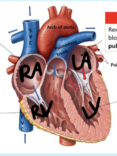
left ventricle
thickest myocardium (3x right ventricle)
contains trabeculae carneae, papillary muscles, and chordae tendineae similar to RV
aortic vestibule: smooth outflow tract to aortic valve
opens into ascending aorta via the aortic semilunar valve
forms apex of the heart
blood flow
body → SVC/IVC → RA → tricuspid valve → RV → pulmonary semilunar valve → pulmonary trunk → lungs → pulmonary veins → LA → mitral valve → LV → aortic semilunar valve → aorta → body
blood flow
right heart → pumps DEOXYGENATED blood to lungs (pulmonary circuit)
left heart → pumps OXYGENATED blood to body (systemic circuit)
valves ensure one-way flow btwn chambers and vessels
fetal circulation
ductus venosus:
connects umbilical vein → IVS
allows oxygenated blood from placenta to bypass the fetal liver
foramen ovale:
shunts blood from RA → LA, bypassing pulmonary circuit
ductus arteriosus:
connects pulmonary trunk → descending aorta
diverts most blood awat from the nonfunctional fetal lungs
umbilical arteries (2):
carry deoxygenated blood from fetus → placenta
after birth:
foramen ovale → fossa ovalis
ductus arteriosus → ligamentum arteriosum
umbilical vein → ligamentum teres hepatis
ductus venous → ligamentum venosum
ausculation points
A: aortic valve- 2nd intercostal space, right sternal border
P: pulmonary valve- 2nd left intercostal space, left sternal border
E: Erb’s Point- 3rd left intercostal space
T: tricuspid valve: 4th left intercostal space, left lower sternal border
M: mitral valve: 5th intercostal space, midclavicular line (apex)
peritoneum- visceral layer
greater omentum
lesser omentum
peritoneum- parietal layer
mesentery
mesocolon
greater omentum
greater curvature of stomach
connects with transverse colon, spleen, and diaphragm
lesser omentum
lesser curvature of stomach
connects to stomach and duodenum of liver
retroperitoneal structures (SADPUCKER)
S: suprarenal glands
A: abdominal aorta/IVC
D: duodenum (2nd & 3rd segment)
P: pancreas (except tail)
U: ureters
C: colon (descending and ascending)
K: kidneys
E: esophagus
R: rectum
peritoneal ligaments- off liver
double layer of peritoneum
connects organ to organ or ro abdominal wall
off liver
falciform ligament: liver to anterior abdominal wall
hepatogastric ligament: membranous part of lesser omentum
hepatoduodenal ligament: free edge of lesser omentum, contains portal triad
hepatorenal ligament: connects liver to area around kidneys
peritoneal ligaments- off stomach
continuous attachment to greater curvature, paer of greater omentum
off stomach
gastrophrenic ligament: to inferior diaphragm
gastrosplenic ligament: cover hilum of spleen
gastrocolic ligament: to transverse colon
subdivisions of peritoneal cavity
greater sac: larger main compartment
omental bursa (lesser sac): sac-like cavity posterior to stomach, lesser omentum, and related structures
allows smooth movement of stomach against posterior structures
omental foramen:
also called epiploic foramen or foramen of Winslow
opening between greater and lesser sacs
boundaries
anterior: hepatoduodenal ligament
posterior: IVC and right crus of diaphragm
superior: liver
inferior: superior duodenum
digestive organs
functions: ingestion, secretion, propulsion, and mixing, digestion, absorption, defecation
digestion can be mechanical or chemical
organs are found in head & neck, thoracic cavity, abdominal cavity, and pelvic cavity
GI Tract vs Accessory Organs
alimentary tract = gastro-intestinal (GI) tract
muscular tube from mouth to anus about 20-30 ft long
true GI organs form the actual tube food passes through
accessory organs either do not touch foos or are unnecessary for digestion, but help by secretions or mechanical action
embryonic regions & vascular supply (CSI)
all 3 regions drain venous blood into the hepatic portal vein (liver)
foregut:
supplied by branches of celiac trunk
forms esophagus, stomach, part od duodenum, liver, gallbladder, and upper pancreas
midgut
supplied by branches of the superior mesenteric artery
forms duodenum to proximal 2/3 transverse colon, including lower pancreas
hindgut
supplied by branches of inferior mesenteric artery
forms distal 1/3 transverse colon to superior rectum
borygmus
gurgling stomach sounds
celiac trunk
3 major branches
L gastric a.
splenic a.
common hepatic a.
superior mesenteric artery
inferior pancreaticduodenal artery: feeds under surface of pancreas
superior mesenetric artery
jejunum: empty portion of stomach (absorption of nutrients)
jejunal arteries:
loops of these are called arcades
anastomic
longer vasa recta (straight parts)
ileum: more arcades, shorter vasa recta
ileal arteries
appendicular artery
ileocolic artery: ileum + colon
right colic artery: feeds R portion of colon
middle colic artery: gives blood to transverse colon
inferior mesenteric artery
left colic artery
several (flexure/bend) sigmoid artery
superior rectal artery
hepatic portal vein
primary formation: superior mesenteric vein + splenic vein
also receives inferior mesenteric vein, gastric vein, jejunal and ileal intestinal, ileocolic, R colic, middle colic, L colic, sigmoidal, & superior rectal veins
drains GI tract to liver
hepatic veins then drain from liver to IVC
portal caval anastomoses
esophageal: L gastric ←→ esophageal veins of azygous system → esophageal varices
rectal: superior rectal ←→ middle and inferior rectal veins → rectal varices/ hemorrhoids
paraumbilical: paraumbilical ←→ superficial epigastric veins → capcut medusae
retroperitoneal: colic veins ←→ renal, suprarenal, gonadal, and paravertebral veins → ascites
intrahepatic: L branch of portal vein → patent ductus venosus/ portosystemic shunt
liver
largest gland, 2nd largest organ
4 lobes: right, left, quadrate, caudate
ligaments: falciform, ligamentum teres hepatis, ligamentum venosum, R and L coronary, R and L triangular
bare area and IVC on posterior liver
portal triad travels in lesser omentum
gallbladder
stores bile on posteroinferior surface of liver near quadrate lobe
pancreas
retroperitoneal gland with endocrine & exocrine function
parts: head, body, tail
about 6in long and sits behind stomach
2 ducts: main pancreatic duct & accessory pancreatic duct
spleen
lymphatic organ, not a digestive organ
highly vascular; recycles blood cells (red pulp) & provides immunity (white pulp)
1, 3, 5, 7, 9, 11 rule: 1×3×5 in, 7 oz, deep to ribs 9-11
bile duct system
R and L hepatic ducts → common hepatic duct
cystic duct comes from gallbladder
common hepatic duct + cystic duct → common bile duct
common bile duct joins main pancreatic duct at hepatopancreatic ampulla & papilla
also called ampulla of Vater with sphincter of Oddi
pancreatic duct system
main pancreatic duct = duct of Wirsung
joins common bile duct at hepatopancreatic ampulla and sphincter of Oddi
empties into duodenum at major duodenal papilla
accessory pancreatic duct = duct of Santorini
empties into duodenum at minor duodenal papilla
4 layers of GI tract
mucosa: forms lumen
submucosa: vasculature for mucosa
muscularis: inner circular + outer longitudinal smooth muscle
stomach adds a 3rd oblique layer
mouth, pharynx, superior half of esophagus, and external anal sphincter are skeletal muscle
serosa= serous membrane from peritoneum; called adventitia in thorax
stomach review
features: cardia with cardiac sphincter, fundus, greater curvature, lesser curvature, pylorus with pyloric sphincter
rugae are folds of mucosa
muscle layers: longitudinal, circular, oblique
small intestines
parts and features
duodenum: shortest segment; receives stomach contents, common bile duct, and main pancreatic duct
duodenum subdivisions: superior, descending, inferior, ascending; superior part includes duodenal bulb
jejunum= middle portion about 2/5 of length
ileum= final segment, 3/5 of length; ends at ileocecal valve
internal features
plicae circulares = circular folds
villi with microvilli increase SA
villi contain capillaries
lymph capillaries= lacteals; absorb lipids
jejunum vs. ileum
jejunum: more plicae circulares & villi, fewer longer arcades & vasa recta, traces of MALT
ileum: fewer plicae circulares and villi, multiple shorter arcades and vasa recta, Peyer’s patches
large intestine
main parts
cecum and vermiform appendix
ascending colon, transverse colon, descending colon, sigmoid colon
sigmoid colon enters pelvic cavity
rectum & anus are in pelvic cavity
key features
ileocecal valve
tenia coli
Haustra formed with plicae semilunares
appendices epiploicae
hepatic flexure and splenic flexure
rectal valves, anal canal, internal sphincter, external sphincter
kidney
location
lateral to vertebral column, deep to 12th rib
superior level about T11, inferior level about L3, hilum at L1-L2
R kidney lies lower than L
primary retroperitoneal structure; overlies transversalis fascia
covering and structure
fibrous capsule & adipose capsule provide padding and protection
adipose capsule helps prevent nephroptosis
hilum is medial indentation for ureter, renal artery, and renal vein
renal sinus contains fat
renal pelvis is expanded upper ureter in hilum
minor calyces drain pyramids; major calyces form renal pelvis
blood supply
renal artery from abdominal aorta; renal vein to IVC
arterial sequence:
segmental → lobar → interlobar → arcuate → cortical radiate → afferent arteriole → glomerular capillaries → efferent arteriole → peritubular capillaries/ vasa recta