Lab 07 Lymphatic & Digestion
1/43
There's no tags or description
Looks like no tags are added yet.
Name | Mastery | Learn | Test | Matching | Spaced | Call with Kai |
|---|
No analytics yet
Send a link to your students to track their progress
44 Terms
lympatic system starts at the
cellular level – close-ended capillary-like vessels within the arteriole/venuole capillary beds and nearby tissues
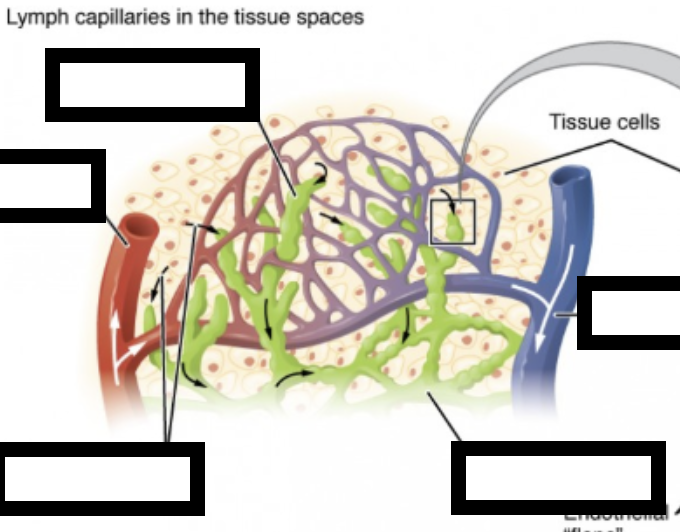
lymph capillary, arteriole, tissue fluid, tissue cells, venule, lympahtic vessell
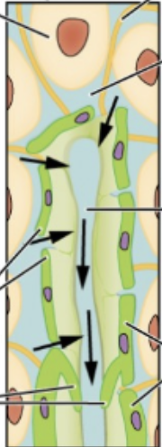
collagen fiber, intersital fluid, lymph, lymph vessel endotheial cells, endothelial flaps, backflow prevention value
start end
lymphatic capillaries, vessels, lg vessels, nodes, lg vessels, trunks, collecting ducts end into r and l subcalvain veins
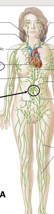
jugular trunk, r lymphatic duct, r brachiocephalic vein, bronchomediastinal trunk, intercoastal trunk, cisterna chyli, internal jugular vein, thoratic duct, subclavian trunk, thoartic duct, intestrinal trunk, lumbar trunk, lymphatic vessels
order
lymphatic capillary, afferent lymphatic vessels, lymph node, efferent lympatic vessel, lymphatic trunk, collecting duct, subclavian vein
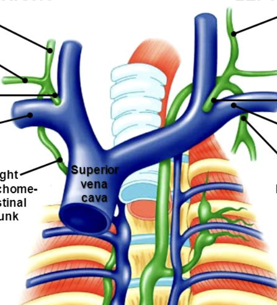
r jugular trunk, r subclavian trunk, r lymphatic duct, r subclavian vein, r bronchomediastinal trunk, l jugular trunk, l subclavian trunk, thoartic duct, l subclavian vein, l bronchomediastinal trunk
lymphatic organ
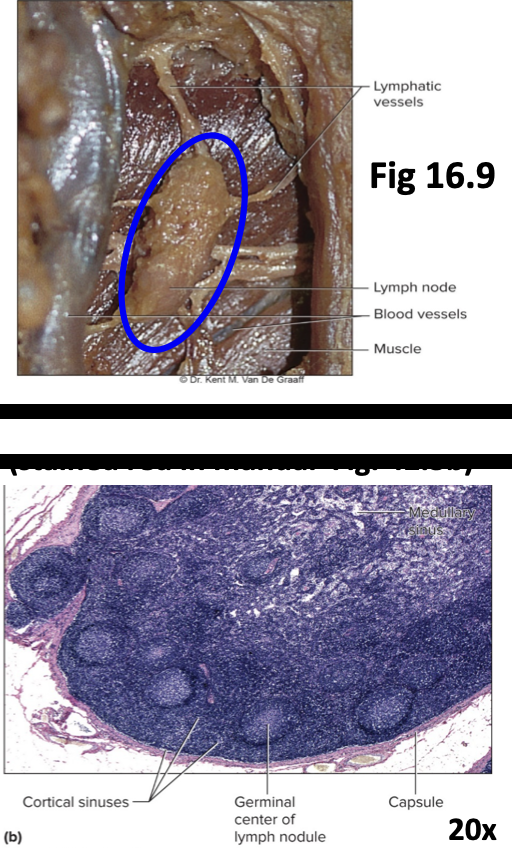
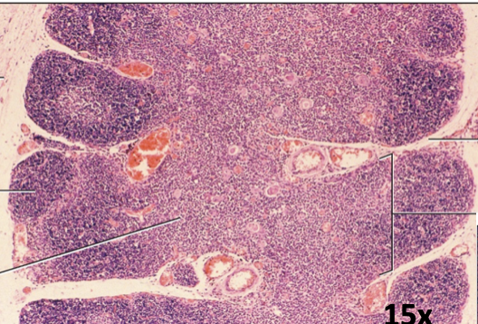
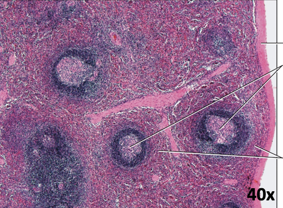
lymph nodes
thymus
capsule, cortex of lobule, medulla of lobule, septum of ct, lobule
spleen
capsule, white pupl, red pulp
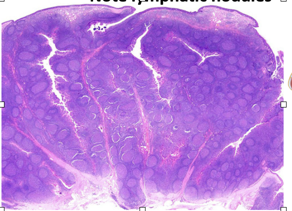
Lymphatic tissue contains lymphoid nodules -
Densely packed lymphocytes in loose connective tissue in mucous membranes.
MALTS
Mucosa-Associated Lymphatic Tissue
Tonsils
large nodules in wall of pharynx
Peyer’s Patches –
grouped nodules in small intestine
Vermiform appendix –
fused nodules in the walls of the appendix
palastine tonisl
pharyngeal tonsil, tubal tonsil, palatine tonsil, linguinal tonsil
digestive organs IDAE
ingestion
digestion
absobation
elimination
2 major parts digestive system
alimentary canal: (GastroIntestinal – GI tract)
( a “tube” from mouth to anus)
(mouth, pharynx, esophagus, stomach,
small & large intestines, rectum, anal canal)
Accesssory digestive organs: salvary glands, gallbladder, liver, pancrease
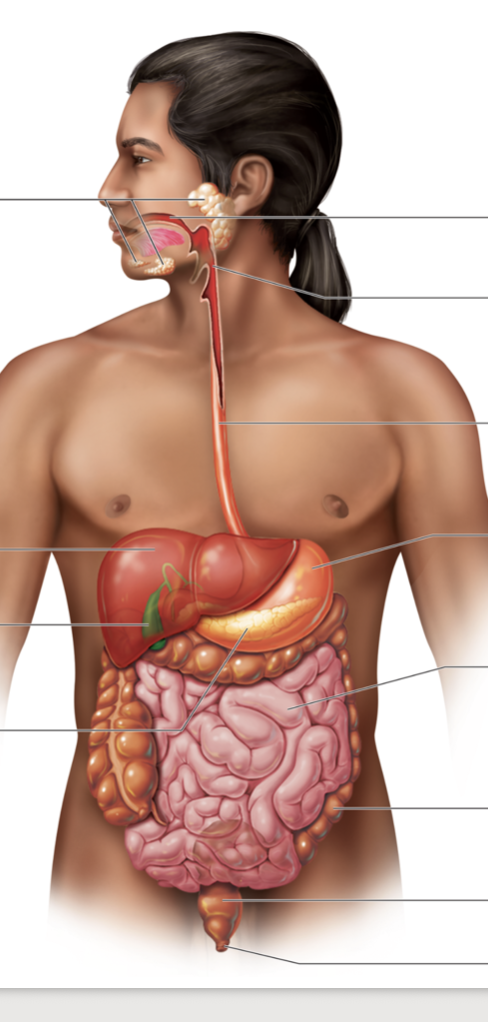
salivary glands, liver, gallbladder, pancreas, mouth, pharnyx, esophagus, stomach, small intenstine, lg intesntine, rectum, anus
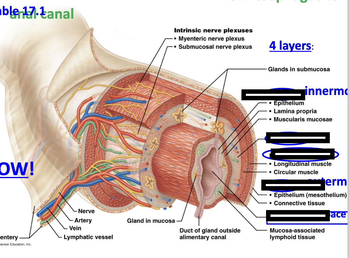
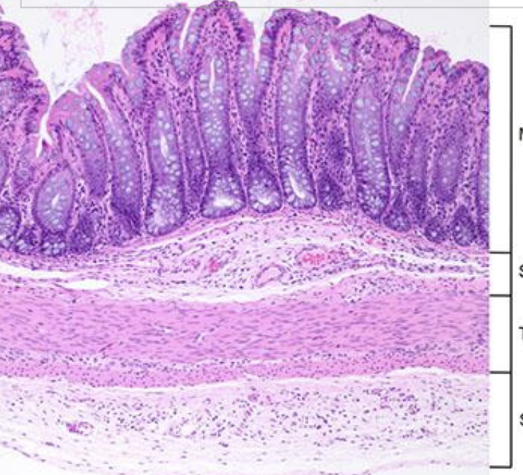
mucosa, submucosa, muscularis externa, serosa, lumen
GI tract Wall
oral cavity
ingest/digest
(Mouth) – [ingest & masticate]
tongue, teeth, salivary glands hard & soft palates, uvula, [palatine & lingual tonsils] - swallowing moves bolus down
- [digestion starts]
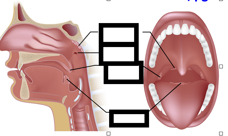
pharanx
3 parts ((naso-) , oro- laryngo-, skeletal muscles initiate wavelike contractions (peristalsis) propelling food down tube
[no digestion]
ingest/digest
esophagus
transports food down to stomach, smooth muscles propel food via peristaltic waves - involuntary [no digestion]
injest/digest
digestion
mechnaical/chemical
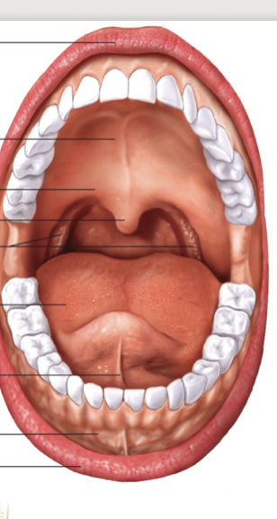
hard palate, soft palate, uvula, palatine tonsisls, tounge, ligiunal frenlum, oral vetsibule
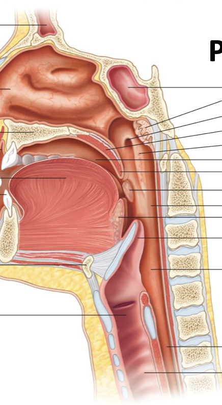
frontal sinus, nasal cavity, hard palate, vestibuke, tounge, tooth, ip, hyoid bone, larnx, sphenoidal sinus, pharngeal tonsil, opening of auditory tube, soft palate, nasophaynx, oral cavoty, uvula, patine tonsil, orophanx, ligunal tonsil, epiglotis, larngophanrx, esophogus, trachea
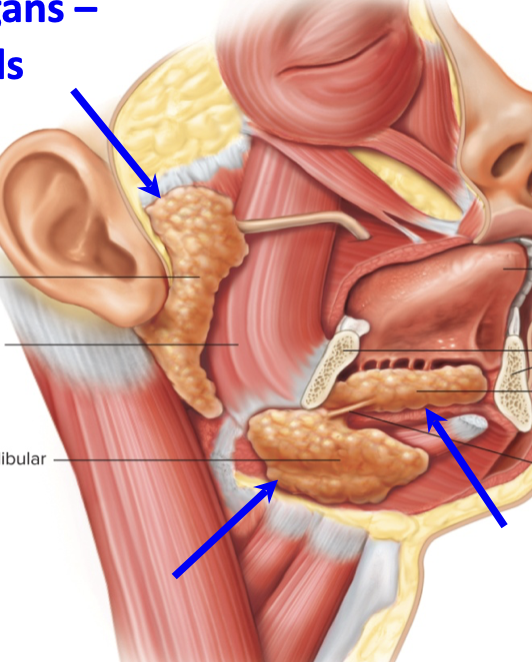
salivary glands, paratoid glands, submandiular gland, sublingual gland
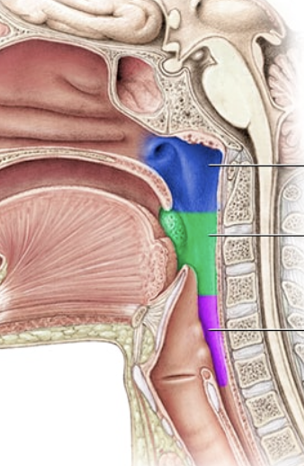
nasopharnx, oropharnx, larnoharnx
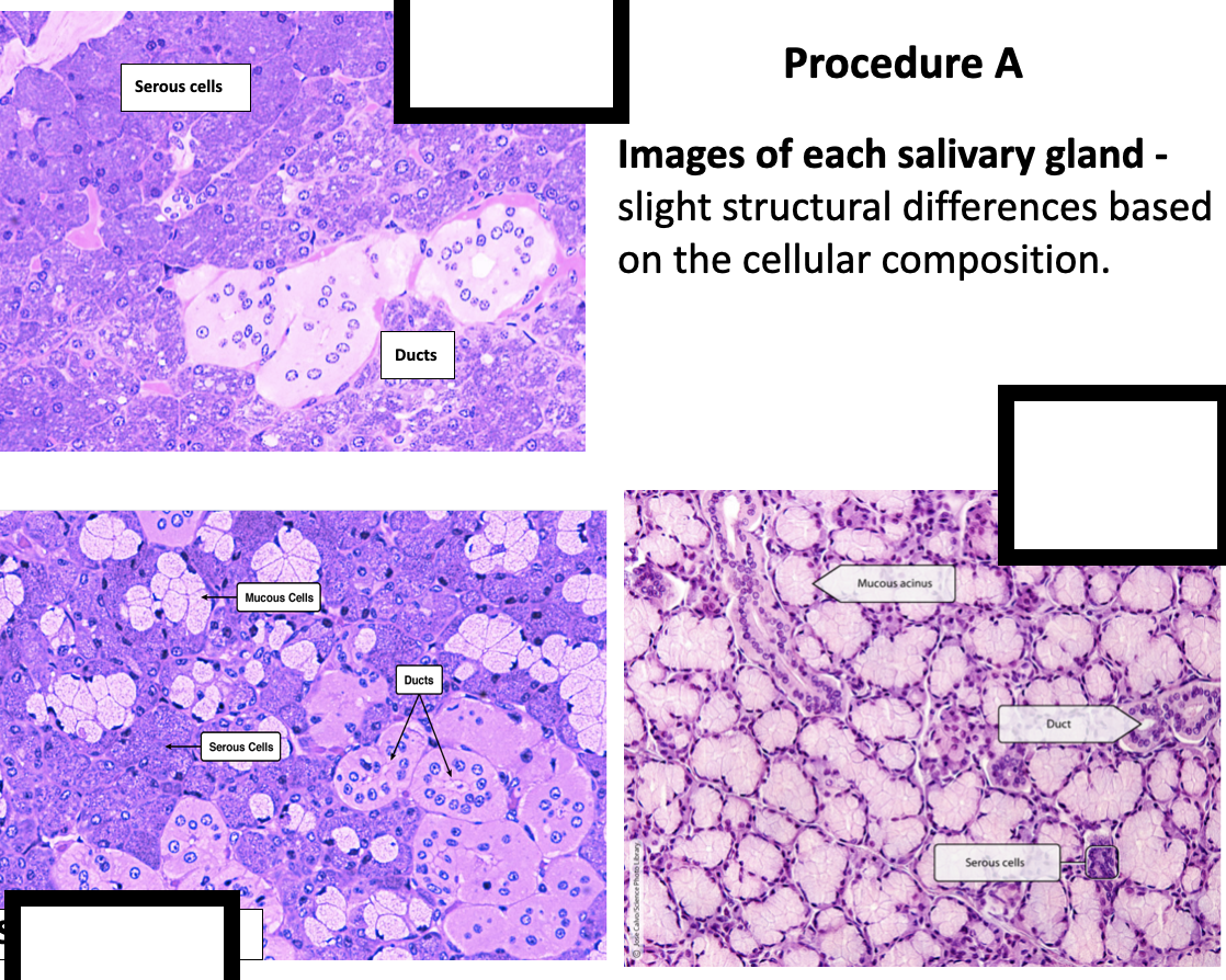
paratoid, submandiular, sublinguinal
digest absorb
lower esophgeal spinicheter valve, stomach, pyloric sphincter valve, small intentsinesm ileoceal sphincter valve
stomach
4 parts (cardia, fundus, body, pylorus), greater & lesser curvatures, rugae (gastric folds, gastric pits, gastric & mucous glands), additional muscle layer - oblique in muscularis externa layer, chyme
[big time digestion & storage]
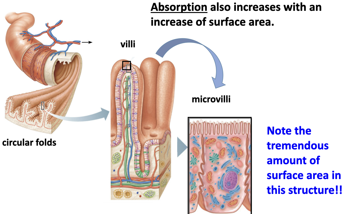
small intestine
duodenum, jejunum, ileum, villi, microvilli pancreatic duct, bile duct,
[final digestion - big time absorption]
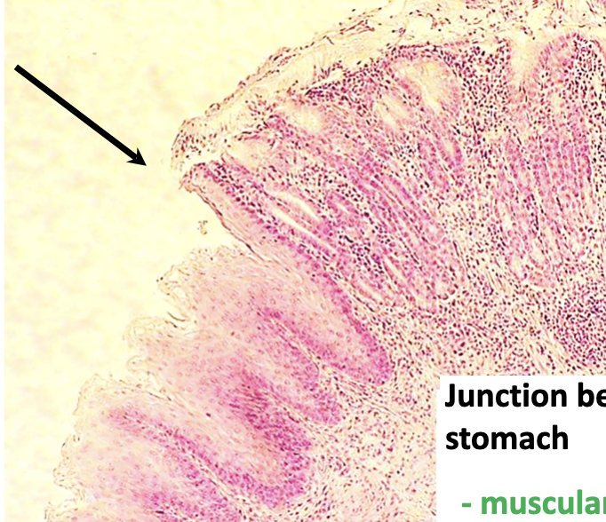
Lower esophageal sphincter (junction)
Junction between esophagus and stomach
- muscularis layer (circular)
always contracted (why
chyme does not go back into
esophagus
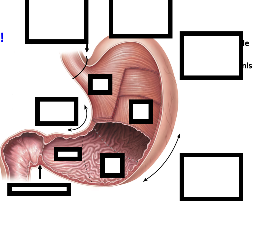
pyloric sphincter, pyloris, rugae, greater curvature, assitional muscle layer in musclaris externis, cardia, body, fundus, lower esophgeal spincter lesser curvature
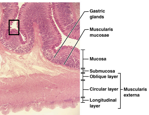
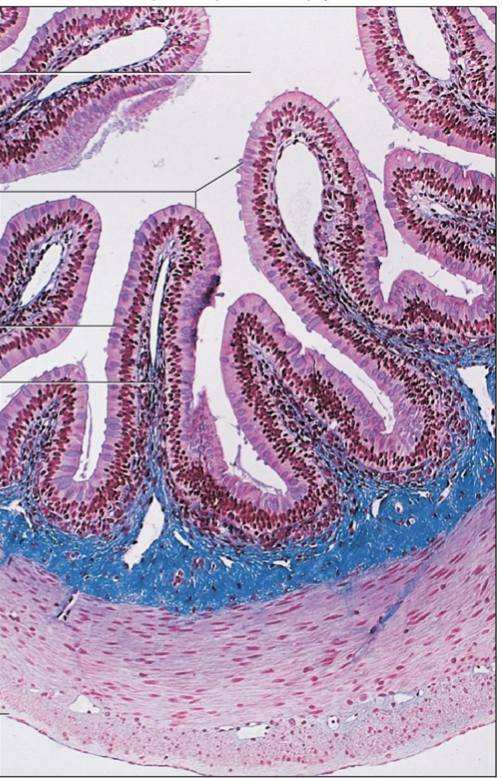
stomach lining
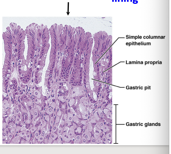
Simple columnar epithelium produce mucous – shed and replaced continuously
Gastric glands – composed of
parietal (hydrochloric acid (HCl)), and
chief (pepsinogen/pepsin) cells
stomach lining
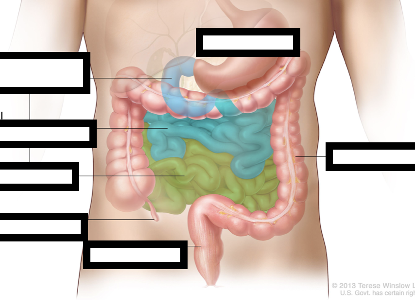
duedenum, jejunum, ilieum, appendix, stomach, lg intestine, rectum
small inestine
needs to be more
alkaline for absorption
Pancreatic juice is alkaline
lumen, vili, simple columnar epithelium, ct in villus, mucoasa, submucosa, circular fibers, longitudal fibers, seorsa
absorb eliminate
Large Intestine – ileocecal sphincter, cecum, appendix,
ascending, transverse, and descending colons,
sigmoid colon, rectum, anal canal, anal
sphincters, anus
large intestine tissue – mucosa dominated w/ goblet cells
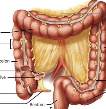
transverse colon, haustrum, acensing colon, illieum cut, illoceal vlave, cecum, appendix, descdedning colon, tanien coli, sigmoid colon, rectum anal canal, externa. anal sphincter
lg intestine
mucosa, submucosa, tunica mescularis, serosa
assorsy digestive organs
Salivary Glands – parotid, sublingual, submandibular,
mucin (mucous), salivary amylase, bolus
Liver –common hepatic duct, bile duct, cystic duct,
hepatocytes, hepatic triad, (bile production)
[hematological & metabolic regulation]
Gallbladder – cystic duct (bile storage)
Pancreas – main pancreatic duct, head & tail
[produces essential digestive enzymes]