Endemic Diseases (Lepto, Salmonellosis, Malignant Catarrhal Fever)
1/40
Earn XP
Description and Tags
Module 21, week 4
Name | Mastery | Learn | Test | Matching | Spaced |
|---|
No study sessions yet.
41 Terms
Leptospirosis
Who does it affect? Zoonotic? Clinical Effects?
worldwide distribution
affects virtually all mammals - zoonotic disease
broad range of clinical effects from mild, to subclinical, to multi organ failure and death - commonly headache, fever, lethargy, malaise
Leptospirosis
Etiology of Leptospirosis
aerobic, gram negative spirochete
fastidious, slow growing, corkscrew like motility
Leptospirosis
What are the cattle host-adapted types of Lepto
USA and much of the world: L. borgpetersenii serovar Hardjo type hardjo-bovis (HB)
Primarily in the UK: L. interrogans serovar hardjo type hardjo-prajitno (HP)
Often referred to simply as Leptospira Hardjo
Leptospirosis
What is the transmission epidemiology of Lepto
sheds in bodily fluids eg urine, milk, vaginal discharge, semen
penetrates mucous membranes (eyes, mouth, nose, genital tract)
persists in environment in moist conditions
chronic carriers - often asymptomatic, intermitted shedding, often seronegative/low titres
Leptospirosis
What are risk factors for lepto epidimiology
open vs closed herd x 2
bulls vs ai x 4
sheep co grace with cattle x 6
cattle have access to waterways x 8
excretion in during grazing, decreases when house and fed silage
Leptospirosis
Pathogenesis of Lepto
where does the bacteria hide?
infection of non immune animals
bacteremia
antibody from day 5
from day 7 limited to immunologically privileged sites: brain, joints, kidney tubules which shed into urine for 18 months, repro tract, seminal vesicles in bull, uterus, placenta, fetus in cow, multiplies in fetus
Leptospirosis
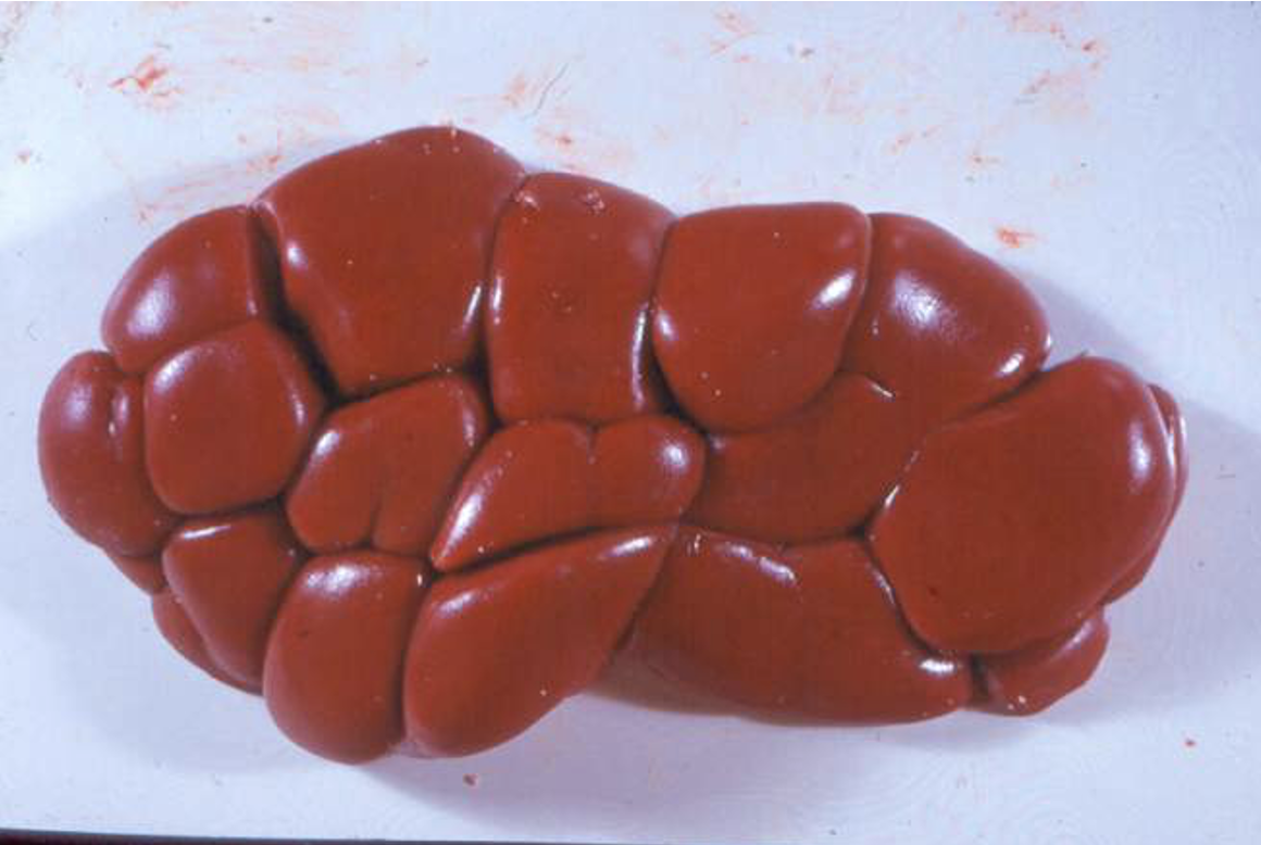
What does the affected lepto kidney look like
small white foci in tubules
Leptospirosis
Clinical signs of Lepto
Infection with host adapted serovars (L hardjo)
acute phase usually subclinical (apart from lactating cows)
repro disease: infertility, low conception rates, abortion, stillbirths, weak calves
infertility: uterus inflammation and embryo death
abortion: 6-12 weeks post infection, usually lasts 3 months of gestation, tend to affect younger cattle more frequently
milk drop: ‘flabby bag’, sudden onset of fever and agalactia, all 4 quarters of udder is soft and flabby producing yellow/orange secretion which may contain small clots, may affect >50% of cows at a time, milk has high leukocyte count therefore high SCC
Leptospirosis
Clinical signs in calves and youngstock (Rare)
often non-host adapted serovars - not the L hardjos
calves under 2 mo:
meningitis, anorexia, severe depression
opisthotonus (muscle spasm causing arching in head/neck), trismus (cant open mouth), muscle tremors, paddling
pyrexia (40.5-41.5)
ophthalmitis, hypopyon (accumulation of inflammatory cells, such as pus, setting in anterior chamber of eye), optic disc edema, congestion of retinal vessels
calves over 2 mo:
anorexia and dullness
rarely pallor, petichiation, jaundice, hemoglobinuria
Leptospirosis
How do we diagnose Lepto
Direct methods: dark ground microscopy (can see them), culture and identification (difficult), PCR, immunofluorescence/peroxidase in tissue
Indirect methods: serology ELISA on Blood or milk
Leptospirosis
Describe the antibody rise/drop of lepto
rise at first and may be associated with clinical disease
then they fall
abortion can take place with low levels of antibodies (up to 12 weeks after infection)
antibody is present in the serum of carriers and vaccianted animals
Leptospirosis
How would you diagnose lepto on a herd basis
easy on herd basis
serology - rising tire in paired samples taken 14 days apart, individual samples with tires >1:100 indicates chronic or active infection
abortion - fetal serology, culture
bulk milk ELISA now regularly used for surveillance
Leptospirosis
What are the 3 aims of lepto treatment
Aim:
to reduce the number of infected animals
to minimize urinary shedding
to reduce spread of organism to other cattle and other species including man
Leptospirosis
What abx do we use to treat lepto
Dihydrostreptomycin 25mg/kg (repeat after 7 days)
off data sheet - problems with milk loss if a whole herd tx
also sensitive to other abx including amoxycillin, oxytetracycline, tilmicosin
Leptospirosis
4 aspects to control of lepto
identification and removal of carrier animals
vaccination
test/treat/vaccinate replacements
hygiene with special attention to water supply
Leptospirosis
What are the 2 types of vaccinations used for lepto
Leptavoid (MSD)
Spirovac (Zoetis) - more common
- Vaccine availability: issues with leptavoid-H, farmers advised to boost with spirovac
Leptospirosis
When do you vaccinate dairy cattle?
Dairy: if in close contact with workers, raise replacements separately therefore heifers are naive, complete vac before breeding, spring booster
Beef: young stock usually acquire some level of immunity
Salmonellosis
Who does it affect? Zoonotic?
wide host range, various animals plus humans ZOONOTIC
only a few are important in cattle
serious economic health and public health implications
can hide from the immune system
Salmonellosis
What are the 3 important types? Which is the most common isolated in british cattle?
S. enterica Dublin - most common
S. enterica Mbandaka
S. enterica Typhimurium
Salmonella Mbandaka
Who does it affect? Clinical signs? Origin?
adults: diarrhea and malaise, also abortion
infected feed origin
mostly larger herds supplementing feed/housing all year (SW)
very rarely seen in humans
Salmonella Typhimurium
Who does it affect? Clinical signs?
affects mainly calves
various clinical signs
carrier animals
Salmonella Dublin
Who does it affect? Clinical signs? Origin?
host adapted
affects both calves and adult cattle
latent or persistent carriers
associated with abortion
infrequent in humans, but potentially fatal
via infected livestock or unpasteurized milk
Salmonellosis
What are the 4 routes of transmission
cattle to cattle: usually a fecal oral route
slurry : persists in slurry for months, soil for a year
Fomites: farm visitors, animals, birds, vehicles, equipment
Feed/water: watercourses and feedstuffs can be contaminated by other stock and wildlife
Salmonellosis
What are the transmission risk factors
buying in cattle/co-grazing
high stocking density, group pens
poor hygiene
concurrent disease - fluke, bvdv?
season
age/status - calves under 3 months, cattle in first 2 weeks of lactation though to be at a higher risk
Salmonellosis
Clinical signs
range of clinical signs - may be overlooked/under-diagnosed, severity may depend on infective dose and age/stage
acute or chronic enteritis
abortion
septicemia
reduced productivity
poor calf health
Salmonellosis - Acute Enteritis
Clinical signs of acute enteritis? Who does it affect?
high fever, severe diarrhea, sometimes bloody, anorexia, colic, abortion
severe dehydration
fatalities can be up to 75%
calves >2week and adults
Salmonellosis - Chronic Enteritis
Clinical signs of chronic enteritis? Who does it affect?
may follow acute enteritis
reduced weight gain, intermittent D+, inappetance
stressors can trigger disease - poor nutrition, long transport times, calving, mixing, crowding
Salmonellosis - Septicemia
Clinical signs of septicemia? Who does it affect?
mainly seen in neonatal calves (<2-3 weeks)
depression, fever, lethargy, labored breathing, nervous signs, rapid death (6-48 hours)
dry gangrene of extremities after initial phase
joint infections
Salmonellosis - Abortion
When does it occur? CS? Abortion storms?
usually 5-8 months of pregnancy
±/- fever and anorexia
retained placenta and reduced lactation
abortion storms: up to 25% of the herd
Don’t forget these things..
poor calf health
pneumonia
poor growth rates
illl thrift
meningitis
Salmonellosis
How do we diagnose on an individual case basis?
fecal culture: fecal sample NOT swab, pooling decreases sensitivity, remember previous use of abx will affect culture
PM: culture a range of tissues, in abortions culture fetal stomach contents
Serology:
best results in calves 3-10 mo
poor seroconverstion <12 weeks
cross reactivity
retrospective due to time taken to seroconvert
Salmonellosis
How do we diagnose on a herd level?
carrier animals: shedding may be intermittent, 3 serology tests over 8 months?
culture: slurry samples or feces of cases
serology: BMT, serology of all animals, serology of a subset of animals (calves, those showing clinical signs, 10 youngest calves over 12 weeks)
Salmonellosis
How do we treat?
early treatment is essential for septicemic salmonellosis - S Dublin usually sensitive to most abxs
controversy regarding the use of antimicrobials for intestinal salmonellosis - carrier status
intestinal cases may cure clinically but not bacteriologically
Salmonellosis
How do we control
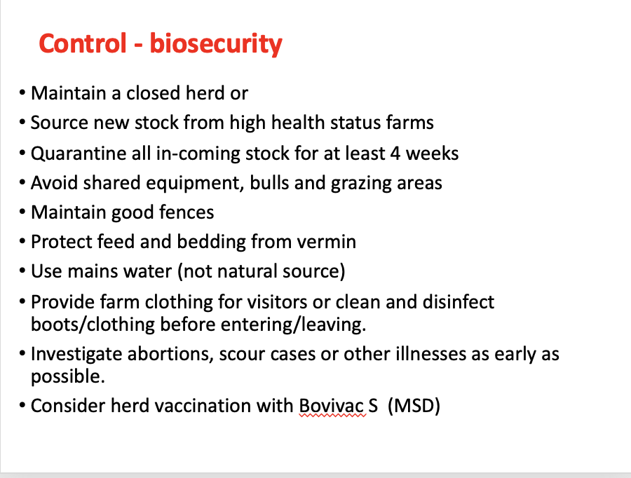
Negative herd = prevent entry (biosecurity)
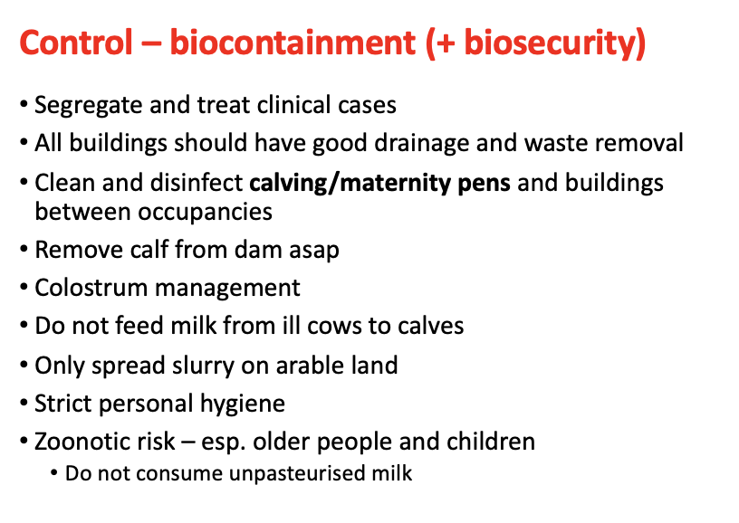
Positive herd = biocontainment (and biosecurity)
Vaccination
Salmonellosis
Biosecurity measures to take
Salmonellosis
Biocontainment measures to take
Salmonellosis
Which vaccinations are licensed for salmonellosis
Bovivac S (MSD) is the only one. contains both S Typhimurium and S Dublin.
inactivated vaccine
Malignant Catarrhal Fever (Snotsiekte)
How is it transmitted
efficiently between individuals of a natural host, inefficiently between other species
direct, aerosol, may be IMH
typically sporadic, multiple cases usually caused by close proximity of lambing ewes to housed cattle
reactivation - calving/lambing time
recrudescence is possible in recovered cattle
Malignant Catarrhal Fever (Snotsiekte)
Clinical Signs (brace yourselves ☹ it’s a lot)
head and eye peracute, intestinal
extreme dullness
anorexia
agalactia
copious mucopurulent oculo-nasal discharge ±/- blood
drooling of saliva
dyspnea and stertorous breathing
loss of condition
usually fatal - survive- 1 week
pyrexia 41C, 106F
congestion of scleral vessels, centripetal corneal edema, hypopyon
corneal neo-vascularization
diffuse oral ulceration with pain extending onto the rhinarium
generalized lymphadenopathy
dermatitis
cystitis +- pyuria
altered fecal consistency
SEE THE LECTURE FOR PICTURES
Malignant Catarrhal Fever (Snotsiekte)
Pathology
lesions in virtually every system
hydropic degeneration, vesicle formation and erosion in stratified squamous epithelium
ulcers coalesce and can become very extensive
vasculitis - perivascular cuffing with lymphoid cells
paracortical expansion in lymphoid tissues
SEE LECTURE FOR PICTURES
Malignant Catarrhal Fever (Snotsiekte)
Diagnosis? Treatment? Control?
Diagnosis:
antibodies in serum or from affected tissues
PCR for virus (tissue / whole blood)
exclude important DDX - mucosal disease, FMD, Bluetongue
Treatment:
euthanasia
supportive therapy
Control
avoid contact with sheep - lambing time