HP LEC 8: Characteristics of Sensory Receptors & Sensory Cells, Ears/Hearing
1/31
There's no tags or description
Looks like no tags are added yet.
Name | Mastery | Learn | Test | Matching | Spaced |
|---|
No study sessions yet.
32 Terms
Intro to sensory receptors (location)
sensory receptors are located in specialized cells (sometimes neurons but not always) that detect & respond to physical & chemical stimuli- “sensory receptor cells”
many sensory receptors cells are located at the surface to detect external stimuli, but others lie within body tissues to monitor internal organ functions & provide crucial homeostatic feedback regulation
Sensory Receptor Functions
can transduce (change) different forms of energy in the “real world” into nerve impulses
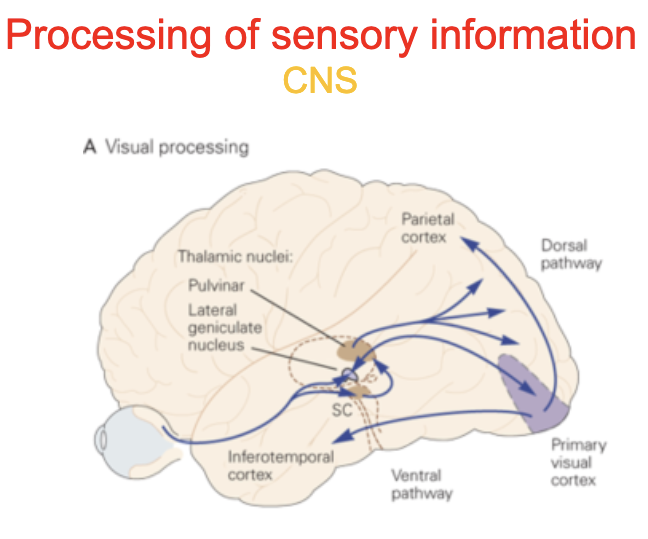
different modalities of sensations (sound, light, pressure, taste. odor) arise from differences in neural pathways & synaptic connections
ex: if the optic nerve delivers an impulse, the brain interprets it as light even though the nature of the nerve impulses (in the form of action potential) is the same as for hearing, taste, olfaction, etc
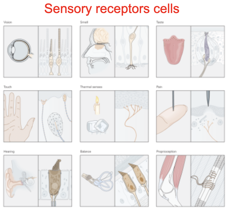
Functional Categories of Sensory Receptors: Type of Signal (5)
According to the type of signal they transduce:
Chemoreceptors: sense chemicals in external (taste, smell) or internal (CO2) environment
Photoreceptors: sense light
Thermoreceptors: respond to various degrees of heat
Mechanoreceptors: stimulated by mechanical deformation of the receptor (touch, hearing)
Nociceptors: sense stimuli that accompany tissue damage (high heat, high pressure acid)
Functional Categories of Sensory Receptors: type of information they deliver to the brain
according to the type of information they deliver to the brain
Proprioreceptors: found in muscles, tendons, and joints - provide a sense of body position & allows fine muscle control
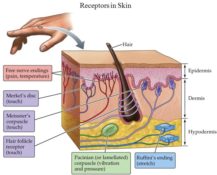
Cutaneous (skin) receptors: touch, pressure, heat, cold, and pain
Special senses: vision, hearing, taste, smell, equilibrium
Functional Categories of Sensory Receptors: origin of the information
according to the origin of the info:
Exteroceptors: respond to stimuli from outside the body; includes cutaneous receptors & special senses
Interoceptors: respond to internal stimuli; found in organs; monitor blood pressure, pH, and oxygen concentrations
Functional Categories of Sensory Receptors: how they respond to stimulus
according to how they respond to stimulus:
Phasic receptors
Tonic receptors
Generator (Receptor) Potential
sensory receptor cells behave very similarly to dendrites of neurons
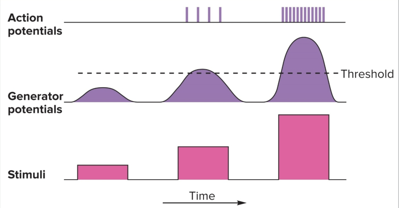
stimuli produce depolarizations called generator potentials
similar to EPSPs: it is a graded response
light touch on a Pacinian corpsucle in the skin produces a small generator potential
increasing the pressure increases the magnitude of the generator potential until the threshold is met and an action potential occurs
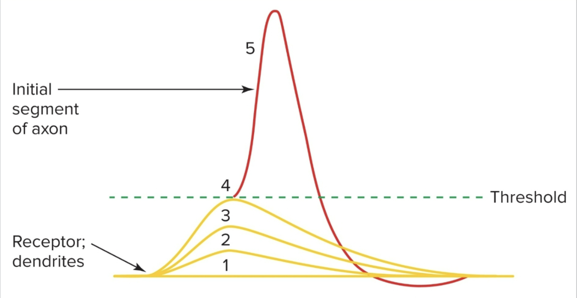
Generator Potential Graph
the generator (receptor) potential is (like the EPSP) proportional to the intensity of the stimulus
increased intensity results in increased frequency of action potential after threshold is reached
Generator Potential further def
stimulated dendrites of free nerve endings, encapsulated nerve endings, and olfactory receptors produce a generator potential which can reach threshold and generate an action potential
is a type of graded depolarization that occurs directly in the sensory nerve endings of afferent neurons, which are the neurons that carry sensory information toward the CNS
Generator Potential Steps & Parts
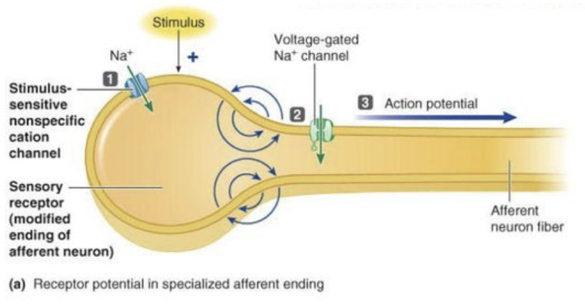
In sensory receptors that are specialized afferent neuron endings, stimulus opens stimulus-sensitive channels, permitting net Na+ entry that produces receptor potential
Local current flow between depolarized receptor ending and adjacent region opens voltage gated Na+ channels
Na+ entry initiates action potential in afferent fiber that self-propagates to CNS
Olfactory sensory neurons (OSN)- nasal epithelium, free nerve endings-skin, pacinian corpuscle-skin
Receptor Potential
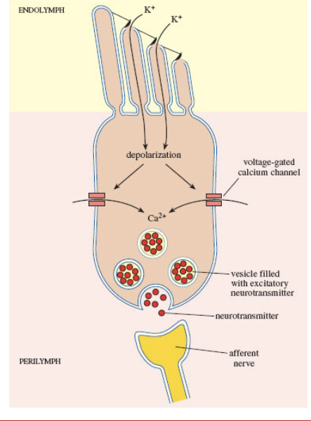
hair cells (ear), taste receptors, and photoreceptors do not generate potentials; their graded potentials tigger NT release & generate PSPs (postsynaptic potentials) in 1st order neurons
a receptor potential is a graded change in membrane potential occurring in specialized sensory receptor cells that are seperate from the afferent neurons; these receptors synapse onto afferent neurons and modulate neurotransmitter release based on the stimulus
Receptor Potential Steps & Parts
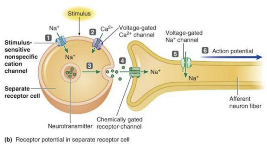
in sensory receptors that are seperate cells, stimulus opens stimulus-sensitive channels, permitting net Na+ entry that produces receptor potential
this local depolarization opens voltage gated Ca2+ channels
Ca2+ entry triggers exocytosis of neurotransmitter
nuerotransmitter binding opens chemically gated receptor-channels at afferent ending, permitting net Na+ entry
resultant depolarization opens voltage-gated Na+ channels in adjacent region
Na+ entry initiates action potential in afferent fiber that self-propagates to CNA
Photoreceptors- retina
hair cells- cochlea
taste cells- taste bud
Categories of Sensory Receptors
Tonic
Phasic

Tonic Sensory Receptors
maintain a high firing rate if the stimulus is applied (they can be no adapting or slow adapting)
ex:
no adapting: pain receptors, and proprioreceptors (A)
slow adapting: Merkel's discs and Ruffini corpuscles (touch and pressure), interoceptors (B)
Phasic Sensory Receptors
respond with a burst of activity when stimulus is first applied but quickly adapt to the stimulus by decreasing response - fast adapting (C)
a) allow sensory adaptation: cease to pay attention to constant stimuli
b) may deliver another burst when stimulus is removed to provide on and off information
c) ex: smell, touch (Pacinian Corpuscles), temperature- can actually occur with all senses (image on ipad)
The sense of hearing
The human ear ( and other animals) are sensitive detectors capable of detecting the fluctuations in air pressure that impinge upon the eardrum
Capable of detecting sound waves with a wide range of frequencies, randing between approximately 20Hz to 20000 Hz (Dolphins can detect frequencies as high as 200,000 Hz) ( Cats can detect frequencies as low as 45Hz and as high as 85000 Hz
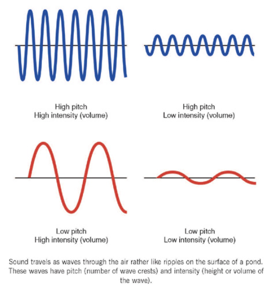
Intensity or loudness, measured in decibels
related to the amplitude of the wave
human optimal range is 0 to 80 dB
Frequency or pitch
the frequency of sound waves is measured in Hertz (Hz), or the # of waves that pass a fixed point in a second
The Ear
Outer Ear
Middle Ear
Inner Ear
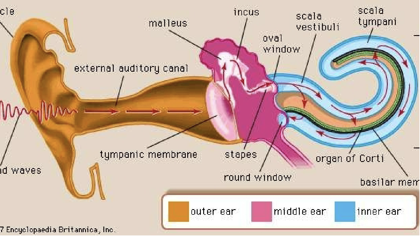
Outer Ear
external ear: Pinna or auricle and external auditory canal- sound gathering
Outer Ear Info
Sound waves enter the outer ear and travel through a narrow passageway called the ear canal (external auditory meatus), which leads to the eardrum or tympanic membrane
The eardrum vibrates from the incoming sound waves and sends these vibrations to three tiny bones in the middle ear
Middle Ear
made of tiny bones- modulation of sound vibrations and transfer to the inner ear
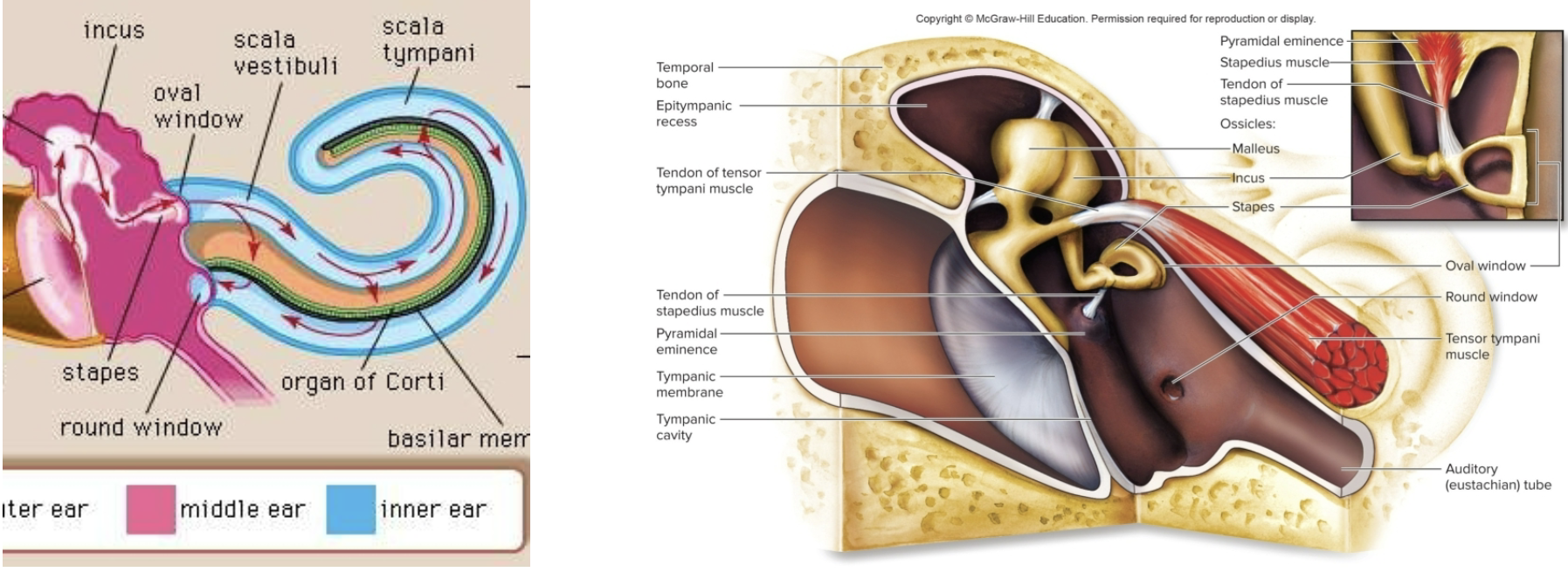
Medial View of the Middle Ear
Air-filled cavity between the tympanic membrane and the cochlea
contains 3 bones called ossicles:
a) Malleus, incus, and stapes
b) vibrations are transmitted and amplified along the bones
c) the stapes is attached to the oval window, which transfers the vibrations into the cochlea in the inner ear
d) stapedius muscle dampens the stapes if the sound is too intense
Inner ear
cochlea- transduction of sound vibration into electrical signals
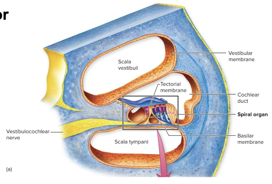
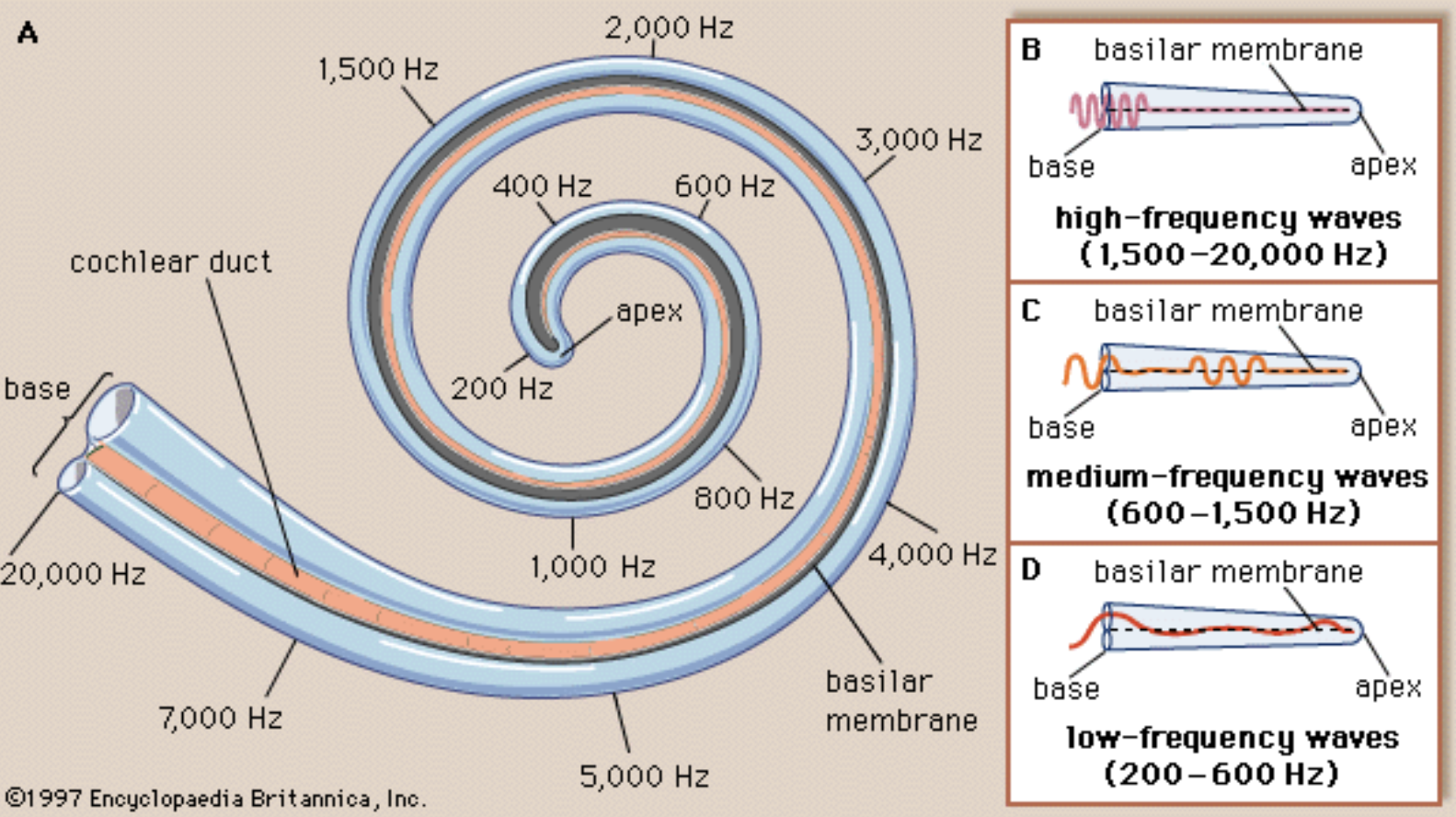
The Cochlea
The cochlea is the hearing part of the inner ear. It has a characteristic snail-shaped structure, it is composed of 3 chambers
Upper Chamber: Scala Vestibuli
Lower Chamber: Scala Tympani- both chambers are filled with perilymph
the cochlea also contains a portion of the membranous labyrinth called the scala media, or cochlear duct, filled with endolymph. This chamber contains the sensitive element in the inner ear the organ of Corti (spiral organ)
Endolymph (high K+) and perilymph vary significantly in their concentration of ions, this difference is essential to the overall function of the cochlea
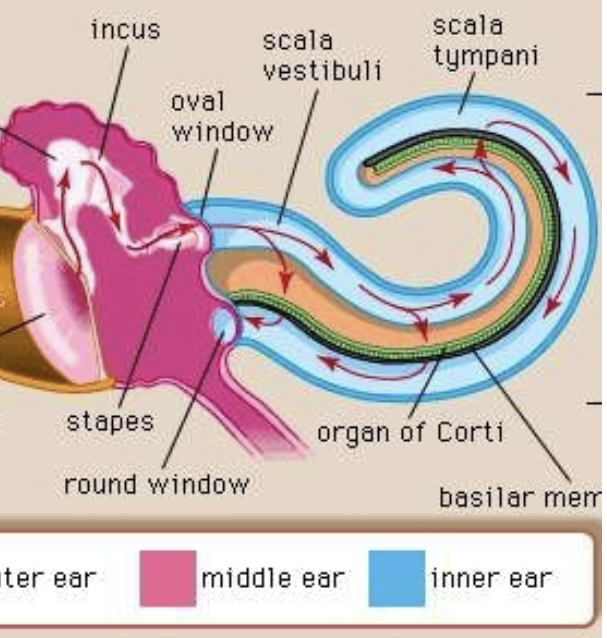
The Cochlea’s journey of sound vibration reaching the organ of the Corti
Vibrations from the oval window of the middle ear displace perilymph in the scala vestibuli
Vibrations pass through the vestibular membrane into the scala media (or cochlear duct) through the endolymph
Next, vibration pass through the basilar membrane into the perilymph of the scala tympani
Vibrations leave the inner ear via the round window
Spiral Organ (Organ of Corti)
The organ of Corti is the sensitive element in the inner ear and can be thought of as the body’s microphone
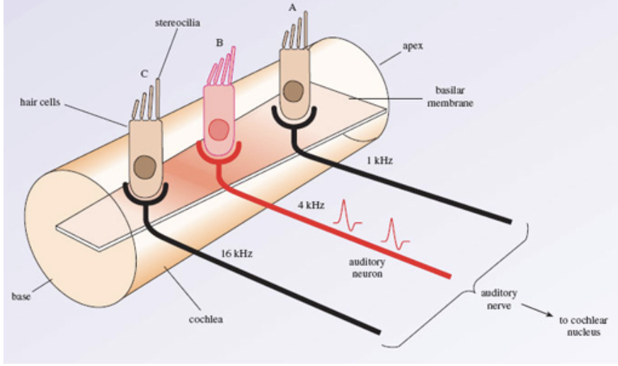
Sensory Hair cells are located on the basilar membrane, projecting into the endolymph (high K+ of the cochlear duct)
Inner hair cells and Outer Hair cells
Sensory Hair cells: Inner & Outer
Inner Hair cells: they transform sound waves into nerve impulses, each is innervated by 10 to 20 sensory neurons of cranial nerve VIII and relay a sound
Outer Hair Cells: their role is mostly to amplify softer sound and sharpen pitch perception by changing their length, such movement are believed to aid the sensory function of the inner hair cells
How Hearing Works
When sound waves enter the scala media, the tectorial membrane vibrates, bending stereocilia of the inner hair cells
1) Opens mechanically-gated K+ channels that are facing the endolymph
2) K+ rushes in, depolarizing the cell- ionic gradients are unique to the endolymph (High K+)
3) The hair cell itself does not fire an action potential, instead the influx of positive ions from the endolymph in the scala media depolarizes the cell, resulting in a receptor potential
4) releases glutamate onto sensory neurons
5) The greater the amt of basilar membrane displacement and bending the stereocilia, the more glutamate is released, producing a greater receptor potential
The Cochlea: Tonotopic Organization
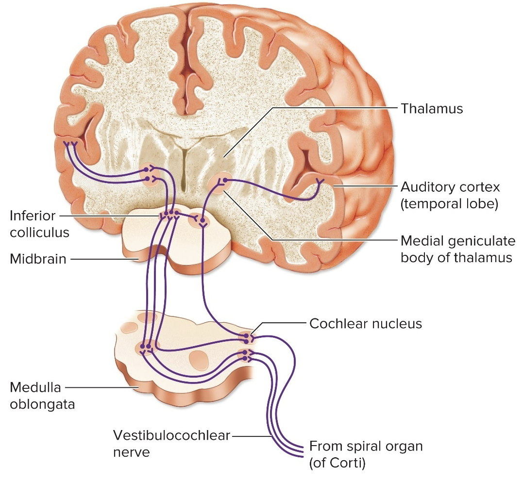
Neural Pathways (5)
Vestibulocochlear nerve (cranial nerve VIII (8th)
Cochlear nuclei in the medulla oblongata & pons
Inferior colliculus of midbrain
Medial geniculate body of the thalamus
Auditory cortex of temporal lobe
Hearing Impairment-1
Conduction deafness: sound aves are not conducted from the outer to the inner ear
may be due to a buildup of earwax, too much fluid in the middle ear, damage to the ear drum, or overgrowth of bone in the middle ear
impairs hearing of all frequencies
can be helped by hearing aids
Hearing Impairment-2
Sensorineural/perceptive deafness: Nerve impulses are not conducted from the cochlea to the auditory cortex
may be due to damaged hair cells (from loud noises)
may only impair hearing of a particular sound frequencies and not others
may be helped by cochlear implants
Hearing Impairment-3
Presbycusis- age related hearing implant