PSC 100 : Final Exam Review
1/282
There's no tags or description
Looks like no tags are added yet.
Name | Mastery | Learn | Test | Matching | Spaced | Call with Kai |
|---|
No analytics yet
Send a link to your students to track their progress
283 Terms
LEC 1: Cellular Neuroanatomy
Lec 1
Neurons and their Fxn
Specialized cells only in NS→Communicates info by chemical/ electrical signals
Collect info from other cells →Send info to other cells
Limitations of a Single Neuron
A single neuron in isolation cannot do too much
Cannot do complex processes needed: Conscious thought, recalling memories, processing sensorty info
Neural Networks
System of cells working together → Mass communications allows brain to do these complex processes
1.1 Q’s: What is the primary function of neurons
Communication
T/F: A single neuron is enough to perform a complex process like recalling memory
False:
Neural networks→ System of cells working together
Draw a typical neuron and label following structures: Dendrites, Cell Body, Axon Hillock, Axon, and Synaptic Terminal
Draw it!!
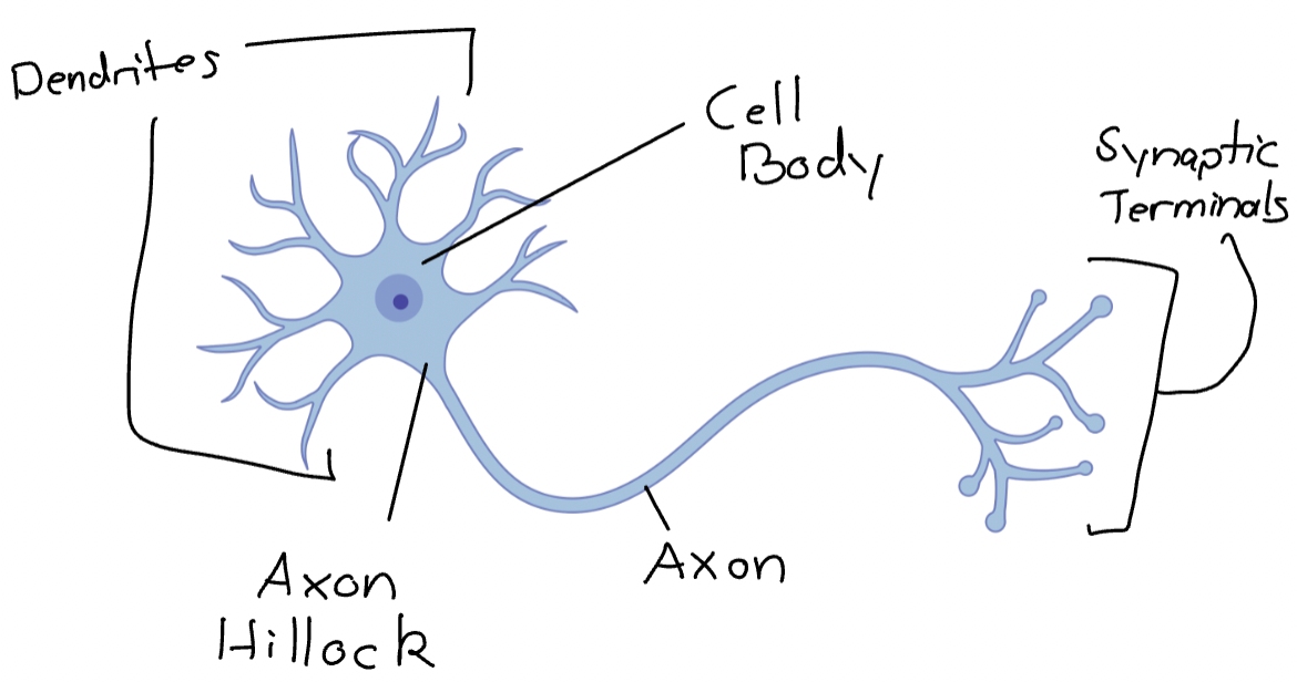
Dendrites
Specialized structures for collecting info from many axons of other cells.
Dendritic Spines: Tiny structures on dendrites for a single axon to connect to
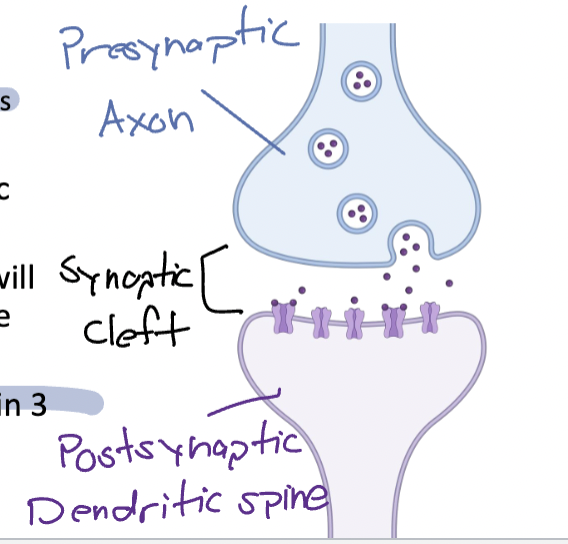
Synapses
Where axons meet dendritic spines
Post synaptic(Top) dendrites receive info from the presynaptic axon
Cell Body
AKA Soma: Contains all organelles allowing cell to fxn/survive(Nucleus, mitochondria, etc)
Most importantly, Location of gene expression and protein production
Axon Hillock
“Little Hill” → Starting location of AP’s
Info from dendrites pass through cell body to reach axon hilock
Filled with Voltage Gated Na+ channels and K+ channels
Axon
Long tube electrical currents travel through (AP’s)
Cord that AP’s travel from cell body → axon terminal
Synaptic Terminal
AKA: Axon Bouton→ End of axon where axon meets its target(Dendrite)
Filled with structures/proteins needed for chemical signaling
Location of Neurotransmitters(NT)
1.3 Electrical Properties
Axons great at conducting electrical signals BUT electrical charge is constantly leaking/ dissipating as it travels dow axon
T/F: Electrical charge constantly leaking is a major issue for short axons
FALSE: Electrical charge constantly leaking is a major issue for longer axons
AP will dissipate before it arrives at the end
Myelin
Layers of fatty tissue that wrap around long axons → Help APs: Travel farther (less dissipation)/ Travel Faster
Nodes of Ranvir
Segmented with small areas of exposed axon, important for AP travel
White Matter
Part of NS densely packed with axons wrapped in myelin → No information processing, just carrying information
Gray Matter
Part of brain w/ little-to-no myelin → Primarily Cell Bodies/Dendrites = All processing happens
Multiple Sclerosis(MS)
Disorder causing myelin in brain/spine to be progressively destroyed -→ APs don’t always reach their targets
Symptoms: Muscle Weakness, Vision issues, Loss of Sensation, Tremors
1.3 Q’s: Why do APs travel farther and faster in myelinated axons when compared to unmyelinated axons
The axon is better insulated, so less electrical charge leaks out the axon
Which axon would you expect to NOT be myelinated?
Axon communicating with the next closest cell
Typically grey matter, cell body/dendrites → All processing happens
T/F: White matter is primarily made from cell bodies and dendrites of many neurons
FALSE! White matter is primarily made of densely packed axons wrapped in myelin→ just carry information
Glial Cells
Specialized glial cells performing diff fxn’s, don’t work in isolation
Astrocytes→ Deliver energy to neurons, Clears excess NT from synapses, Filters blood before reaching the neuron, Structural support, Promote synaptic information
Myelinating Glia → Forms/wraps myelin around the axon of neurons
Oligodendrocytes: CNS:Brain and Spine
Schwann cells: PNS: Everywhere else
Microglia → NS “white blood cells” , immune cells clearing out debris, destroying invaders, support healing to damage
Astrocytes
Wide range of fxn’s:
Deliver energy to neurons
Clears excess NT from synapses
Filters blood before reaching the neuron
Structural support
Promote synaptic information
“Star Cell” → Blood-brain Barrier (Wrap around blood vesicles in brain filtering contents)
Allow in good stuff, blocks out bad (Cannot filter out everything)
Myelinating Glia
Forms/wraps myelin around the axons of neurons
2 classes of glia:
Oligodendrocytes → CNS: Brain and Spine
Schwann cells → PNS: Everywhere else
Microglia
NS immune cells , “White blood cells”
Migrate around brain to:
Clear out debris, Destroy invaders, Support healing to damage
1.4- Glia Q’s: Select all fxn’s that are done by astrocytes:
Promotes synaptic formation, Filters blood before it reachers the neurons, Clears neurotransmitters from synapse
Which cells form myelin in the CNS?
Oligodendrocytes → CNS
Schwann cells → PNS
LEC 2: Gross Neuroanatomy
Lec 2
NS Intro
NS organized/ complex, thoroughly named and subdivided:
2 major branches
Central Nervous System (CNS)
Peripheral Nervous System (PNS)
CNS
Comprised of Brain/ Spinal Cord → Where all processing happens, housed in boney structures for protection (Skull/Spine)
PNS
Everything outside of the CNS→ Connects CNS to all sensory receptors, muscles, and organs
2.1 NS Branches: What is something that the CNS and PNS don’t have in common?
Encased in bone
T/F: The brain is part of the Central Nervous System
TRUE!! CNS → Brain/Spinal Cord
T/F: The spinal cord is part of the PNS?
FALSE!! The Spinal Cord is part of the CNS, PNS is everything else
PNS
Dived based on which direction information is moving
Afferent (Sensory) System
Info is coming from the body INTO the CNS
Efferent (Motor) System
Info is coming from the CNS out to the body
The Afferent (Sensory) System
AKA: Sensory Pathway
Sensory info (Touch, pain) travels along axon into the CNS → Touch receptors activated send AP along a nerve to arrive at the spine
The Efferent (Motor) System
AKA: Motor Pathway
APs from CNS travel along axons to stimulate muscles → Further divided based on type of muscle the axons communicate w/:
Somatic
Connects to all skeletal muscles, voluntary control, just carries info from the CNS
Autonomic
Somatic Motor System
Connects to all skeletal muscles (AKA: Muscles used to move)
All voluntary control → PNS system doesn’t decide what movements to do, just carries the info from CNS
Autonomic Motor System
Connects to all smooth muscles—AKA: Muscles making up organs( Heart, blood vessels, intestines, pupils)
All involuntary control ( 'Autonomic’ name)
Further subdivided
Sympathetic- Fight or Flight
Parasympathetic- Rest or Digest
The efferent division of the PNS carries information:
To muscles
Which pathway carries information for “fight or flight” responses?
Sympathetic
S = Stressed → Fight or Flight
Info about controlling muscles travel along which division of the PNS?
Somatic
CNS Collaboration
Brain areas all across CNS working in tandem all the time to accomplish highly complex processes
No one brain area works in isolation
Still processes we don’t understand in each of these areas
CNS Structures
Spinal Cord
Brain Stem
Cerebellum
Subcortical Structures
Thalamus, Hypothalamus, Basal Ganglia, & Hippocampus
Cerebral Cortex
Frontal, Parietal, Temporal, & Occipital Lobes
Spinal Cord
Where all info passes through bw brain to PNS
Primarily a dense cord of axons carrying info → Doesn’t do any processing , only carries info bw brain and rest of body
Spinal Cord- Organization
Organized based on:
Direction info is moving
Where info is going
Type of info traveling
Info passes in and out in each vertebra
Less info in spinal cord the further down you are
Spinal Cord- Damage
Extremely imp/delicate structure → Protected by spine
Damage to spinal cord greatly influences info traveling to/from body
Impacts more significant based on how close it is to the brain
Midback Injury
Possible loss of motor control/sensation to legs
Mid-shoulder Injury
Possible loss of motor control/sensation to arms, torso, & legs
Neck Injury
Complete paralysis or death
Brainstem
Top of spinal cord
Almost all info passes through the brainstem
First site of processing → Sends out commands
Controls foundational physiological processes:
Heart Rate, Breathing, Blood Pressure, Maintaining consciousness
Some foundational processing of auditory info:
Where sounds are coming from
Brainstem-Damage
Extremely impactful → Strokes in brainstem can be fatal (Damages areas responsible for breathing/heart rate)
Locked-in Syndrome
No info in or out → Conscious, but can only move eyes
Cerebellum
“Little Brain” → Importance in fine motor movement/ fact-checking movements
BUT! Doesn’t initiate motor movements
Cerebellum- Damage
Struggle with fine motor function/ adapting to changes
Such as: Smooth movement of limbs
Unable to error check as they do a movement
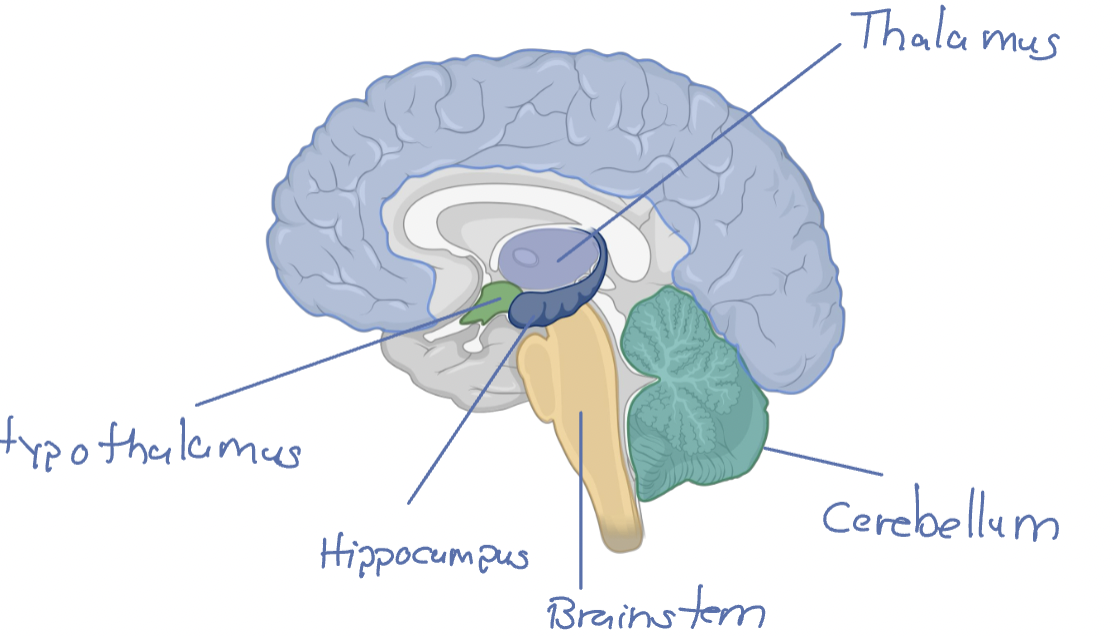
Subcortical Structures
Multiple distinct areas found in innermost region of brain
Thalamus, Hypothalamus, Basal Ganglia, and Hippocampus
Located under(sub) the outer most region (cortex) and above the brainstem
Thalamus
Relay station for info in brain
In and out of the brain / Between brain areas
Filtering and regulating flow of info
Thalamus- Damage
Strokes in thalamus may have issues with:
Arousal and pain regulation
Sensory Experiences
Motor Language fxn
Cognitive fxn, mood, and motivation
Hypothalamus
Hypo= Below → Below thalamus
Important for internal regulation/ homeostasis
Body temp
Appetite
Circadian Rhythms
Primarily responsible for processes controlled by hormones
Pituitary gland is connected → Master gland controlling hormone regulation
Hypothalamus- Damage
Hypopituitarism:
Under stimulation of pituitary gland
Issues w/: Metabolism, Stress Regulation, Puberty, Growth
Hypothalamic Obesity
Uninhibited eating → Brain never receives signal that person is “full”
Basal Ganglia
Regulates motor control → Highly connected with frontal cortex
Basal Ganglia- Disorders
Parkinson’s Disorder
Hypokinesia: Reduced movement
Huntington’s Disorder:
Hyperkinesia: Uncontrollable increase of movement
H for Huntington’s = Hyperkinesia
Possibly: Obsessive Compulsive Disorder(OCD):
Similar to Hunington’s → Instead of uncontrolled movement, is uncontrolled cognitive proceses
Hippocampus
“Sea Horse” → Importance for Memory Consolidation ( Short-term → Long-term)
Also important for spatial navigation
Hippocampus-Damage
Anterograde amnesia
Unable to create new declarative memories
Label Subcortical Structures!
2.3 Q’s: Huntington’s disease is associated with a malfunction in which brain area?
Basal Ganglia → Regulates motor control
Huntington’s disease= Hyperkinesia → Uncontrolled increase of movement
Every brain location where all info coming and going from the PNS passes through:
Spinal Cord, Brain Stem, & Thalamus
A person suffered from a stroke and no longer has the ability to feel satiated/full, even though they just ate a full meal. They also now experience difficulty with their sleep cycle. Which brain area was most likely affected by the stroke?
Hypothalamus
Cerebral Cortex
Responsible for things aware of:
Thinking, Self Awareness, Motor control, Memory, Learning, Language
All share the same general structure:
Flat sheet of cells
Constructed of 6 layers
Each layer made of many cells
Flat sheet’s crinkled to fit inside skull in a space efficient way
Cause of folds/wrinkles → AKA gyri and sulci
MORE FOLDS= MORE AREA FOR PROCESSING
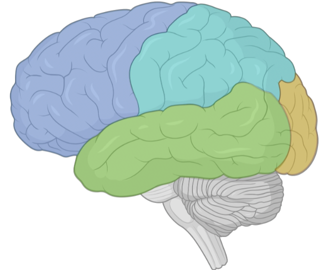
Subregions of Cerebral Cortex
4 lobes
Frontal
Parietal
Temporal
Occipital
Frontal Lobe
Frost most region → Responsible for Abstract processes/ high-level processes
Functions:
Executive Fxn (EF)
Working Memory(WM)
Planning Movements
Controlling movements
Parts of language processing and production
Parietal Lobe
Upper back portion → Wide range of fxn’s:
Spatial Info
Touch
Attention/ Eye movements
Temporal Lobe
Lower-lateral portion → Diverse in its fxn
Auditory processing
Object categorization
Support hippocampus for memory
Occipital Lobe
Back most region → Almost completely dedicated to Visual Processing
Receives direct connections from eyes
VERY visual organisms→ Dedicated great deal of cortex towards processing visual info
Label the 4 Lobes
2.4 Q’s : T/F: All regions of the cortex has 6 layers and those layers are equal thickness across all areas
FALSE!! All regions of the cortex have 6 layers but the thickness varies across all areas
Following a traumatic brain injury(TBI) to the back of the head, a person is having difficulty directing their attention, processing spatial info , and controlling eye movements. Which brain area was most likely affected by the TBI
Parietal Lobe
T/F: Most severe neurological damage is irreversible
TRUE!! Neurons don’t regenerate → Most severe neurological damage is irreversible
LEC 3: Development
Lec 3
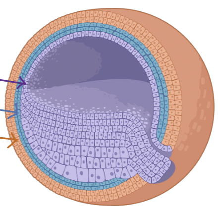
Gastrula
Stage of development where precursors of major organ systems begin to be created
Named after creation of cavity that will become the gastrointestinal (GI) tract: AKA - Stomach, colon
3 major layers of cells
Endoderm (Inner)
Mesoderm (Middle)
Ectoderm (Outter)
Ectoderm
Precursor cells for : Skin cells/ NS
Nearly identical to each other → Develop into specific cells based on external chemical signals
Cell Fate: Determine what type of cell a stem cell will develop into
Chemical Signals
Cellular processes start/stop when chemical signals come in contact w/ cell surface
Chemical Signals- Noggin
Chemical signal determining which ectoderm cells become the NS
Ectoderm cells → Presence of Noggin → NS cells
Ectoderm cells → No Noggin → Skin cells
Naturally produced by cells along back side of gastrula pore
T/F: All cells that come in contact with noggin will become the NS
TRUE!! Presence of noggin→ NS
Neurulation
Ectoderm cells exposed to Noggin pinch closed to form neural tube
Neural tube develops over time to form brain/spinal cord
Exposing other side of gastrula with Noggin
Second NS will be created → Creates a new head (AKA Noggin)
3.1 Q’s: Which of the following is not a layer of the gastrula : Hypoderm, Endoderm, Mesoderm, Ectoderm
Hypoderm
Endoderm (Inner)
Mesoderm (Middle)
Ectoderm (Outter)
If an ectoderm cell is not exposed to Noggin, what type of cell will it develop into?
No noggin → Skin cell
Sonic Hedgehog(SSH)
Chemical signal determines axis
AKA: What is front / what is back of NS
Secreted by the base of the neural tube
VERY high concentration at base
Very low concentration at top
Amount of exposure to SHH will determine cell fate
Top= Low conc. → Back of NS
Medium conc. → Middle/ sides of NS
Base = VERY high conc → Front of NS
3.2 Q’s:SHH is released by which region of the neural tube
Base of neural tube
Which factor of SHH would determine a cell’s fate?
Concentration
Low conc. at top of neural tube= Back of NS
High conc. at base of neural tube= Front of NS
Growth Cone
Tip of axon will have a specialized structured called a growth cone → Network of Filopodia (“Little Feet”), feel around/grab onto surfaces
Will literally PULL the axon forward → Axon grows longer as growth cone pulls it forward
Growth Cones-Chemical Signals
3 types of "Cues”:
Permissive Cues
Allow growth cone to attach and survive → W/o them axons wouldn’t have nothing to latch onto→ No access to nutrition for survival
Attractive Cues
Encourage axon to grow towards them → Growth cone follows cue up conc. gradient ( Moves to where there is more of the cue) → Follows cue to final destination
Repulsive Cues
Encourage growth cone to grow AWAY from chemical signal → Travel down conc. gradient to get away from it
Dynamic Chemical Cues
Same chemical signal can be a repulsive cue for one growth cone, but an attractive cue for another
How millions of axons find correct loc. and not get mixed up w/ wrong axons
Pioneering axons
Only first connections depend on growth cones/chemical signals → All relevant axons grow along pioneering axon
3.3 Q’s: Which structure is used to navigate an axon to its final destination?
Growth cones
A growth cone will cause an axon to grow ___ an attractive cue and __ from a repulsive cue
Towards attractive cue, Away from a repulsive cue
How do growth cones navigate to their final destination ?
Guided by chemical cues
Synaptic Refinement
NS sends out more axons than it actually uses → More efficient to send out more than needed than to refine down to best connections
Developing axons in constant competition w/each other.
Synaptic Refinement- EX
Cell A and B both have axons that terminate onto dendrites of Cell C
Cell C responds better from cell A signals: More in sync
Cell C doesn’t respond well to cell B signals: Less in sync
Cell C dedicates more resources to reinforce synapse w/ Cell A → Fewer resources for synapse w/ Cell B
Cell A synapse will get stronger
Cell B synapse will die away
Hebbian Plasticity
Synapse is strengthened bc pre/post synapse cells are co-activated
“Cells that fire together, wire together”
“Use it or lose it”
Experience Based Plasticity
Some neurons that are more active due to experience will stay, while under stimulated neurons will die away
Cat ODC ex
One eye sutured close for first 2.5 months of life → Brain only retained connections from the active eye .
Axons of uncovered eye will communicate to ODCs (Strengthen its synapses)
Axons of covered eye will not communicate as much w/ODCs: Synapses won’t be strengthened → Leading to them dying away
Cat ODC-Critical Period
ONLY happens during early development → Cat w/ eye covered after 1 year of normal exposure will not have the same degree of change
Critical Period: Limited time window the brain is extremely sensitive to a specific stimuli