Lecture 61: Nervous System Pathology 5
1/79
There's no tags or description
Looks like no tags are added yet.
Name | Mastery | Learn | Test | Matching | Spaced | Call with Kai |
|---|
No analytics yet
Send a link to your students to track their progress
80 Terms
primary neoplasms of the CNS in domestic animals occur most frequently in
dogs
embryonal tumor
originate from primitive cells in the CNS
described sporadically in cattle, horses, and dogs
found most commonly in young animals and are aggressive biologically
what type of embryonal tumors are most common in young animals?
medulloblastomas
medulloblastomas
arise in the cerebellum, usually in close proximity to the vermis, and invade into adjacent structures
macroscopic lesions for medulloblastomas
well circumscribed, soft, gray to pink masses
usually do not cause hemorrhage and/or necrosis or form cysts
expansile growth can compress the fourth ventricle causing obstructive hydrocephalus
metastasize throughout the ventricular system
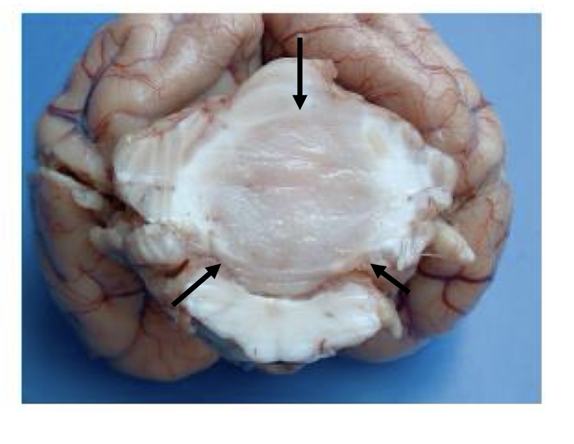
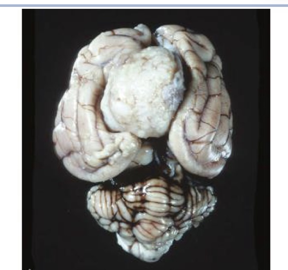
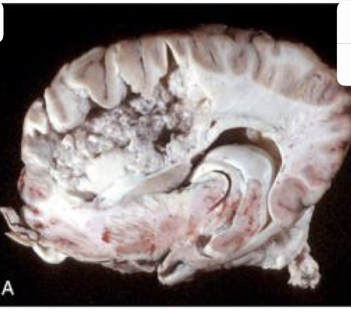
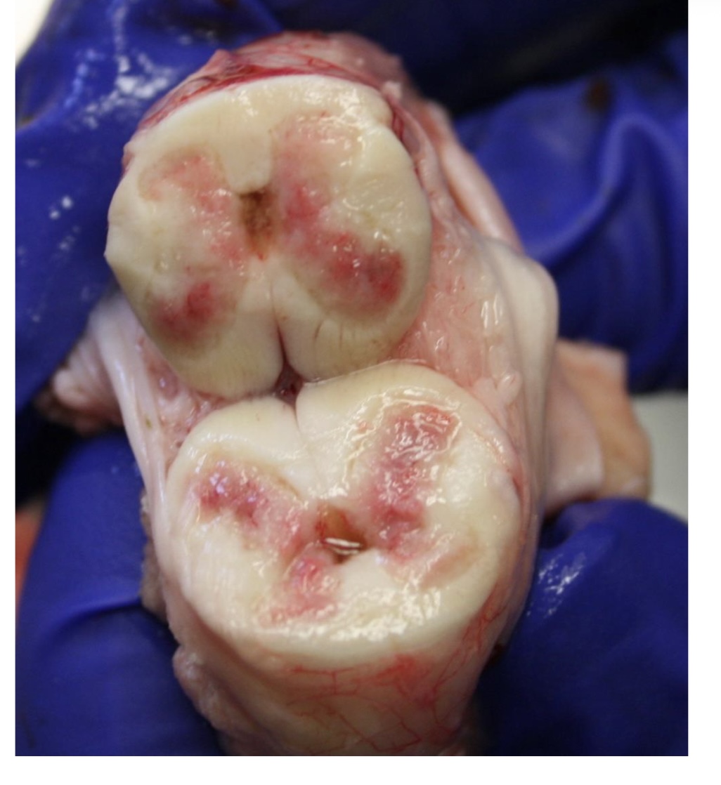
what is this?
medulloblastoma → white to tan, homogenous mass that compresses the cerebellum and brainstem
astrocytoma
reported most commonly in dogs and cats, rarely in horses, cattle, and pigs
brachycephalic breeds are the most commonly affected and have peak incidence from ages 5-11
common sites include the cerebral hemispheres
true or false: astrocytomas are 20-30% incidence of all gliomas.
true
lesions of astrocytomas
often displace normal tissue
poorly defined, firm and white to pink (lower grade)
regions of hemorrhage, necrosis, cavitation (higher grade)
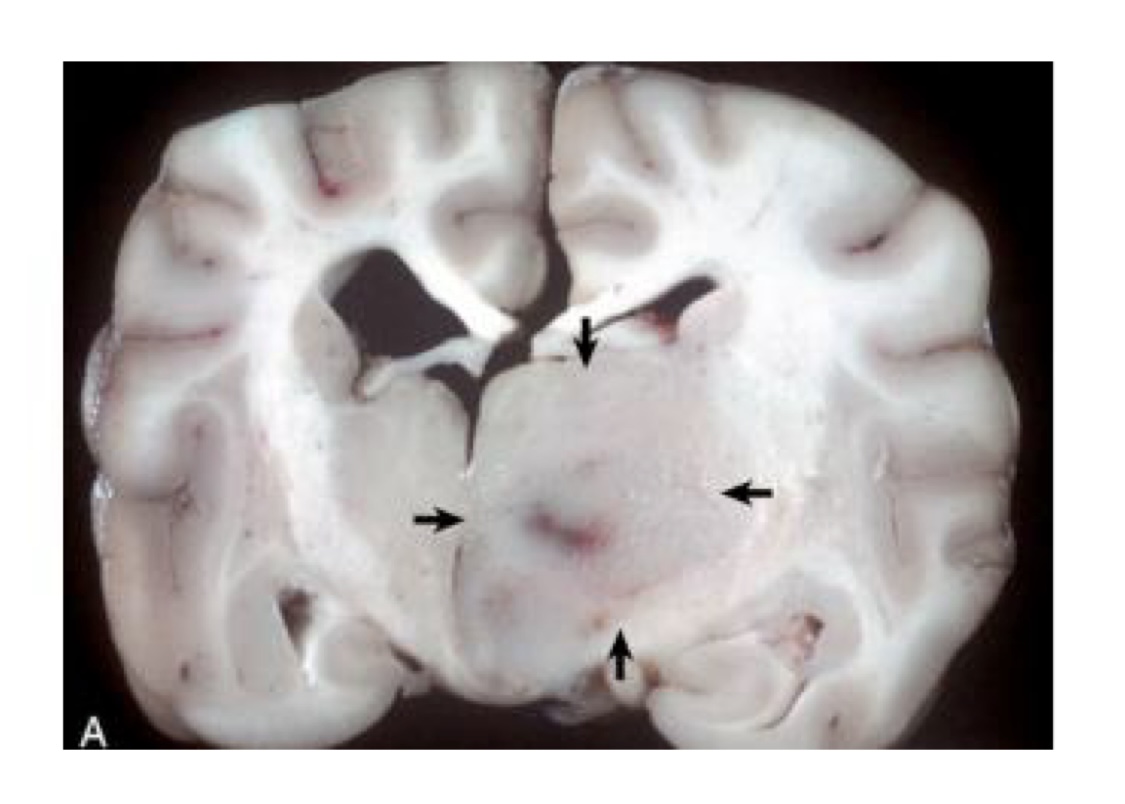
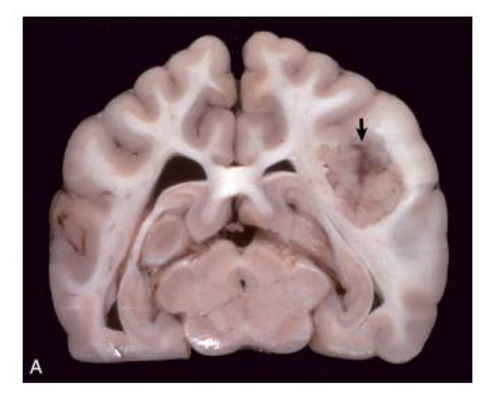
what is this?
astrocytoma
right hemisphere contains a poorly demarcated, nonencapsulated, expansile mass
lesion has displaced the midline to the left and compressed the right lateral ventricle
oligodendroglioma
most common in dog
most common glioma
predilection for brachycephalic breeds
age range 5-11
occur in all areas of the cerebrum and brainstem, especially in close proximity to the lateral ventricles
what is the most common glioma?
oligodendroglioma
oligodendroglioma lesions
well-demarcated mass of variable size
gray to pink-red and soft to gelatinous with areas of hemorrhage
larger tumors, the central area may be cystic
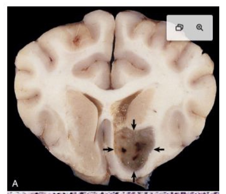
what is this?
oligodendroglioma → tumor is arising in a periventricular location and is gray, soft, and gelatinous
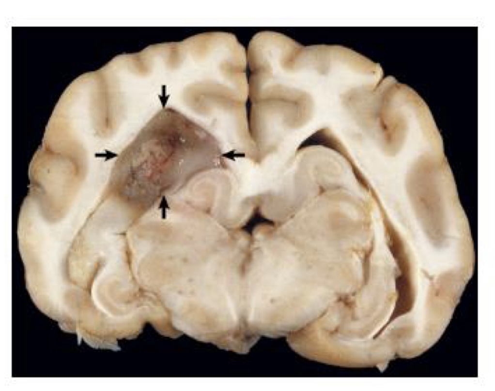
what is this?
oligodendroglioma → tumor is gray, soft, and gelatinous
ependymoma
most common in the cat
involve the lateral as well as third and fourth ventricles
can occur in central canal of spinal cord
can metastasize to subarachnoid space due to flow of CSF
lesions for ependymoma
large expansile intraventricular masses with generally well-demarcated margins
soft and gray-white to red, depending on blood content
smooth cut surface in dogs
more granular cut surface in cats
aggressive tumors show invasion into the normal tissue at its margins
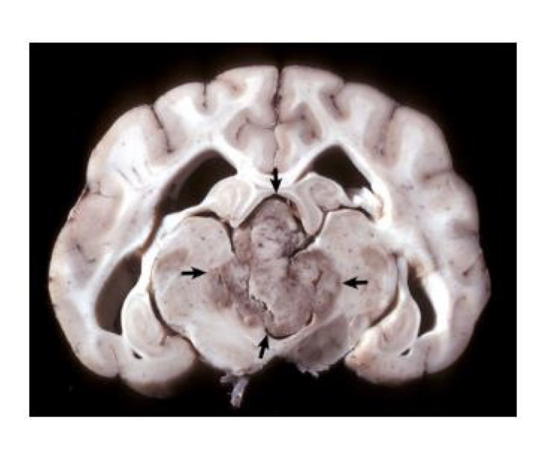
what is this?
ependymoma → third ventricle contains a moderately well demarcated expansile mass that has invaded normal tissue ventral to it; moderate hydrocephalus in both lateral ventricles
choroid plexus tumors
most common in dogs
5-10% of all primary brain tumors in middle aged to older dogs
breed predilection reported for golden retrievers
occurs most frequently in fourth ventricle
lesions of choroid plexus tumor
well-defined, expansive, granular to papillary growth located within ventricular system
gray-white to red and compresses the adjacent nervous tissue
can develop to hydrocephalus due to obstruction
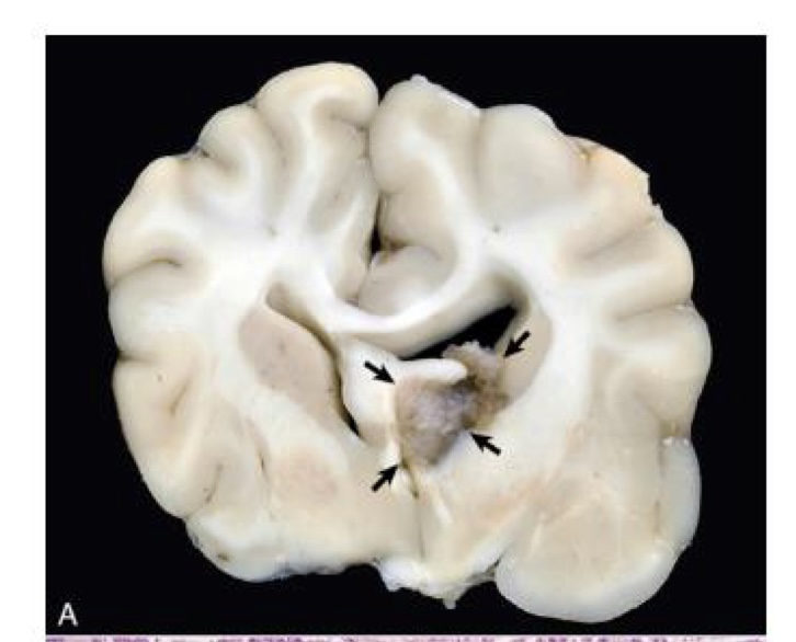
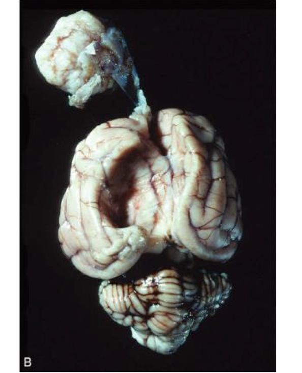
what is this?
choroid plexus carcinoma → lateral ventricle contains an expansile mass that has partially filled the ventricular lumen and is associated with midline shift
what is this?
choroid plexus carcinoma → 3rd ventricle has expansile mass with metastasis
what is the most common neoplasm of the CNS in dogs and cats?
meningiomas
meningiomas
occurs in dogs between 7 and 14 years of age and in cats 10 years old or older
dog → basal and lateral area of the brain, surface of the spinal cord
cats → cerebral hemispheres, over the cerebellum
where do meningiomas arise from?
the arachnoid cell layer and project into subdural space
since meningiomas grow slowly, what can they cause?
pressure atrophy of the adjacent nervous tissue
lesions of meningiomas
solitary and vary in size
well defined, spherical, lobulated, or plaque-like in shape
firm, encapsulated, gray-white
cut surface has soft, red, brown, or gray areas of hemorrhage and necrosis
lesions of meningiomas in cats
most always compressive and not invasive → can be shelled out
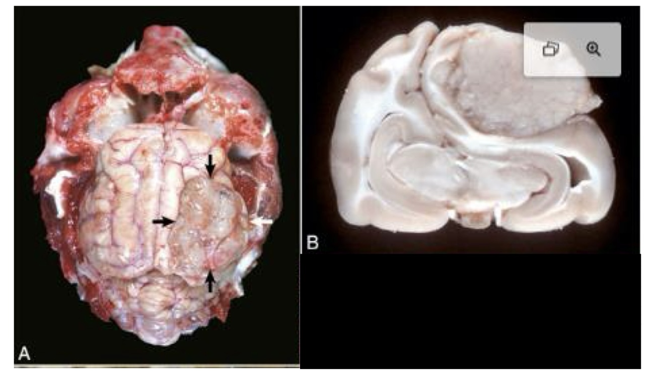
what is this?
meningioma → surface of the right parietal cortex is a mass that has compressed and distorted the adjacent parenchyma
what is this?
meningioma → occupies the longitudinal fissure and displaces the left and right cerebral hemispheres laterally
what is this?
meningioma with the neoplasm shelled out
lymphosarcoma
primary CNS lymphoma occurs in all domestic animal species → most common in dog and cat
in cattle, there can be manifestations of bovine leukemia virus that can result in
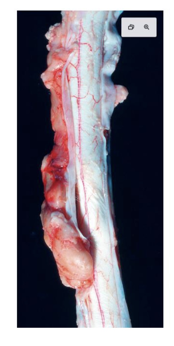
lymphosarcoma → presents as an extradural white to yellow lobulated compressive mass in spinal canal
what is this?
lymphosarcoma → beige mass within the meninges on the left side of spinal cord
histiocytic sarcoma
hematopoietic neoplasm that can present either as a primary CNS tumor or as part of disseminated disease
primary tumor presents as a meningeal mass that compresses and invades the underlying neuroparenchyma
metastatic tumors
most common in dogs
mammary gland carcinoma and pulmonary carcinoma in dog occur most frequently
what is one of the most common metastasizing sarcomas in the dog and the most common metastasizing tumor to the brain?
hemangiosarcoma
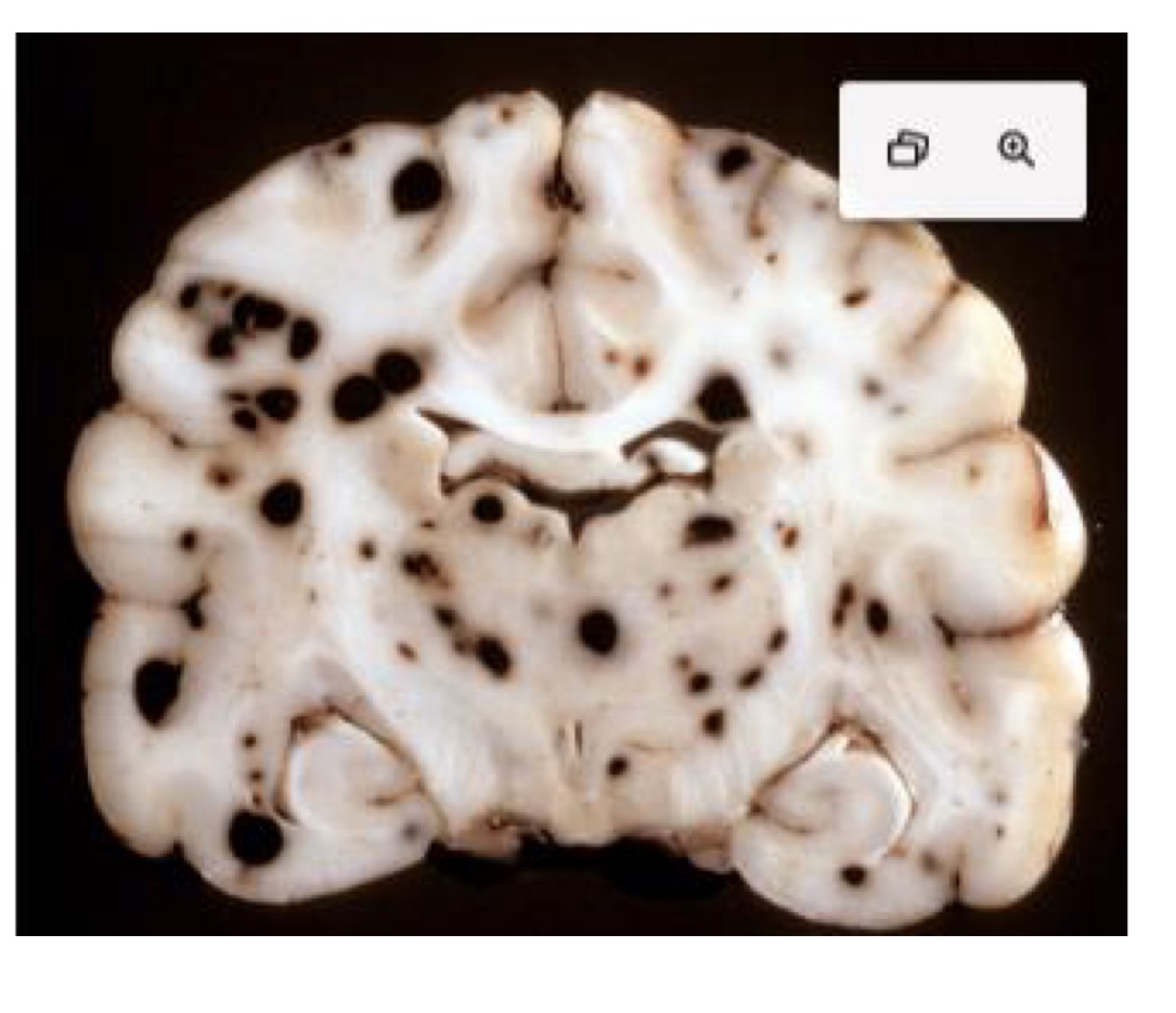
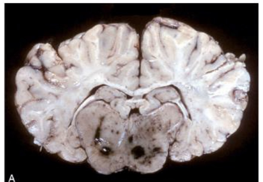
what is this?
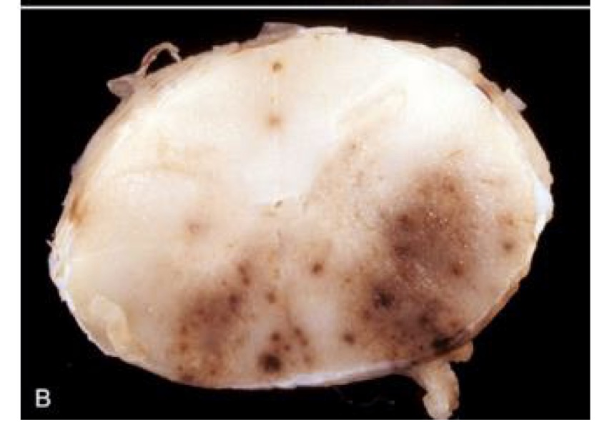
metastatic hemangiosarcoma → prominent hematogenous metastases, which appear as black nodules of various sizes distributed throughout the brain
in an unfixed specimen, what color would the nodules of metastatic hemangiosarcoma be?
red to dark red
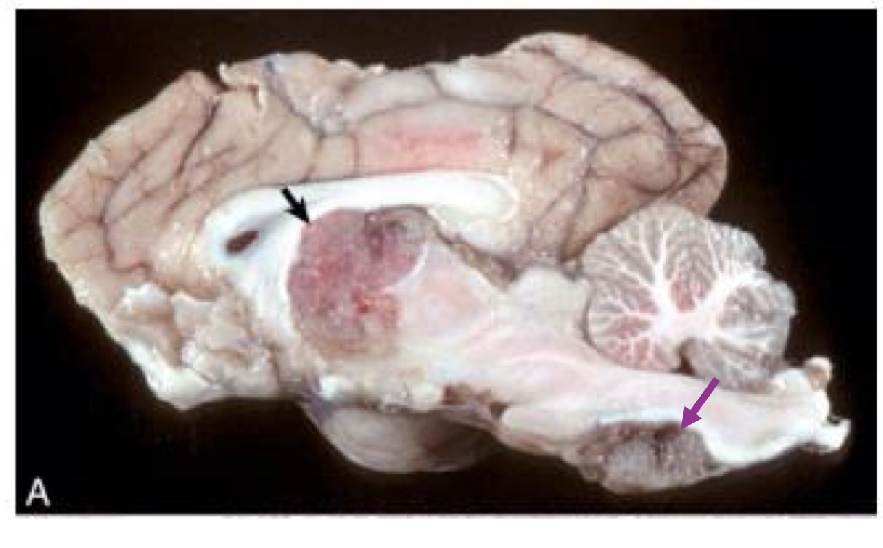
what is this?
metastatic mammary carcinoma → right cerebral hemisphere contains a well-demarcated mass which has caused enlargement of the right cerebral hemisphere and compression of right lateral ventricle
the leptomeninges of animals with heavily pigmented skin can have
melanin
true or false: congenital meningeal melanosis produces no clinical impairment in affected animals.
true
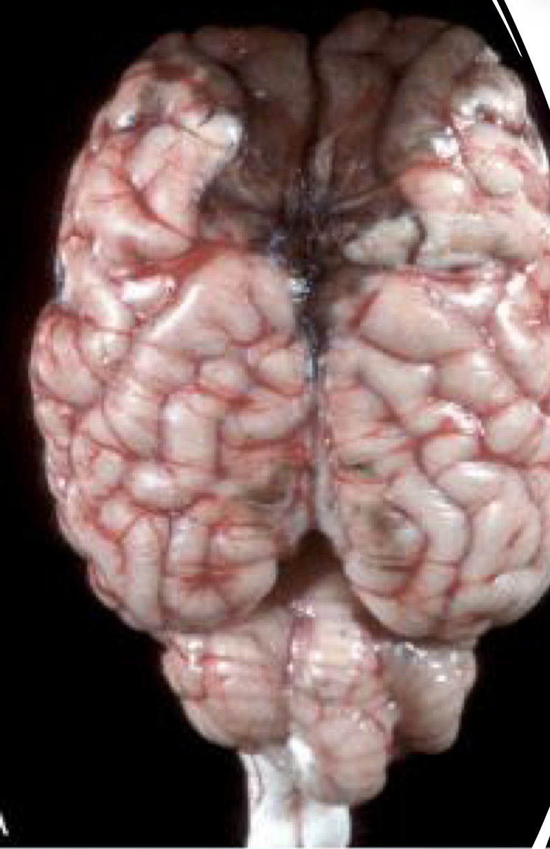
what is this showing?
black pigmentation of the leptomeninges overlying the olfactory poles and dorsal aspect of the frontal lobe
equine encephalomyelitis
infection occurs via mosquitoes
primary target cell for infection and injury is neurons
inflammation in gray matter of brain and spinal cord
EEE, WEE, and VEE
induce an encephalomyelitis that has similar characteristics
lesions for equine encephalomyelitis
uncommon and nonspecific
cerebral hyperemia, edema, petechiation, focal necrosis, and increased CSF in subarachnoid space
gross lesions found in gray matter
what is this?
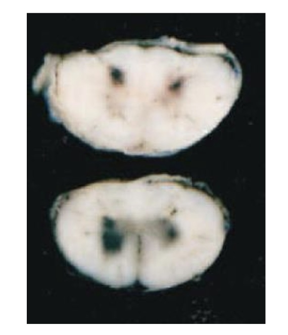
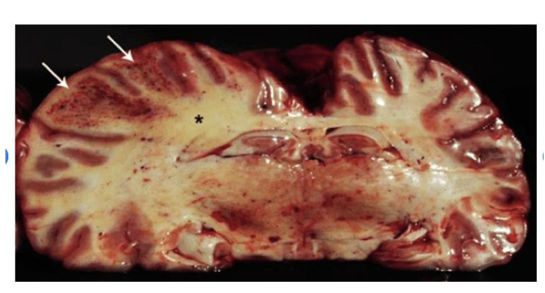
equine encephalomyelitis → thalamus has dark red to black discoloration as a result of congestion and hemorrhage
what is this?
equine encephalomyelitis → red to brown discoloration of the gray matter in the dorsal and ventral horns; focused more heavily on the gray matter
west nile virus
mosquito-borne
causes acute polioencephalomyelitis in humans, birds, and horses → targets gray matter of spinal cord
ubiquitous in lower 48 states of US
lesions of west nile virus
usually involve gray matter
hyperemia and petechiation to prominent hemorrhage → brain stem and gray matter of thoracolumbar spinal cord
what is this showing?
west nile virus
equine herpesvirus
important cause of equine abortion, perinatal foal infection and death, and rhinopneumonitis, in addition to myeloencephalitis
neurologic form can affect zebras
vasculitis is the principle lesion → endotheliotropic
how does equine herpesvirus cause vasculitis?
inhalation of virus
virus infects epithelial cells of the nasopharynx and spreads to local lymphoreticular tissue, where it infects lymphocytes and macrophages
transferred to endothelial cells of CNS
vasculitis
what is the characteristic lesion in the CNS caused by EHV-1 infection?
vasculitis affecting endothelial cells of small blood vessels with thrombosis and resulting in focal CNS necrosis
lesions of EHV-1 occur in both
gray and white matter of the spinal cord and brain with regions of hemorrhage and necrosis
what is this?
EHV-1 → focal or multifocal areas of hemorrhage and/or necrosis
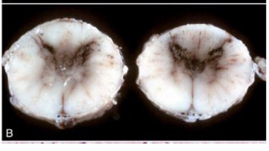
equine protozoal myeloencephalitis
caused by protozoa sarcocystis neurona, with less common isolates of neospora hughesi
pathogen enters body through ingestion of sporocysts
protozoa shed in opossum feces
lesions of equine protozoal myeloencephalitis
variably sized dark red-brown foci of necrosis and hemorrhage
white and gray matter with random distribution
more common in spinal cord
brain → most common in brainstem
what is this?
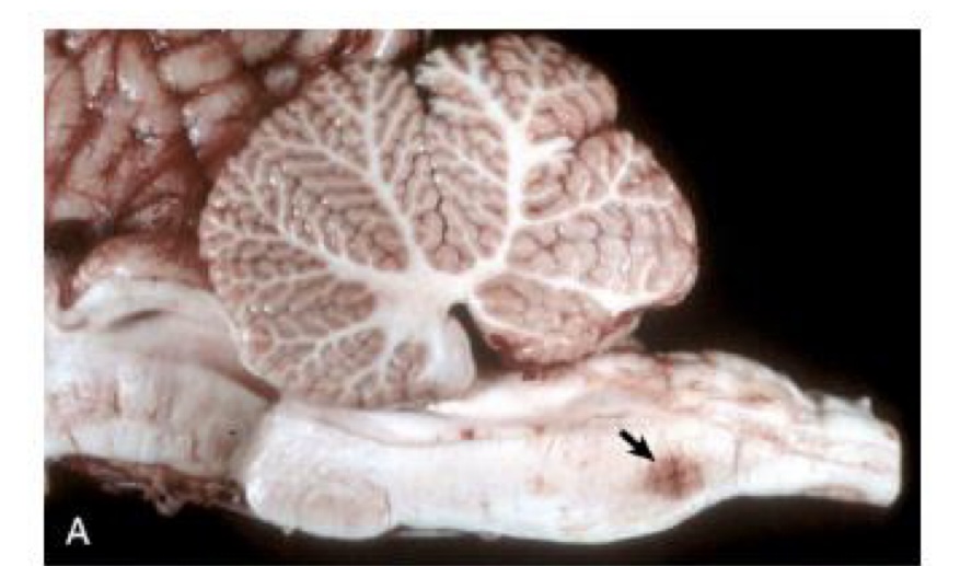
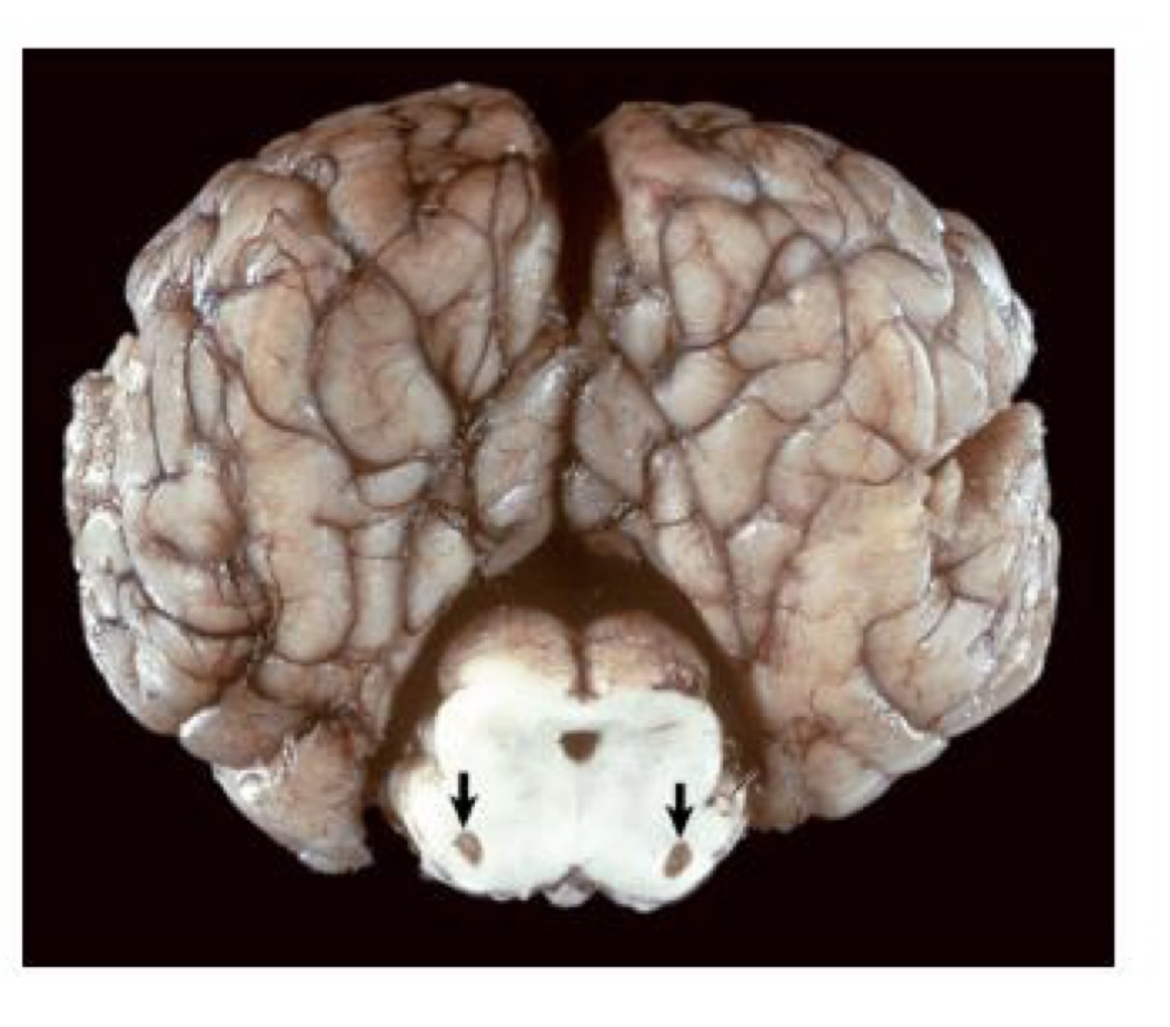
equine protozoal myeloencephalitis → large focus of hemorrhage and necrosis in the caudal brain stem caused by sarcocystis neurona
what is this?
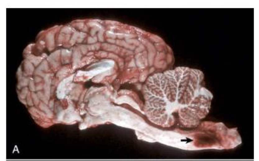
equine protozoal myeloencephalitis → prominent focal hemorrhage and necrosis are present in the right lateral funiculus and in the right and left lateral ventricle
halicephalobus gingivalus
free-living rhabditiform nematode that can infest the nasal cavity, CNS, and kidneys of horses
tan-white, multifocal to diffuse foci
results in foci of granulomatous and eosinophilic inflammation with associated nematodes
what is the primary predisposing factor of equine degenerative myeloencephalopathy?
dietary insufficiency of alpha-tocopherol (vitamin E)
equine motor neuron disease
associated with a vitamin E deficiency
develops in middle-aged to older horses
lacks a known genetic component
affects animals sporadically within a herd
severe neurogenic muscle atrophy
what can be used to support the diagnosis of equine motor neuron disease?
biopsy of the sacrocaudalis muscle to look for neurogenic muscle atrophy
what are clinical signs of equine motor neuron disease?
short-striding gait, muscle fasciculations, and narrow-based stance
leukoencephalomalacia
softening of white matter of brain
ingestion of moldy feed composed of corn or corn by-products contaminated with fusarium verticillioides
primary toxin is fumonisin B1
lesions of leukoencephalomalacia
white matter of the cerebral hemispheres most common
can impact brainstem and cerebellum
edema, brain swelling is marked with flattening of cerebrocortical gyri
gross lesion at time of death is malacia and liquefaction of the affected white matter
gray matter is spared
what can be the sole manifestation of leukoencephalomalacia?
hepatotoxicity
what is this?
leukoencephalomalacia → white matter of the frontal and parietal lobes is malacic due to necrosis, gray matter is not affected
what is this?
leukoencephalomalacia → hemorrhages concentrated on white matter and yellow discoloration of white matter (soft)
horses grazing on yellow star thistle or russian knapweed for 1 month or longer during hot months develop a disorder called
nigropallidal encephalomalacia
lesions of nigropallidal encephalomalacia
sharply demarcated foci of yellow discoloration and malacia in the globus pallidus and substantia nigra
lesions are bilateral and vary in severity
affected horses with nigropallida encephalomalacia
have persistent chewing movements
have difficulty in prehension of feed and drinking water
what is this?
nigropallidal encephalomalacia → lesion caused by yellow star thistle poisoning
cholesteatoma
form in the choroid plexus of the lateral ventricles in horses as an aging change
lesion thought to result from edema and minor but repeated hemorrhages within the choroid plexuses, which result in cholesterol deposits
masses are incidental but can cause acquired hydrocephalus
lesions of cholesteatoma
tan to yellow-brown firm masses with a smooth, often glistening surface
occasionally the masses are mineralized
can have associated granulomatous response
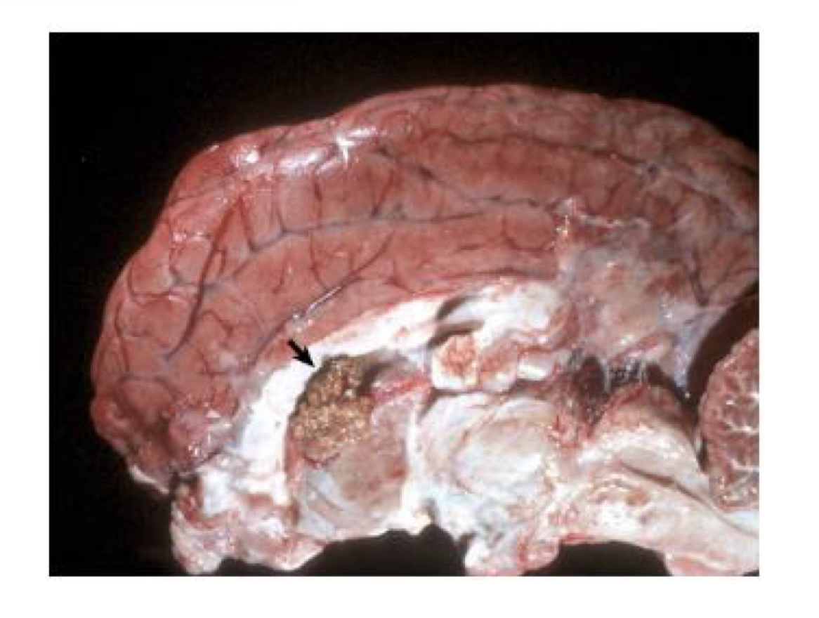
what is this?
cholesteatoma → choroid plexus of lateral ventricle contains an expansile mass consisting of cholesterol and a granulomatous inflammatory response
postanesthetic myelopathy
a hemorrhagic myelopathy secondary to general anesthesia in which the animals are placed in dorsal recumbency
after surgery, they are unable to stand
hemorrhage throughout gray matter of spinal cord
lumbosacral area most common
corresponds to neuronal necrosis
postanesthetic myelopathy lesion
develops secondary to the weight of the animals blocking normal venous drainage from the vertebral sinuses, leading to an infarctive lesion in the spinal cord
what is this?
postanesthetic myelopathy