THIS IS ONLY UP TO PART 4. for trials and hsc, add part 5 in!!
Mechanisms of Reproduction at3
Individuals do not live forever. The continuity of life from generation to generation is the result of
reproduction.
● Mechanisms: Organisms must be able to pass on their genes, either by replicating themselves [asexual],
or mating with another individual [sexual] to produce fertile* offspring.
● Reproduction - one of the distinctive characteristics of living organisms
*Because what is the point if the offspring is NOT fertile? It would be the END of your genes being passed on!
REMEMBER: we are trying to ensure continuity of life (i.e. survival).
NOTE: organisms in the same species are able to produce fertile offspring. Like a chihuahua and a pomeranian.
NOTE: You don’t need to know/memorise MRS GREN but it is a nice way to remember the requirements of life
There are TWO main methods of reproduction:
Asexual Sexual
● Involves ONE parent
● There is NO fertilisation or new genes
● Offspring are genetically identical to
parents - because there is no crossing over,
independent assortment or random fertilisation
○ This is good if the environment
DOESN’T change
● Involves TWO parents
● Each parent provides half of the genetic
material [haploid*] for the offspring in the
form of a gamete
● Two gametes fuse together during
fertilisation to form a new cell [zygote=humans]
● Offspring are similar to parents but NOT
identical
*Half the genetic material so that they combine together to form a new cell which is diploid (in humans it’s a zygote)
NOTE: DO NOT use the word ‘zygote’ for plants or some other animal
Continuity of Species - SUPER IMPORTANT!!! at3
Individuals have a finite lifespan, so in order for a population or species to survive, genetic material
must be passed from one generation to the next. This ability to reproduce is known as the reproductive
success of the individual.
● In evolutionary terms, reproduction is less significant for individual success and more important for the
continuation of the species.
NOTES: Do not say ‘community’ (different species together) and do not say ‘ecosystem’ (includes abiotic stuff).
Significance of this image is that these are all the same species but are of different populations
● Animals: advantages of external and internal fertilisation [NOTE: check worksheet 5111]
In animals, the union of male and female gametes can occur outside the body (external fertilisation) or inside
the body (internal fertilisation). e.g. Most fish like Salmon do external fertilisation
NOTE: You MUST be able to compare internal and external fertilisation.
external and internal fertilisation at3
External Fertilisation
● Occurs in aquatic or moist terrestrial environments, to prevent dehydration
of gametes e.g. ocean, river
● Gametes must be produced in large numbers to ensure success
○ Chances of fertilisation are low because gametes can be destroyed by
environmental factors which is why gametes must be released in large
numbers
● Occurs in invertebrates and vertebrates (fish and amphibians)
● NOTE: You still need a mate, but you DON’T need to expend energy in mate
attraction and copulation. Less energy expended on gestation
NOTE: Less energy expenditure with external fertilisation as they are NOT having sex. BUT
DO NOT TALK ABOUT ENERGY - there is a large room for error.
Internal Fertilisation
● Occurs inside the body of the female
● Involves mate attraction and copulation*, which require energy investment and put the organisms at risk
of predation, but fewer eggs need to be produced
○ In internal fertilisation, there are few female gametes however there is a large number of male
gametes are involved
● Occurs in invertebrates (insects and snails) and vertebrates (reptiles, mammals and birds)
● Chances of offspring survival is higher than for external fertilisation, however only a few offspring can be
produced at once
Parts of a flower to know:
General parts:
● Petals - attracts pollinating agents
● Leaf
● Sepal - the little thing that cups it a bit. Looks like a tiny leaf.
It’s next to the petals.
● Stem
● Roots
Female parts:
● Pistil made up of multiple carpels[NOTE: “carpel” & “pistil” can be used interchangeably]
● Carpel:
○ Stigma
○ Style
○ Ovary
Male parts:
● Stamen:
○ Anther
○ Filament
at3
asexual reproduction at3
Fungi: budding, spores
at3
Asexual Reproduction
When conditions are good*, fungi reproduce ASEXUALLY** either through budding or producing
spores***.
● In budding, part of the adult organism starts dividing by mitosis and produces a small bud which
separates from the parent and grows into a new individual.
*They don’t need to change (no need for natural selection as conditions are good) - variation can still occur through
mutations but this is more randomised
** Asexual reproduction is faster as they don’t need to find a mate (fungi cannot physically move to find a mate)
***NOTE: Spores can also be used to reproduce sexually
NOTE: Yeast (unicellular species of fungi) reproduces asexually → most by the process of budding (which is identical)
NOTE: People get binary fission and budding mixed. Note that when fungi are budding, the buds are NOT THE
SAME SIZE! Whereas in binary fission, offspring are identical.
Fungi Spores
at3
Multicellular fungi like bread mould are made of threads called hyphae
which forms the mycelium (fungal body)
Structures called sporangia produce very large numbers* of spores
which are light and easily dispersed**, travelling long distances by
wind.
*Large numbers means a higher chance of survival
**So that they can travel/spread and do not have to compete with a parent
Spores: tiny, light, unicellular reproductive cells that are produced in
great numbers by organisms such as fungi (e.g. moulds and
mushrooms)
● Spores effectively expand the distribution of the species, and
the species can colonise new environments
⭐️[vv important] → ENSURES THE SURVIVAL OF THE
SPECIES
Sexual Reproduction
at3
When conditions are BAD, fungi reproduce SEXUALLY*
● Spores from different fungi combine to form a new fungus
● This INCREASES genetic variation and the species’ chances of survival
*for Natural Selection → VARIATION comes from a new combination of genes by sexual reproduction
Bacteria: binary fission [Lowkey similar to Mitosis]
at3
Binary Fission is the most common form of asexual reproduction in bacteria and involves the
division of a unicellular organism into TWO
Some bacteria can double their numbers every twenty minutes wOW!
Steps:
1) A cell grows to TWICE its size
2) DNA replicates
3) DNA separates
4) Protein* accumulates at cleavage site
5) The cytoplasm divides
6) A new wall is synthesised
7) Two identical offspring are formed
*Both cells need proteins
Protists: binary fission, budding
at3
Asexual Reproduction
Protists: Eukaryotic, single cell organisms which do not
classify as animals, plants or fungi. e.g. Amoeba
● Reproduce either by binary fission (like bacteria) or
budding (like fungi) depending on the species
advnatges + disadv of sexual repro
at3
Analyse the features of fertilisation*, implantation and hormonal control of pregnancy
and birth in mammals
*Fertilisation happens in the fallopian tube
**Implantation in uterus (if it happens in the fallopian tubes, it is an ectopic pregnancy where the embryo will not survive
as miscarriage will occur)
Pregnancy and Birth in Mammals
Mammal: An organism that is warm blooded, has a back bone and is able to produce milk and has hair
● Placental mammals* develop a placenta at implantation e.g. Humans
● Marsupials rely on a pouch to protect the embryo as it develops e.g. Kangaroo
● Monotremes rely on an egg to protect the embryo as it develops e.g. Platypus
*NOTE: Placental mammals are the main focus
Fertilisation [fusion of male and female gametes]
In mammals, the male ejaculates sperm into the female’s reproductive tract for internal fertilisation in the
oviduct*
● During fertilisation, the haploid** sperm nucleus fuses with the ovum nucleus. The ovum provides a
single set of chromosomes, and the sperm provides a complementary set of chromosomes. → Produces
a Zygote - single cell
● The ovum*** also provides nutrients for the growth of the embryo and regulatory factors that control
early development → as in, the ovum has all the other organelles (e.g. mitochondria. [RECALL: mitochondrial DNA],
sperm only gives half genetic information
○ Makes sure that it develops properly
*Oviduct = fallopian tubes
**Half genetic information (Haploid)
***Ovum = egg
STEPS:
1) The female’s body releases an egg from the ovary (ovulation).
a) In humans it happens about once a month around day 14 (middle of the cycle)
2) Egg travels through the oviduct which is where fertilisation will occur
3) The male releases semen (a fluid containing sperm) into the vagina during sex. Sperm swims through
the cervix and the uterus, to the oviduct where the egg is waiting. Here the nuclei of a sperm cell and the
egg fuse to form a zygote
4) The zygote forms a strong membrane to stop other sperm from entering
a) Otherwise there will be TOO MANY CHROMOSOMES - we only want ONE set
Hormonal Control [can see page 16 of blitzing for extra info]
The Hypothalamus (a region of the brain) which acts as a control centre - secreting Gonadotrophic Releasing
Hormone (GnRH)*.
GnRH acts on the Pituitary Gland (located in the brain) causing the pituitary gland to secrete other hormones
which travel through the bloodstream** to the reproductive organs.
*NOTE: ‘Gonad’ = sex organ
**Hormones usually use the bloodstream to travel
The Menstrual Cycle
Lasts (on average) 28 days
● First 14 days before ovulation are known as the follicular phase
● Second 14 days after ovulation are known as the luteal phase (a.k.a. lutinising phase)
1. Follicular Phase
1) The Pituitary Gland in the brain secretes Follicle Stimulating Hormone (FSH) which travels in the
bloodstream to the ovary. It causes an egg to mature and stimulates the ovaries to release oestrogen
2) Oestrogen stops FSH being produced (so only one egg matures per cycle), repairs and thickens the
uterus lining and stimulates the pituitary gland to release lutenising hormone (LH)
2. Luteal Phase
3) LH triggers ovulation [there is also a small rise in FSH at ovulation]
4) After ovulation, the follicle forms a corpus luteum that produces high levels of progesterone. This
maintains the lining of the uterus and inhibits the production of FSH (because you only want one egg).
5) Two things that could happen:
a) If a fertilised egg implants, progesterone levels remain high
b) Without implantation, the corpus luteum degenerates and the lining of the uterus breaks down
again
Hormonal Control - Pregnancy
If a Blastocyst* implants in the uterus lining, the placenta secretes the following hormones to maintain
pregnancy:
1) Progesterone
2) Oestrogen [Stops FSH and repairs uterus lining]
3) Human Chorionic Gonadotrophin (HCG) [is the hormone pregnancy tests test for - maintains corpus luteum]
The corpus luteum** degenerates after about three months, and the pituitary gland does not secrete FSH until
after birth.
*Blastocyst consists of an outlet layer of cells (which develops into the placenta) and an inner mass of cells that will
develop into the embryo and fluid-filled cavity
**produces progesterone, but it is okay because the placenta is releasing that too
Hormonal Control - Birth
Relaxin is produced during pregnancy [labour] to loosen uterine muscles and ligaments in the pelvis.
● In the period before birth, the uterus and cervix become more sensitive to oxytocin, the hormone that
causes uterine contractions
● The fetus usually has moved with its head low on the pelvis, and the pressure on the cervix causes
further release of oxytocin and labour begins
● After the baby is delivered, the uterine contractions are maintained by oxytocin until the placenta is
pushed out and the uterus starts shrinking back to normal size
● Oxytocin also promotes the protective mothering instinct and works with prolactin to stimulate
lactation for feeding the newborn baby
NOTE:
● ‘Stimulate’ - produce
● ‘Inhibit’ - stop
Hypothalamus releases GnRH
GnRH acts on pituitary gland to stimulate the release of FSH
FSH travels in the bloodstream to the ovaries, stimulating them to
release oestrogen
Oestrogen stops FSH being produced and stimulates the pituitary
gland to release LH
LH trigger ovulation
AFTER OVULATION - follicle forms a corpus luteum which
produces high levels of progesterone
YES BABY:
Corpus Luteum degenerates and
lining of uterus breaks down
NO BABY:
Progesterone levels remain high
Hormone Summary Table
5.1.3. Evaluate* the impact** of scientific knowledge on the manipulation of plant and animal
reproduction in agriculture***
*Make a judgement based on criteria
Usually 4-9 mark questions [usually a combination of this and another module]
● RTQ carefully!!
● Think about the question during reading time
**BIODIVERSITY!!
***Need to know >1 example for each!
Setting out the Question
4 Marker
1) Judgement
a) Put this first because this is what the
marker is looking for
2) Pro (2 points)
3) Con (2 points)
4) Definition
5) Example
9 Marker
1) Judgement
2) Pro (2 points) + example
3) Con (2 points) + example
4) Definition (>1 definition)
5) Example
Agriculture
Agriculture: the growth of crops and animals for human needs
● Provides us with almost all out food and clothing so that we don’t need to produce these things
ourselves
○ Food e.g. Corn/rice/chicken/egg
○ Clothing e.g. Linen/cotton/wool/silk [i.e. natural clothing - NOT polyester]
Biotechnology: The use of biological systems, processes and organisms in the creation of new products and
technologies
➢ Reproductive technologies: the use of technology to assist and improve reproduction
→ Selective breeding is almost as old as the practice of farming itself [original/old method - still used to
this day]. It allows for improved quality* and yield** of crops and stock.
*Quality e.g.
● Seedless watermelon
● Grapes that are sweeter (cotton candy grapes)
● Carrots that are orange, not brown
**Yield e.g.
● Plant: Apple tree that produces more apples
● Animal: Chicken that produces more eggs
Steps for Selective Breeding:
1) Determine the desired trait*
2) Interbreed parents who show the desired trait
3) Select the offspring with the best form of the trait and breed these offspring
4) Continue this process until the population reliably reproduces the desired trait
*Select trait wanted so that there is a higher chance of offspring having that desired trait
🌱Plant Reproduction in Agriculture
● Over 10, 000 years of cultivation, wheat has developed from natural and artificial selection (selective
breeding)
○ Natural selection (what NATURE does)
1) Environment changes
2) The one with the trait that best suits the environment survives and reproduces
○ Artificial selection (i.e. selective breeding)
■ Humans choose the trait we want reproduced, NOT nature
● William Farrer pioneered Australian wheat research when he systematically used cross-breeding
(hybridisation) to improve bread wheat. He initially used his wife’s hairpins to transfer the fine grains
of pollen by hand until he acquired a pair of forceps.
○ This is sexual reproduction, artificial pollination: choosing the pollen from which plant to put on the
stigma of whatever plant you choose
NOTE: Hybrid Vigour
● Hybrids are generally more resilient because of a larger gene pool
e.g. Horse + Donkey = Mule (these are two different species, mule is sterile)
We don’t want the recessive trait being presented - by increasing the gene pool, it decreases the chance of homozygous recessive
traits that are harmful being expressed
🌱Plant Reproduction in Agriculture // cont.
Vegetative reproduction through using runners etc. can be used to clone plants
● Grafting and growing calluses are other techniques used
○ Grafting: chop part of plant off, put into water, let grow root, plant in soil
Advantage Disadvantage
● Guaranteed genotype of offspring ● Low genetic diversity [at risk if environment
changes]
e.g. The cavendish bananas are at risk of Panama Disease -
they have no genetic diversity [genetically identical]
🐮 Animal Reproduction in Agriculture
Cows have been selectively bred to improve milk quality and yield
● Genetic techniques can now be used to improve stock animals, including artificial insemination
○ Artificial insemination: the injection of male semen into the vagina of a female without sexual
intercourse using an insemination gun [must use this exact wording]
Advantages and disadv of Artificial Insemination:
Bull does not get hurt travelling
Cow/bull don’t get hurt during sexual intercourse
● Chance of offspring being produced is higher as the semen is directly injected into the female
reproductive system
● Increased control over mating
● Better ability to record accurate pedigrees
Disadvantages of Artificial Insemination:
● Inbreeding if same source is used too often
● Needs special equipment and training to perform chicken
Part 2: Cell Replication
at3
Most cells in the body contain a nucleus
➢ Inside the nucleus, chromosomes can be found.
➢ Chromosomes are composed of protein and DNA (together
this is called a chromatin - a fibre of packed nucleosomes)
➢ Each DNA strand is made up of about 50 genes per
millimetre- it is the genes that control the inheritance of
genetic characteristics; these are passed on from parents to
offspring
➢ Each inherited characteristic is controlled by at least one pair
of alleles
5.2.1. Model the processes involved in cell replication, including but not limited to:
DNA structure diagrams
at3
cell replication
at3
DNA Structure
DNA (Deoxyribose nucleic acid) is made up of repeating units called
nucleotides (a basic unit of DNA)
Nucleotides consist of:
1. A phosphate
2. A sugar (deoxyribose)
3. A nitrogenous base
DNA Chemistry
The carbon atoms in the DNA sugar (deoxyribose) are numbered from 1’ to 5’.
The phosphate is always attached to the 5’ carbon atom
Basically:
● 1’ connects to the base
● 3’ is by itself
● 5’ connects to the phosphate
The phosphate and the sugar forms the
backbone of the DNA molecule (a.k.a.
phosphate- sugar backbone)
A double stranded DNA molecule is made up of two single-strands of DNA
that are held together by hydrogen bonds between complementary base
pairs.
There is always direct pairing:
● Adenine (A) pairs with Thymine (T) by two weak hydrogen bonds
● Cytosine (C) pairs with Guanine (G) by three weak hydrogen
bonds
This ensures that A-T bond, and
C-G bond - oOh very cool :)
18
Gabriella Chen
For the bases to form hydrogen bonds, one strand of DNA will run
antiparallel. The two strands will then coil into a spiral shape, forming
a double helix.
A way to remember the nitrogenous base pairs: Gabby Chen, Annie Tan
DNA has Direction
The two strands in a double-stranded DNA molecule run in opposite
directions - they are antiparallel.
dna replication diagrams
at3
DNA Structure: The Watson and Crick Model
at3
Watson and Crick discovered the structure of DNA in 1953
- This discovery was built on many years of earlier investigations and technological developments by
other scientists
- 1940 - Chargaff discovered that the amount of nitrogenous bases A
and T were always the same and the amount of G and C were also
the same, BUT A-T and G-C may be present in different amounts
- 1950 - Franklin and Wilkins used X-ray crystallography to produce
images of the DNA structure
Using both of the above findings, Watson and Crick made adjustments to their 3D
modelling of DNA, positioning the bases inside the helix.
prokaryotes and eukaryotes
RECAP: Prokaryotes and Eukaryotes
Prokaryotes
‘Pro’ = before ‘Karyon’ = nucleus
● Prokaryotic cells lack membrane bound organelles and therefore
have no nucleus.
● Usually unicellular
● They are composed of:
○ Cell membrane
○ Ribosomes
○ Cytoplasm
○ Genetic material - found in the cytoplasm
● Includes:
○ Archaea
○ Bacteria
20
Gabriella Chen
Eukaryotes
‘Eu’ = true ‘Karyon’ = nucleus
● Eukaryotic cells have a membrane bound nucleus which
contains the genetic material of the cell
● All of the internal structures of eukaryotic cells are
membrane bound and are known as organelles
○ Each organelle has a specific function within the cell
● They can be unicellular (protists and some fungi) or
multicellular (plants, animals, and some fungi)
● Includes:
○ Animals
○ Plants
○ Protists
○ Fungi
DNA Replication (at3)
DNA replication: the production of two identical DNA strands from one original
double helix molecule
- The process of DNA replication ensures that the genetic material is copied
exactly
- DNA replicates BEFORE cell division- during interphase, so that each cell can
receive one full copy of the coded instructions that control the basic life
functions of the cell
Features of DNA that help with DNA replication:
★ Weak hydrogen bonding between pairs
★ Anti-parallel
★ G-C together
★ A-T together
Helicase: Unzipping enzyme
DNA Polymerase: replicates DNA molecules
Primase: Makes primer- helps DNA polymerase
Ligase: glues stuff
Step 1: Unwinding and Unzipping
● An enzyme called helicase causes the DNA double stranded helix to progressively uncoil
● As DNA uncoils, the weak hydrogen bonds between complementary base pairs of the nucleotides on
opposite strands break, creating a replication fork
● Single-stranded binding proteins (SSBs) bind to and stabilise the newly separated single-stranded DNA
Step 2: Adding Primers
● Each separate strand of the new DNA molecule acts as a
template for the production of a new strand of DNA
● For synthesis* to be initiated, DNA primase (the initializer)
makes primers: a short strand of RNA, and attaches this to
the DNA to show DNA polymerase where to start
*Basically the creation of DNA
Remember!
The two strands in a double stranded DNA molecule run in opposite directions - they are
antiparallel.
One strand runs from 5’ to 3’ and the other 3’ to 5’
Step 3: Adding Nucleotides
● The enzyme, DNA polymerase, adds complementary nucleotides to the DNA strand
● It picks up free nucleotide units (sugar-phosphate-base) floating in the fluid in the nucleus and inserts
them opposite their complementary base partner, using the existing strands as templates
● There are TWO DNA polymerase enzymes working at the same time (one on each strand), DNA
polymerase only works in one direction, it makes a new DNA strand in the 5’ to 3’ direction, this means
it starts reading the existing DNA strand from the 3’ end.
● On one strand of DNA, nucleotides are added in a long chain,
growing in the same direction as the replication fork opens
up. This is called the leading strand and replication is
continuous.
● On the other strand (the lagging strand), nucleotides are
added in ‘chunks’ (called Okazaki fragments), from the
replication fork backwards. Replication in this strand is
therefore discontinuous
23
Gabriella Chen
The minimum: The enzyme, DNA polymerase, adds complementary nucleotides (Adenine to Thymine, Guanine to
Cytosine) using the existing strands as templates.
Step 4: Completing the lagging strand
As replication on the lagging strand is discontinuous, the Okazaki fragments are joined
up by an enzyme called ligase, forming one continuous strand.
Step 5: Proofreading
The base pairing is checked constantly by DNA polymerase which ‘edits’ any incorrect additions to ensure
accuracy.
Incorrect base pairings will result in a change in the DNA base sequence known as a mutation
The Results
- The replicated DNA molecules coil into the double helix
conformation
- The process of DNA replication in cells is termed
‘semi-conservative’, as each resulting double-stranded DNA
molecule has one new strand and one ‘old’ or conserved strand
- Each double strand is identical to each other, and to the original
molecule from which they formed
dna replication diagrams
dna replication diagrams
mitosis and meiosis at3
DNA replication using the Watson and Crick DNA model, including nucleotide composition,
pairing and bonding
● Mitosis and Meiosis
Cell Replication/Division: the process by which cells replicate their genetic material and divide to form new
cells.
Cell Division
- In UNICELLULAR organisms, cell division is by asexual binary fission: where one organism becomes
two; no gametes (sex cells) are involved
- In MULTICELLULAR organisms, cell division by mitosis leads to the formation of two new identical
cells that contribute to the growth of an organism. This occurs in the somatic (body) cells
Mitosis - makes body cells [https://youtu.be/L61Gp_d7evo]
Mitosis: a type of cell division where one parent cell divides once to produce two identical daughter cells for
asexual reproduction, growth, and replacement of old or damaged cells.
Mitosis plays an important role in:
● Growth of multicellular organisms
● Repair of damaged tissue and replacement of worn-out cells
● Asexual reproduction e.g. growing plants from cutting and cloning
● Genetic stability - mitosis ensures the precise and equal distribution of chromosomes
to each daughter nucleus, so that all resulting cells have the same chromosome
number and genetic information as each other
○ i.e. A cell will not function properly without a full set of correct genes
○ e.g. Down Syndrome (a.k.a. Trisomy 21) is caused when abnormal cell division
results in the 21st pair having an extra chromosome (3 chromosomes)
Cell Cycle
- The cell cycle: the life cycle of a cell, involving growth, replication of DNA and
division of cells to produce two identical daughter cells.
Has three main phases:
1. Interphase (cell spends most of its time in this phase)
2. Mitosis
3. Cytokinesis
process of mitosis at3
INTERPHASE: Preparing for Mitosis
- During interphase, the cell continues to carry out its daily cell functions,
it grows and the DNA is replicated; a single- stranded chromosome
becomes a double stranded chromosome
PROPHASE
‘Pro’ = before
- The chromatin material (protein and DNA) shortens and thickens by
coiling, and the DNA separates out into chromosomes which are now
visible with a light microscope. This process is called condensing.
- The nuclear membrane begins to break down (nuclear membrane → the
membrane that encloses the cell nucleus)
METAPHASE
M for ‘Middle’
- The nuclear membrane has completely broken down and the chromosomes
line up across the equator (middle) of the cell
- Spindle fibres attach to the centromere of each chromosome
26
Gabriella Chen
ANAPHASE
A for ‘Away’
- The spindle fibres contract and the chromosomes are pulled by their
centromere, causing the sister chromatids to separate and move to opposite
poles (sides) of the cell; they are now termed ‘daughter’ chromosomes
TELOPHASE
T for ‘Two’
- The daughter chromosomes gather at opposite poles of the cells where the
spindle fibres break down and the nuclear membrane begins to reform
“After Telophase, the cell division ends with Cytokinesis where the cytoplasm
cleaves to form two identical daughter cells.”
CYTOKINESIS
C for ‘Cytoplasm’
- Cytokinesis is the division of the cytoplasm
- This marks the end of cell division, where two identical daughter cells have
been formed which are identical to the parent (original) cell
The DNA goes from Chromatids (in Telophase) to uncondensed (in Cytokinesis)
→ It is packaged so it is easier for them to split, THEN they unwind and Interphase occurs (the cell does cell
functions), then the DNA condenses again in Prophase.
Mitosis Summary:
- Growth, replacement of old and worn out cells asexual reproduction
- I PMAT
- Two identical daughter cells
mitosis diagram at3
meiosis at3
Meiosis: One original parent cell divides twice to produce four non-identical daughter cells (aka gametes, or,
sex cells - sperm in males and ovum in females)
Karyotype: a visual depiction of the number, size and shape of
chromosomes in an individual
- It is lined up in homologous pairs
The number of chromosomes varies between species.
The karyotype for humans →
27
Gabriella Chen
Humans have 46 chromosomes (23 pairs). One of these 23 pairs of chromosomes is the sex chromosomes, XX
in females and XY in males. The remaining 22 pairs are called autosomes*.
*People always forget this. So, dON’T.
Homologous Chromosomes
● Chromosomes occur in pairs.
○ One of the pair is inherited from the father (the paternal
chromosome) and the other from the mother (the maternal
chromosome).
○ These pairs of chromosomes are called homologous pairs.
Homologous Chromosomes are similar (same size and shape) but not identical. Each carries the same genes in
the same order, but the alleles for each trait may not be the same.
e.g. X and Y Chromosomes ARE homologous despite being different sizes. Homologous autosomal chromosomes are the
same size
Same gene: eye colour
Different allele: different eye colour
Meiosis
● Meiosis starts with a germ line cell* - A germ line cell contains a full
set of chromosomes and is said to be diploid (2n)
● After meiosis has occurred, it produces gametes (egg/sperm cells) -
Gametes contain half the number of chromosomes as is said to be
haploid (n).
As meiosis results in half the number of chromosomes, it is often referred to
as ‘reduction division’.
*Egg and sperm cells (gametes) are MADE FROM germ line cells
There are TWO stages of Meiosis:
1. Meiosis I : a diploid cell divides into two haploid
cells
2. Meiosis II : the two haploid cells divide again,
resulting in four haploid daughter cells
a. ALL are different to each other
Each daughter cell has HALF the original number of
chromosomes that the parent cell had and are NOT
identical to each other nor to the parent cell
meiosis 1at3
//Meiosis I
INTERPHASE: preparing for division
During interphase, the cell continues to carry out its daily cell functions. It grows
and the DNA is replicated (duplicated); a single stranded chromosome becomes a
28
Gabriella Chen
double stranded chromosome Organelles are also replicated but u dont really need to write that
PROPHASE I
‘Pro’ meaning ‘before’
The chromatin material shortens and thickens by coiling and the DNA separated out
into chromosomes, which are now visible with a light microscope. This process is
called ‘condensing’.
The nuclear membrane begins to break down and the homologous chromosomes pair
up, which can result in crossing over.
Crossing Over
Crossing over is the exchange of genetic information between
homologous pairs to make new combinations of genes, introducing
genetic variation (important for natural selection).
Genes that occur on the same chromosome are said to be linked.
Crossing over ensures that not all linked genes on a chromosome are
inherited together. The exchange of genes during crossing over
causes mixing of paternal and maternal genes which results in an
increased number of combinations of genes* that may be transmitted to the offspring.
Every crossover is different which increases the number of combinations of genes
*Mrs Seo likes this
29
Gabriella Chen
METAPHASE I
‘M’ for Middle
The nuclear membrane has completely broken down, and the chromosomes line up
across the equator (middle) of the cell; they do this randomly, meaning maternal and
paternal chromosomes do not line up on the same side of the midline.
This is called independent assortment.
Unlike mitosis, the chromosomes line up in homologous pairs
Spindle fibres attach to the centromere of each chromosomes
(See in the picture) the paternal and maternal aren’t lined up on the same side
ANAPHASE I
‘A’ for Away
The spindle fibres contract and the homologous pairs separate from one another and
move to opposite poles (sides) of the cell. This separation of maternal and paternal
chromosomes not only halves the chromosome number in gametes but also leads to
genetic variation, depending on which chromosome of each pair ends up in which
daughter cell.
This is called random segregation*
*Random segregation occurs due to independent assortment
TELOPHASE I and CYTOKINESIS
‘T’ for Two
The spindle fibres break down and the nuclear membrane begins to reform
At the end of this first division of meiosis, there are two daughter cells with the
chromosome number halved (there are now two haploid cells)
diagram of meiosis 1 at3
meiosis 2 at3
PROPHASE II
The nuclear membrane begins to break down and the spindle fibres start to form.
Unlike Prophase I, the DNA does NOT duplicate and crossing over does NOT
occur. This is because the DNA has already replicated and there are no longer
homologous chromosomes in each cell (they have been separated), so crossing
over cannot occur
METAPHASE II
‘M’ for Middle
The chromosomes line up across the equator (middle) of the cell. Unlike Meiosis I, the
chromosomes in Meiosis II line up in a single file.
Spindle fibres attach to the centromere of each chromosome
30
Gabriella Chen
ANAPHASE II
‘A’ for Away
The spindle fibres contract and the chromosomes are pulled by their centromere, causing the
sister chromatids to separate and move to opposite poles (sides) of the cell
TELOPHASE II and CYTOKINESIS
The daughter chromosomes gather at opposite poles of the cells where the spindle
fibres break down and the nuclear membrane begins to reappear.
Cytokinesis is the division of the cytoplasm. This marks the end of cell division,
where the four non-identical daughter cells have been formed, with half the original
chromosome number (haploid cells)
Fertilisation
After meiosis, fertilisation occurs; the fusion of a sperm cell and an egg cell.
Remember that each of these gametes carry half the number of chromosomes, so
each parent contributes only half of his/her chromosomes to the new offspring,
We now have a zygote, a diploid cell containing a full set of chromosomes
Variety
Many combinations of chromosomes are possible in gametes as a result of
both crossing over and independent assortment that occurs during meiosis.
Further variation is introduced during fertilisation, which involves the random
meeting of any two gametes. Another source of variation is mutation, which
may arise at any point in the process, but most commonly occurs during DNA
replication
//Summary: Mitosis and
Meiosis
● Somatic cells go under mitosis
5.2.2. Assess the effect of the cell replication processes on the continuity of species
The Continuity of Species
All living organisms arise from other living organisms (Cell Theory). The continuity of species refers to the
ongoing survival of a species as a result of characteristics being passed on from parents to offspring in a
continuous lineage. This inheritance of characteristics from ancestors to current organisms relies on two things:
1. The passing on of consistently accurate genetic information (i.e. genetic stability)
a. You want a full set of genetic information → if you don’t have the full set, the cell can’t function properly
2. The introduction of variation (mutation) of some genetic information
a. Talking about GOOD variation → you DON’T want faulty/bad mutations
Both genetic stability and variation play a role in ensuring the continuity of the species
Genetic Continuity [basically, Mitosis]
Genetic Continuity: A way of preserving genetic information across generations and is dependent on:
1. When a cell divides by mitosis, the resulting two daughter cells must have the same number and type of
genes as the original cell
2. When two sexually reproducing organisms breed, the resulting offspring must have the same number of
genes as the parent organisms
a. e.g. a Zygote has the full 46 chromosomes → if a gamete has a different number of chromosomes, it can
lead to things such as Trisomy 21/Down Syndrome happening
Genetic continuity ensures continuation of a species, because it ensures that new cells or organisms have all the
genes they need to survive. A lack of genetic continuity results in disease and sometimes death and extinction
Genetic Stability [basically, Meiosis]
For continuity at a species level, successful desirable traits must be passed on, along with some random errors
(mutations). This allows a species to evolve if an environmental change occurs. Natural selection acts so that
individuals in a population that are best suited to the environment survive and reproduce, passing on their genes
to their offspring. This mixing of parent genes during sexual reproduction, including mutations, increases
genetic diversity and helps maintain continuity of the species.
Ensuring Continuity of a Species [memorise - COFFEE MIMN, like ‘coffee mum’]
Mechanisms that have evolved to ensure genetic continuity and survival and continuity of a species include:
● Consistent replication of DNA prior to cell division
● Orderly distribution of chromosomes when cells divide and gametes form*
● Fertilisation methods** that ensure individuals of the same species breed successfully (i.e. internal
fertilisation)
● Methods to ensure embryo survival (i.e. protection and nourishment from parents)
● Increased variation (independent assortment, crossing over, random fertilisation of gametes)
● Mutation
● Natural selection (survival of the fittest)
*The opposite of this: nondisjunction (the failure of the chromosomes
to separate, which produces daughter cells with abnormal numbers of
chromosomes)
genetic error
**Internal fertilisation > external fertilisation because survival rate is higher though fewer offspring are produced
Genetic Error*
Genetic errors (mutations) can threaten the continuity of species. During DNA replication, enzymes proofread
the DNA strand and bring about DNA repairs.
In humans, a range of diseases have been linked to the reduced ability of cells to repair DNA during or after
replication. This reduced ability to repair DNA can mean humans are more susceptible to cancer, it may also be
responsible for accelerated ageing or give rise to neurodegeneration.
Natural selection is a mechanism that ensures these genes are removed from the populations so that the
continuity of species is not at risk.
*could be good OR bad mutations
5.3.1. Construct appropriate representations to model and compare the forms in which DNA exists in
eukaryotes and prokaryotes
5.3.2. Model the process of polypeptide synthesis, including:
● transcription and translation
● assessing the importance of mRNA and tRNA in transcription and translation
● analysing the function and importance of polypeptide synthesis
at3
Polypeptide Synthesis
DNA holds the original copy of all instructions that an organism needs. It is too large to leave the nucleus* and
therefore, in order for a cell to make a particular protein an intermediate molecule called messenger RNA
(mRNA) is created. Not all the DNA is needed; only the relevant instructions** for those proteins are accessed
in the DNA nucleotide sequence.
Once the mRNA is made, it leaves the nucleus moving into the cytoplasm where it meets a ribosome.
Ribosomes are the cells ‘machinery’ that translates this message into a protein.
*This is why in Prophase it is important for the nuclear membrane to break down [Mod 5 Pt 2]
**This is called a gene = the recipe to make polypeptide
NOTE: It is the same for prokaryotes, however it happens in the cytoplasm
RNA
RNA is a nucleic acid that is made up of a chain of nucleotides
but differs from DNA in the following ways:
1. Single stranded (NOT double)
2. Contains ribose sugar (NOT deoxyribose)
3. Has nitrogenous base uracil (U) (instead of thymine (T))
34
Gabriella Chen
Three types of RNA:
1) Messenger RNA (mRNA): functions as an
intermediate molecule, carrying information from
DNA in the nucleus to the ribosomes in the cytoplasm.
2) Transfer RNA (tRNA): acts as a temporary carrier of
amino acids, bringing the appropriate amino acid to
the ribosome based on the mRNA sequence [Brings
complementary amino acids to Ribosomes]
3) Ribosomal RNA (rRNA): forms a structural part of
ribosomes [literally just a Ribosome]
part 1 transcription at3
Part 1: Transcription
The purpose of transcription is to produce a single-stranded mRNA
molecule
Transcription occurs in three steps:
1. Initiation [Start]
2. Elongation
3. Termination [Finish]
1. Initiation: Unravelling the DNA
An enzyme, RNA Polymerase, binds to a part of the DNA called
the promoter (a section of DNA before the start codon*) and the
DNA unzips (the hydrogen bonds between the complementary base
pairs break and the strands separate over a short length, just in the
part of the DNA that holds the gene to be used)
*codon: 3 bases
2. Elongation [Elongates the strand it needs]
Only one strand of DNA contains the genetic information to
make a protein, this strand is called the ‘non-coding strand or
template strand’. (The other strand, which is NOT used, is called
the coding strand or non-template strand’)
RNA polymerase travels down the non-coding strand, joining
free RNA nucleotides together forming a complementary
single-stranded mRNA molecule (that is, DNA is transcribed into
mRNA). The mRNA strand is the same as the DNA coding
strand, except that it has a U instead of T.
NOTE: If calling one ‘coding’, call the other ‘non-coding’. If calling one ‘template strand’, call the other
‘non-template strand’
35
Gabriella Chen
3. Termination
Transcription is terminated (stopped) when the RNA
polymerase reaches the stop triplet code (stop codon)
at the termination site. The RNA polymerase then
detaches and the two DNA strands come together
4. RNA Processing
The mRNA that is produced is called pre-mRNA.
This contains coding sequences of nucleotides,
called exons, which will be translated into a
polypeptide and non-coding sequences, called
introns, which do not code for amino acids.
Therefore, the mRNA is spliced (edited) and the
introns are removed by a molecule called
spliceosome. The end result is called mature-mRNA and is ready for translation (part 2).
i.e. They copy the fragment required to make the proteins however not all of the fragment is required to make the specific
protein wanted - therefore they identify introns and exons inside the fragment copied into mRNA, introns get cut out and
stay INside the nucleus, and exons combine together to form mature mRNA which will EXit the nucleus to a ribosome
Moving to the Cytoplasm
The mature mRNA moves out of the nucleus (through a nuclear pore) and into the cytoplasm, where it
encounters a ribosome
part 2 translation at3
Translation: The process in which the codons on the mRNA strand are
translated into a sequence of amino acids resulting in a polypeptide.
mRNA
Every three nitrogen bases on the mRNA strand is called a triplet or a codon.
This codon codes for a particular amino acid
*exception: STOP codon does not have
an amino acid
36
Gabriella Chen
Translation
1) The ribosomes move along the mRNA molecule and, as they do so, they attach
tRNA molecules by temporarily pairing the bases of the tRNA anticodons with
their complementary triplets of bases (codons) in the mRNA
2) Only two tRNA molecules can bind to the mRNA at any one time. Once two
tRNA’s are bound, their amino acids join together with a peptide bond.
3) The tRNAs move away from the mRNA, leaving the growing chain of amino
acids and move back into the cytoplasm where they can pick up another amino
acid and be reused. The ribosome moves along the
mRNA strand one codon at a time. This movement is
called translocation.
4) Translation is repeated until the polypeptide is
complete. The completion of that process is signalled
by a stop codon (UAA, UAG, UGA). These do not
correspond with any tRNA, but signal the ribosome to
detach from the mRNA.
5) The polypeptide chain is then folded into the correct
shape for its functioning, forming the final protein end product. The mRNA is broken down into its
individual nucleotides which can be reused.
Importance of Polypeptide Synthesis at3
Genes that are expressed* determine which proteins are produced, giving the cell its functionality and
characteristics. Gene expression** is the process through which information from a gene that is switched on is
used to synthesise a specific functional gene product - a polypeptide***.
Everyday examples of proteins that are produced include:
● Growth hormone proteins are produced in a growing child but are switched off at other times
● Plasma B-Cells of the immune system are regulated to only produce antibody proteins when they are
required to fight an infection
● Enzyme proteins are recycled but new ones will be synthesised to replace those that have reached the
end of their useful life
*expressed = working/shown
**gene expression = which protein is being produced → Use this definition for protein synthesis
***MEMORISE THIS
● assessing how genes and environment affect phenotypic expression [*Phenotypic Expression = what
is shown] at3
Gene: a portion of DNA; it is the smallest unit of heredity. It is
made up of linear sequences of nucleotides that code for a cell
product, i.e. A polypeptide chain
Locus: is the position of a gene on a chromosome
● Usually same for same product
● Location Locus
Genome: the total amount of genetic material that an organism has
in each of its cells
● ALL of the genetic information
Alleles: alternate forms of the same gene
● Alleles are found at the same locus on a chromosome
aA } same gene, different allele a.k.a. Same trait, different versions
Phenotype and Genotype
Phenotype: an organism is often defined as its physical appearance, but today we know that it includes the sum
of all gene products. Hence, phenotype includes the structure, behaviour and physiology of an organism.
E.g. enzymes as well. Some people have lactase (can digest milk) - some people don't (therefore are lactose intolerant)
Genotype: set of genes in an organism's DNA that is responsible for a particular trait. The genotype of every
cell is the same, yet each cell becomes a specific type of cell. This specialisation of cells is brought about by
controlling the expression of genes within the cells. Gene expression is the switching on and off of genes to
make the required proteins. [Use this definition for specialisation]
E.g. hair follicle cells produce Keratin to make hair so for cheek cells gene for keratin is turned off
40
Gabriella Chen
Variation
Some variations in organisms are genetically determined,
whereas others are influenced by the environment. However,
many variations arise as a result of an interaction between the
two - the environment can influence how genes are expressed.
Environment Affects Phenotypic Expression
● The acidity or alkalinity of the soil influences the colour of hydrangeas. Hydrangeas growing in acidic
soil develop blue flowers whereas those grown in alkaline soil develop pink flowers.
● A change in temperature or pH causes a change in the shape of the active site of an enzyme, increasing
or decreasing its binding with the substrate and therefore affecting its functioning
Human height and infant birth weight have a genetic basis but the lack of nutrients or the presence of toxins
(i.e. cigarette smoking) can restrict growth and therefore a human may not grow to their potential
5.3.3. Investigate the structure and function of proteins in living things
Proteins are the most abundant organic molecule in cells. They are responsible for forming the basic structure*
of cells and for carrying out all the work in cells**, by controlling every chemical reaction***.
*e.g. structural proteins: keratin, collagen
**e.g. proteins that carry out work: haemoglobin
***e.g. proteins that control chemical reactions: enzymes
Chemical Structure of Proteins
Shape is important!
→ Amino acids are the building blocks of proteins. They are made up of carbon, hydrogen, oxygen, nitrogen
and sometimes sulfur.
→ Amino acids are joined together in a linear sequence by chemical bonds called peptide bonds, forming a
polypeptide (which can be up to 300 amino acids). [Can be quite long!]
→ One or more polypeptides can be folded into a particular shape, making a protein. The sequence of amino
acids (there are 20 different types) and the shape of the protein determines its function.
proteins
Types of Proteins in Cells
Different types of tissues contain different proteins.
There are two groups:
1. Fibrous Proteins: Stringy/hairlike
Form structural components in cells and tissues
They’re long and soluble in water
e.g. Collagen, found in skin
2. Globular Proteins: Honey-like, globby
Usually spherical in shape and are compact and
soluble in water
They are often transport proteins
e.g. Haemoglobin which transports oxygen around
the body
Function of Proteins
There are many different types of proteins in organisms. Each protein has a different function, and each plays a
vital role in the regulation*, functioning and maintenance of both individual cells and entire organisms. The
specific structure** of each protein enables it to carry out its own function.
Proteins can be classified according to their function [S.E.C.T.S]:
● Structural proteins for support and movement
● Enzymes which control biochemical reactions
● Proteins for cell communication, cell signalling and biological recognition
● Proteins for transport and storage
● Sensory proteins which respond to stimuli
*e.g. DNA Polymerase: regulates DNA to make sure it is the correct sequence
** Amino acid sequence
Structural Proteins
● Often fibrous
● Found in connective tissues such as skin, bone and ligaments
● Provide support by forming the structural components of cells and organs, e.g. collagen in
skin or assist in contractile functions such as muscle, e.g. myosin (protein that causes
muscles to contract)
Enzymes
Enzymes are protein molecules involved in all biochemical aspects
of cellular metabolism.
e.g. anabolic enzymes such as DNA Polymerase which catalyses the
formation of hydrogen bonds
cell communications, transport and storage, sensory proteins, immunity proteins
Cell Communication
Hormones are proteins that act as a chemical messenger between cells. They communicate messages to a cell
about the environment around it and trigger responses in the target cell’s functioning.
e.g. Insulin
Transport and Storage
Transport Proteins: transport substances by acting as carrier molecules. e.g.
Haemoglobin transports oxygen around the body.
Storage Proteins: store chemicals for use by the organisms. e.g. Ferritin stores iron
Sensory Proteins
Assist the cell in responding to chemical stimuli. e.g. Opsins are proteins in the retina of the eye that detects
light. They absorb light energy and transform it into electrical and chemical signals that can be interpreted by
the brain.
Immunity Proteins
e.g. Antibodies are proteins involved in the immune response. They react with
antigens (any molecule that the body recognises as foreign and that triggers an
immune response) to help remove them from the body.
5.4.1. Conduct practical investigations to predict variations in the genotype of offspring by modelling
meiosis, including the crossing over of homologous chromosomes, fertilisation and mutations
The Human Karyotype
Karyotype: a visual depiction of the number, size and shape of chromosomes in an individual
Human body cells contain 46 chromosomes (or, 23 pairs)
- First 22 pairs are called autosomes: chromosomes that code for general traits
- Pair 23 are the sex chromosomes: carry genes that determine sexual traits and therefore influence
whether they are biologically male or female
- X/Y → male, X/X → female
Genes & Alleles
Genes: a segment of DNA on a chromosome which specifies a
particular characteristic*. Each cell contains two copies of every
gene, one inherited from each parent. Different variations of the same
gene are termed alleles.
e.g. the gene for height has two alleles, tall (T) and short (t)**
These versions of the same gene are found in identical positions
(locus) on a pair of homologous chromosomes
*Codes for a cell product like a polypeptide
**Capital means dominant allele, lowercase means recessive allele
5.4.2. Model the formation of new combinations of genotypes produced during meiosis, including but
not limited to:
● interpreting examples of autosomal*, sex-linkage, co-dominance, incomplete dominance and
multiple alleles
at3
punnett square
● A key
● The parent’s genotype (or the “parent”)
● Keep paternal on one side and maternal on the other
● Genotype → TT: 2Tt; tt
● Phenotype → 75% Tall: 25% Short OR 3 Tall: 1 Short
○ Put ratio, then percentage if they ask for chance
Autosomal Inheritance
- Proposed as a result of mendel’s experiments with garden pea plants
- Carried on autosomal chromosomes
- Investigated their breeding patterns to determine the inheritance of a variety of characteristics
- Traits he studied were typical examples of the autosomal inheritance pattern we know today
- If two alleles of a gene are present in a population, one allele is dominant (shown in
phenotype) and the other allele is recessive
Mendel’s Experiments
Mendel carried out a pure breeding cross; that is he crossed two parents (P) who were homozygous for a
particular trait. This resulted in all of the F1 (First generation) being heterozygous, thus displaying the dominant
phenotype.
Mendel then cross-bred two F1 generation organisms (heterozygous) and their offspring (F2 generation) gave a
phenotypic ratio of 3:1 ratio (3 dominant : 1 recessive) a.k.a. The Mendelian Ratio
NOTE: Old HSC syllabus has a lot of questions about Mendel so take care when looking at old questions. Know more on
technique rather than history.
Hereditary Laws
It was from Mendel’s experiments that he derived the following laws:
Law of Segregation
Two alleles coding for the same trait separate
during gamete formation
These alleles would unite after fertilisation, with each
parent contributing one allele to the offspring
Law of Independent Assortment
Genes for different traits assort independently of
one another in gamete production
Explanation:
Different genes are inherited separately. For example,
the gene which codes for seed colour is inherited
separately from the gene which codes for pod colour
UNLESS the genes are on the same chromosome
*Every chromosome has multiple genes because
chromosomes are made up of a huge chain of DNA
NOT TO BE CONFUSED WITH RANDOM SEGREGATION AND INDEPENDENT ASSORTMENT IN
MEIOSIS
Incomplete Dominance [A MIX]
Sometimes pairs of alleles do not show dominance of one allele over the other.
Incomplete dominance: where neither allele is dominant and therefore both alleles are expressed and
contribute equally to the phenotype; there is a blending of the features of the two alleles, giving a hybrid that is
intermediate.
✨SPECIAL NOTATION✨ A letter is chosen to represent the gene e.g., C for colour. The alleles are written as
superscripts next to the gene, e.g., C
R = red and C
W = white.
Codominance [C for Cow]
Codominance: Where both alleles are expressed, creating a new phenotype.
In a heterozygote where two different alleles for the same gene are expressed as separate, unblended phenotypes
and so they are termed co-dominant (co=together; that is, BOTH alleles behave as dominant alleles because they
are both expressed).
multiple alleles, multi-allelic genes at3
Multiple alleles
● Individuals usually have only two alleles for each gene (or one if it is sex-linked)
● Within a population, however, there may be three or more alleles for a single gene trait. This is termed
‘multi-allelic’.
● e.g. In humans, the gene for blood type has three alleles in the population: A, B & O
Blood cells have molecular markers on their surfaces and these play an important role in allowing a
person’s own body cells to be recognised by the immune system as ‘self’’
Multi-allelic genes
● Alleles A and B are codominant, as they each produce a molecular marker on red blood cells. The genes
for these are written as I
A and I
B
. If BOTH alleles are present, the blood cells have BOTH markers.
● The O allele produces NO molecular marker on the red blood cells and is RECESSIVE to both A and B.
The gene for O allele is written as i.
multiple alleles, multiple genes, linked genes, sex determination at3
Multiple Alleles and Multiple Genes
Phenotypic trait that has multiple alleles ≠ Phenotypic traits that are coded by multiple genes
Linked Genes
Although many traits are inherited in accordance to the law of independent
assortment, it is not always the case. The exception occurs when two or
more genes are located on a single chromosome and are inherited
together. This is known as linkage.
Human Karyotype: (Left: Female, Right: Male)
Sex Determination
Sex Determination: the way in which sex
chromosomes separate during meiosis and recombine
during fertilisation to determine whether the offspring is
male or female.
During meiosis, sex chromosomes segregate (like other
homologous chromosomes) where only one of each
chromosome pair passes into a gamete.
A zygote that inherits an X chromosome from both the
mother and father will be female (XX). A zygote that
inherits an X chromosome from the mother and a Y
chromosome from the father will be a male (XY).
Depends on male for the sex of the of spring
Genes and Sex Linkage
Genes carried on sex chromosomes (X and Y) are called sex-linked genes.
X-chromosomes are larger than the Y and carry more genes.
Sex-linked genes in males and females tend to differ in their inheritance
patterns, since males lack one X chromosome and therefore have only one
allele (rather than a pair of alleles as present in females). Since males only
have one allele, whether it is dominant or recessive, males will express this
trait in their phenotype.
✨SPECIAL NOTATION✨: SEX LINKED SYMBOLS
When a sex-linked gene is being inherited, the alleles of that gene, as well as the type of chromosome on which
it is carried (X or Y) must be shown.
e.g. Haemophillia is a recessive, sex-linked disorder.
NOTE: Only X chromosome expresses the trait, Y
does not have an allele [and even if Y did carry it, you
would ‘see’ it as an autosomal trait as you can’t differentiate
between sex-linked and autosomal here]
pedigrees at3
Pedigrees (a.k.a. Family Trees)
● If the traits expressed in a family over several generations are observed, a pedigree chart can be
constructed to record phenotypes. This may be used to work out the genotypes of individuals.
● A pedigree is a graphical representation of the inheritance patterns of a particular trait and is used to
study heredity patterns and to make predictions about the expected phenotypes and genotypes of
future offspring. i.e. shows how traits are passed on
● ✨SPECIAL NOTATION✨
You need a key - if they don’t give you one, make one.
MAKE SURE TO ALWAYS CHECK THE KEY THEY GIVE IN A QUESTION. IT MAY BE DIFFERENT TO THE ONE YOU ARE
USED TO.
autosomal receissive, dominant at3
Autosomal Recessive
Likely if two parents DON’T have a particular phenotype, but one or more of
their offspring DOES.
EXPLANATION: Both parents (II-3 and II-4) contribute one allele each to their
offspring III-3, so both parents must carry the allele responsible for the trait.
Since the parents are both unaffected, both must be heterozygous. The trait is
therefore autosomal recessive.
Autosomal Dominant
Likely if BOTH parents SHOW the trait, but one or more of their offspring DO
NOT show the trait.
EXPLANATION: Both parents (II-3 and II-4) contribute one allele each to their
offspring III-1, so both parents must carry the allele for the unaffected phenotype.
Since the parents are both affected, both must be heterozygous. The trait is
therefore autosomal dominant.
NOTE: III-1 must have two recessive alleles as they do not have the trait, parents must be heterozygous.
Look for parents that are the SAME with a kid who is DIFFERENT
sex-linked inheritance, x-linked recessive
Sex-linked Inheritance
When the inheritance pattern is sex-linked, that means the trait affects males and females differently. This is
because females have two X chromosomes (XX) and males only have one (XY).
To determine whether a trait is sex-linked or autosomal, check these inheritance patterns:
● Differences in the incidence of the trait between males and females
● The frequency of the trait across generations
X-Linked Recessive Inheritance [males more affected]
Inheritance of colour blindness is X-linked recessive.
So, males are affected more frequently than females
(males only have one X chromosome). No colour-blind
females are observed in this particular pedigree.
NOTE: If they do not show the partner (like II-2), they do not
have the trait i.e. they are not affected
Males are more affected as there’s a higher chance of a man
inheriting one faulty X chromosome compared to the chances
of a woman inheriting two faulty chromosomes. This is because if she has a dominant, normal allele, it can mask the effects
of the faulty recessive allele.
53
Gabriella Chen
X-Linked Dominant Inheritance
Traits that are X-Linked Dominant are rare and affect more females than males
Evidence of X-linked dominance is seen in a pedigree in which affected males have daughters who are ALL
affected and sons who are NOT affected. This is because daughters inherit their father’s only X Chromosome,
while sons inherit their father’s Y Chromosome.
/
Ruling out X Linked Recessive
In order for a trait to be X-Linked Recessive,
affected mothers must have affected sons.
II-8 has two sons, one affected and one
unaffected. For the trait to be X-linked recessive,
the mother would be X
hX
h
in order to be affected.
The mother contributes one of these
chromosomes to her offspring, so both sons
would have received an affected allele. But, one
son does not show the trait. Therefore the trait is
not X linked recessive.
Ruling out X Linked Dominant
In order for a trait to be X-linked dominant, every daughter of
an affected male must be affected.
Daughters receive an X-chromosome from their father
(reminder, the father only has one X-chromosome).
I-1 has the trait, but his daughter II-6 does not. Therefore, NOT
X-Linked Dominant - it is autosomal.
Y-Linked Recessive
Only males are affected and affected fathers pass the trait on to
all their sons.
In this pedigree, only males are affected and the trait is present
in the offspring of all affected males. Y-linked inheritance of
disorders is relatively rare.
NOTE: II-7 is not affected as he is not related. Also, look out for a lot of black squares
y-linked recessive
Only males are affected and affected fathers pass the trait on to
all their sons.
In this pedigree, only males are affected and the trait is present
in the offspring of all affected males. Y-linked inheritance of
disorders is relatively rare.
NOTE: II-7 is not affected as he is not related. Also, look out for a lot of black squares
rules of thumb for pedigrees
Autosomal Dominant Autosomal Recessive X Chromosome Recessive
An affected individual ALWAYS
has at least one affected parent
Two affected parents can have an
unaffected child
An affected individual may have
unaffected parents
Two affected parents only have
affected children
Occurs more often in males
Male inheritance can skip a
generation e.g. grandfather to
grandson
All sons of an affected female are
also affected
5.4.3. collect, record and present data to represent frequencies of characteristics in a population, in
order to identify trends, patterns, relationships and limitations in data, for example:
● examining frequency data → See 6.1.6.
● analysing single nucleotide polymorphism (SNP)
Single Nucleotide Polymorphisms
Polymorphism: individuals with different phenotypes from the normal
● Usually arises as a result of a mutation (an error in DNA replication)
● A single nucleotide polymorphism (SNP) is like a typing error in DNA, where one nucleotide is
replaced by another
● SNPs usually arise during DNA replication, where a single nucleotide is incorrectly inserted, creating an
error in the DNA sequence at a particular location on a chromosome
● To be classed as an SNP and not just a mutation*, this altered DNA sequence must occur in at least 1%
of the population *SNPs are point mutations
5.5.1. Investigate the use of technologies to determine inheritance patterns in a population using, for
example:
● DNA sequencing and profiling (generally will compare these to each other)
Most cells contain the entire organism’s DNA, known as its genome. We can isolate fragments of DNA and
study a single gene or determine the base sequence of an entire genome. DNA sequencing and DNA profiling
are two technologies that allow us to study our DNA and use them to determine inheritance patterns in a
population.
DNA Sequencing
DNA Sequencing: the process to determine the exact order of the nucleotides* (A, T, C, G) of a gene on a
chromosome or for the entire organism’s genome.
NOTE: Sequencing = order → So, order of the nucleotides [this determines the allele, or DNA product]
*Exact order of the NUCLEOTIDES, not amino acids
the OG method, the most time consuming method and expensive one (there are other methods) + will probably not need but must learn
anyways
sanger method
The Sanger Method
1) DNA is extracted from cells
2) DNA is amplified (replicated) via polymerase chain reaction (PCR) → we now have lots of copies* of
the DNA
3) The double strand of DNA is separated into single strands by heating
4) There are four reaction mixtures, each including the following:
a) Template DNA
b) Free nucleotides (A, T, C, G)
c) DNA Polymerase
d) Primers
e) Buffers
f) A chain terminating nucleotide (there are four types: A, T, C, G) which stop the reaction (no
further nucleotides can attach)
5) DNA Polymerase reads the template strand creating a new complementing DNA strand. This continues
until a chain-terminating nucleotide is randomly added (tells DNA Polymerase to STOP and determines the
length of DNA fragments). The process is repeated many times until every position on the template strand
has been identified with chain-terminating nucleotides. We now have DNA fragments of varying
lengths
6) The DNA fragments are then separated using gel electrophoresis by their size. The smaller, lighter
lengths of DNA migrate further to the bottom, while the longer, heavier lengths of DNA migrate shorter
distances*. The migration of the DNA fragments creates bands in the gel.
7) The sequence is read from bottom to top (shorter fragments to longer fragments)
tldr sanger method
gel electrophoresis
● Gel Electrophoresis: A technique used to separate fragments of DNA
● The DNA fragments are invisible, so before loading the DNA samples in the wells, they are mixed with
a fluorescent dye
● An electric current is applied which causes the DNA to move through the jelly-like substance (agarose
gel)
● The gel has tiny pores throughout it, where smaller (shorter) fragments can fit through, hence travelling
further. While, larger (longer)
fragments cannot fit through the
pores, hence not travelling as far*.
uses of dna sequencing
1) Genetic Testing
To determine if a patient is at risk of a genetic disease*. These tend to be associated with the presence of
a particular gene**.
**It is easier to find certain genes rather than a full pedigree (i.e. DNA sequencing > Pedigree)
*E.g. Used to find whether someone is at risk for Cystic Fibrosis, Sickle cell Anaemia, Huntington’s Disease -
Also, can be used to find different strains of COVID (e.g. Delta, Omicron)
2) Evolution
DNA tells us about where an organism came from, in evolutionary terms. We can use this to determine
inheritance patterns and to study how different organisms are related and how they evolved.
How closely related organisms are
3) Molecular Biological Research
Study the human genome and the proteins they encode. This is important in determining the locations of
genes and the distances between them.
How genes affect people → certain genes are a precursor for things such as personality traits etc.
4) Identification
To identify and compare people; find someone’s biological parents or identify a person at a crime scene.
However, sequencing isn't always necessary in this instance and it's very expensive and time
consuming. Because of this, DNA Profiling is usually used for identification.
dna profiling
DNA Profiling: determining the identity of an individual.
Even though 99.9% of the genome is identical between
different people, there are some differences. These
differences lie in sections of our DNA called short tandem
repeats (STRs).
STRs: sections of non-coding DNA (doesn’t code for proteins).
● Are a string of repeating nucleotide units
REMINDER:
Exons: Exit the nucleus
(codes for proteins)
Introns: Stays in the
nucleus (does not code
for proteins)
There are lots of different STRs throughout
the genome. Everyone has the same
repeating unit in the same position in the
genome, but the number of units varies
between people, therefore their STRs are
different lengths. The different lengths can
be used to make a person’s DNA profile.
dna profiling process
uses of DNA profiling
1) Determining the maternity and/or paternity of a child
People inherit half their DNA from each parent, so a child’s DNA Profile will consist of a combination
of their parent’s DNA.
NOTE: Will usually ask for paternity as baby swapping is quite uncommon
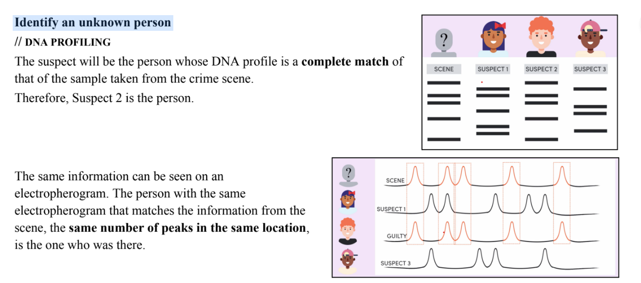
2) Identify an unknown person
In forensic investigations, the identity of the suspect of the crime or unrecognisable victim may need to
be determined
Determining the maternity and/or paternity of a child // DNA PROFILING
Since we receive half our DNA from our mother - it can be assumed that the
bands which match the mother (who is known) must have come from her
[blue].
Thus, all the remaining bands present in the child’s profile must come from
her dad [orange].
Therefore, ‘Male 1’ must be the father.
If there is ever an extra band that isn’t from mum/dad, it could either be due to a mutation, or
perhaps it’s someone related to the ‘father’ (e.g. the father’s cousin, brother).
Identify an unknown person
population genetics
the study of genetic variation within a population, including changes in the frequency*
(amount) of genes and alleles within a population over time. *the proportion
Gene Pool: the alleles of all the genes in a particular population
● Large gene pool (MORE variation)
● Small gene pool (LITTLE VARIATION)
Population Genetics [good morning, shaking my damn nuts]
61
Gabriella Chen
Factors that affect the variation of all the genes in a particular population:
● Size of the population
● Mutation
● Natural Selection
● Genetic Drift*
● Diversity of the Environment**
● Migration Pattern***
Genetic differences between species can be used to determine the evolutionary history of populations; those
with the most similar gene pools are most closely related.
● the use of population genetics data in conservation management
Conservation Management
● Conservation genetics involves the understanding of how genes are inherited in a population. The aim
of this is to avoid extinction* of a species by applying conservation methods to ensure the maintenance
of biodiversity.
● Genetic data is gathered for biodiversity conservation and to make informed decisions about protecting
endangered populations
● Genetic variation is measured by sampling multiple individuals for DNA analysis**
● Through this analysis, scientists are enabled to identify segments of the genome that are essential for the
organism’s adaptation to the environment. They can determine relationships and identify individuals
that could be reintroduced into a population (e.g. captive breeding programs)***
* Avoiding extinction by increasing diversity
**e.g. Sampling Red Panda DNA from Australia, Tokyo and USA → seeing which have the most differences → setting up
on a blind date ∴ diversity!
*** NOT just for animals, also for PLANTS
● Population Genetics Studies used to determine the inheritance of a disease or disorder
Determining the Inheritance of a Disease
Genetic testing has enabled the diagnosis of genetic diseases and the
probability of any offspring inheriting these conditions. Before genetic
testing, pedigrees were used. This is more accurate & convenient in comparison to pedigrees.
In humans, the most common genetic differences in the genome are single
base pair differences, called single nucleotide polymorphisms (SNPs).
Genetic testing for the presence of the SNPs that are unique to a particular
disease can be carried out quickly and relatively easily.
*Polymorphisms means change
e.g. The Newborn Screening Program
In NSW, the newborn screening program provides free genetic tests for all
newborns for the SNPs associated with cystic fibrosis, congenital hypothyroidism,
urea cycle disorder and many more congenital* diseases.
The program has had a significant impact on individuals through early detection
and improved treatment options.
They also generate data about the frequency of the genetic condition in the
population.
*Congenital = (of a disease or physical abnormality) present from birth.
Also, just remember one of these diseases → probably, Cystic Fibrosis (as it is easiest to
remember)
Population Genetics relating to human evolution
Human (Homo Sapiens) Evolution
DNA analysis, fossil evidence and other archaeological evidence can be used to determine human origins and
the evolutionary pathways.
As well as sharing many anatomical and behavioural features with all great apes, modern humans share 98.8%
of their DNA with chimpanzees, our closest living relative.
DNA Evidence
Researchers have used mitochondrial DNA (mtDNA)* to look at human origins. mtDNA was chosen because
it is only maternally inherited, therefore simpler to analyse than nuclear DNA.
A wide sampling of mtDNA from humans currently living was analysed and it was discovered that the most
recent common ancestor - that is, the woman from whom all current human mtDNA were inherited, could be
traced to Africa around 200, 000 years ago. This woman is referred to as mitochondrial Eve.
This provides evidence that a population of Homo Sapiens migrated out of Africa approximately 100, 000 years
ago.
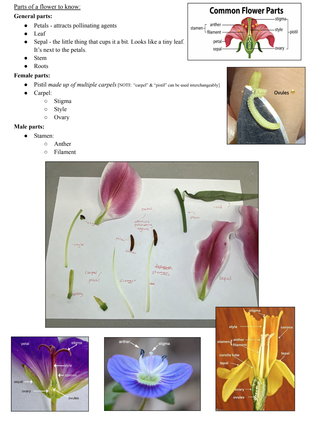
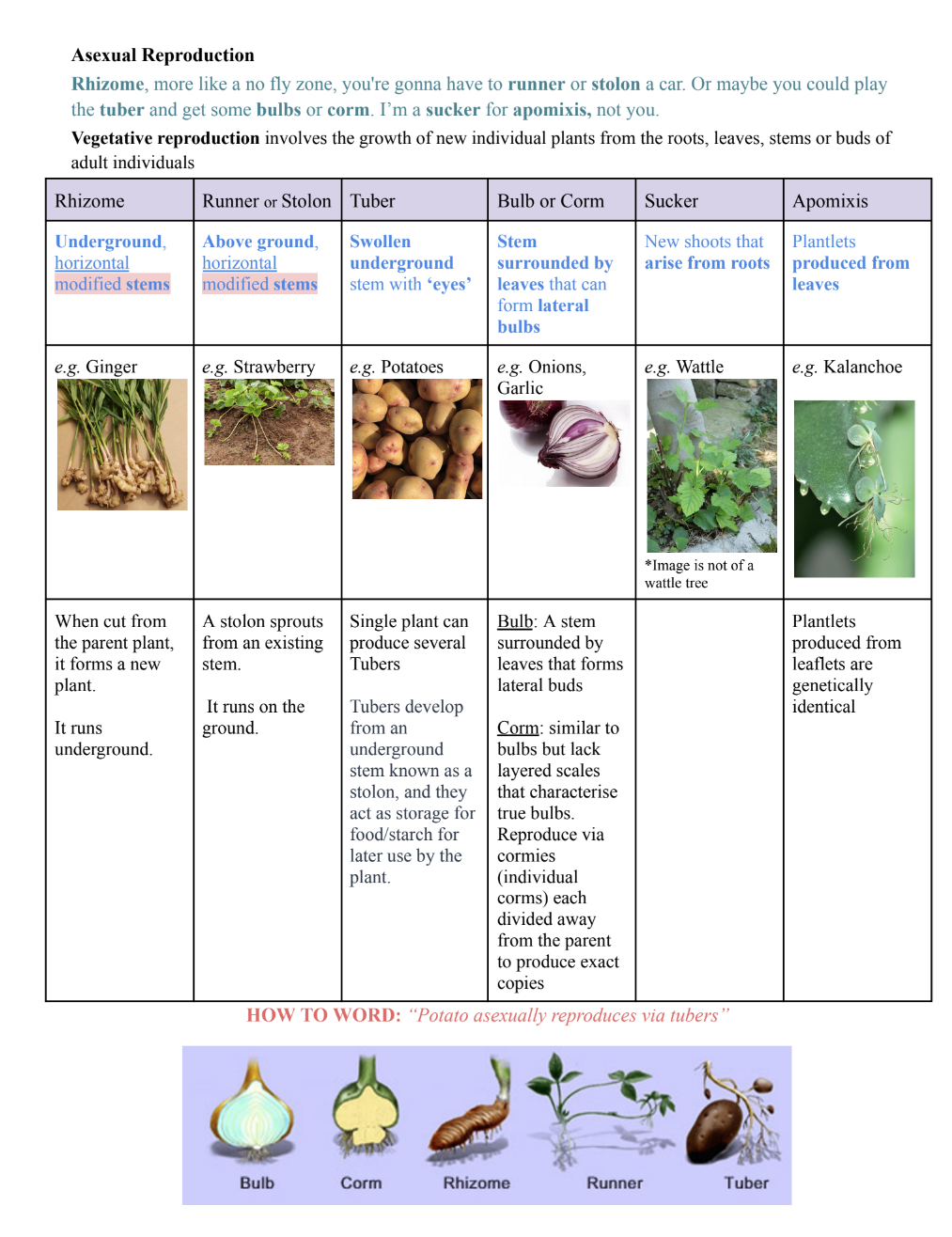
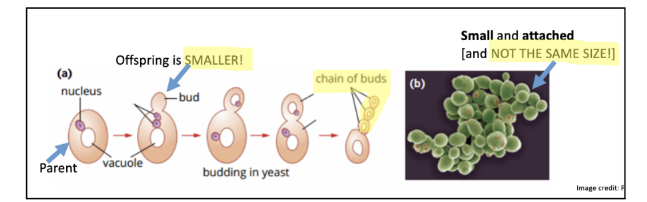
![<p>Multicellular fungi like bread mould are made of threads called hyphae</p><p>which forms the mycelium (fungal body)</p><p>Structures called sporangia produce very large numbers* of spores</p><p>which are light and easily dispersed**, travelling long distances by</p><p>wind.</p><p>*Large numbers means a higher chance of survival</p><p>**So that they can travel/spread and do not have to compete with a parent</p><p>Spores: tiny, light, unicellular reproductive cells that are produced in</p><p>great numbers by organisms such as fungi (e.g. moulds and</p><p>mushrooms)</p><p>● Spores effectively expand the distribution of the species, and</p><p>the species can colonise new environments</p><p><span data-name="star" data-type="emoji">⭐</span>️[vv important] → ENSURES THE SURVIVAL OF THE</p><p>SPECIES</p><p></p>](https://knowt-user-attachments.s3.amazonaws.com/fcd691fd-3000-4b93-ad16-049ae22aa352.png)
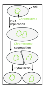
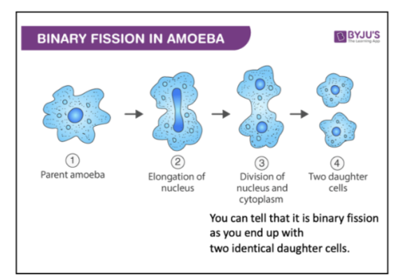
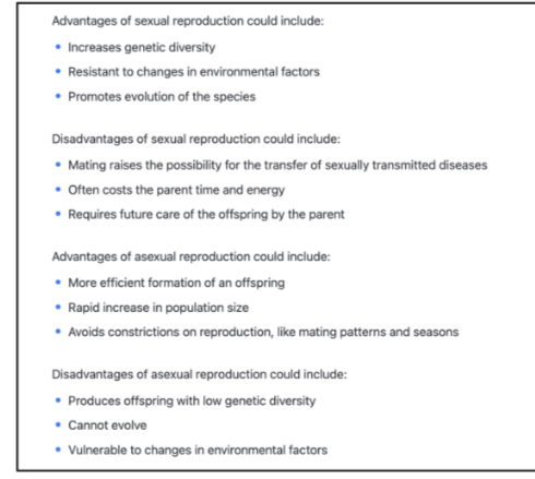
![<p>*Fertilisation happens in the fallopian tube</p><p>**Implantation in uterus (if it happens in the fallopian tubes, it is an ectopic pregnancy where the embryo will not survive</p><p>as miscarriage will occur)</p><p>Pregnancy and Birth in Mammals</p><p>Mammal: An organism that is warm blooded, has a back bone and is able to produce milk and has hair</p><p>● Placental mammals* develop a placenta at implantation e.g. Humans</p><p>● Marsupials rely on a pouch to protect the embryo as it develops e.g. Kangaroo</p><p>● Monotremes rely on an egg to protect the embryo as it develops e.g. Platypus</p><p>*NOTE: Placental mammals are the main focus</p><p>Fertilisation [fusion of male and female gametes]</p><p>In mammals, the male ejaculates sperm into the female’s reproductive tract for internal fertilisation in the</p><p>oviduct*</p><p>● During fertilisation, the haploid** sperm nucleus fuses with the ovum nucleus. The ovum provides a</p><p>single set of chromosomes, and the sperm provides a complementary set of chromosomes. → Produces</p><p>a Zygote - single cell</p><p>● The ovum*** also provides nutrients for the growth of the embryo and regulatory factors that control</p><p>early development → as in, the ovum has all the other organelles (e.g. mitochondria. [RECALL: mitochondrial DNA],</p><p>sperm only gives half genetic information</p><p>○ Makes sure that it develops properly</p><p>*Oviduct = fallopian tubes</p><p>**Half genetic information (Haploid)</p><p>***Ovum = egg</p><p>STEPS:</p><p>1) The female’s body releases an egg from the ovary (ovulation).</p><p>a) In humans it happens about once a month around day 14 (middle of the cycle)</p><p>2) Egg travels through the oviduct which is where fertilisation will occur</p><p>3) The male releases semen (a fluid containing sperm) into the vagina during sex. Sperm swims through</p><p>the cervix and the uterus, to the oviduct where the egg is waiting. Here the nuclei of a sperm cell and the</p><p>egg fuse to form a zygote</p><p>4) The zygote forms a strong membrane to stop other sperm from entering</p><p>a) Otherwise there will be TOO MANY CHROMOSOMES - we only want ONE set</p><p></p>](https://knowt-user-attachments.s3.amazonaws.com/b953ad77-a18b-49b3-bdc8-cf3ad0fdc334.png)
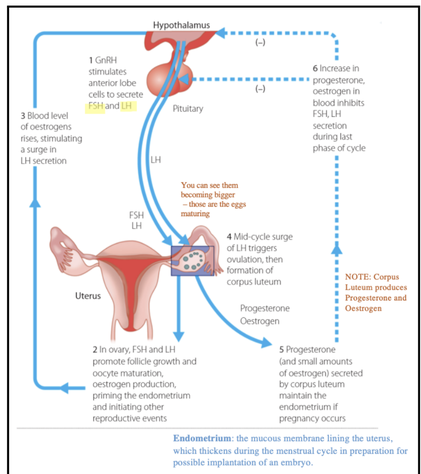
![<p>Lasts (on average) 28 days</p><p>● First 14 days before ovulation are known as the follicular phase</p><p>● Second 14 days after ovulation are known as the luteal phase (a.k.a. lutinising phase)</p><p>1. Follicular Phase</p><p>1) The Pituitary Gland in the brain secretes Follicle Stimulating Hormone (FSH) which travels in the</p><p>bloodstream to the ovary. It causes an egg to mature and stimulates the ovaries to release oestrogen</p><p>2) Oestrogen stops FSH being produced (so only one egg matures per cycle), repairs and thickens the</p><p>uterus lining and stimulates the pituitary gland to release lutenising hormone (LH)</p><p>2. Luteal Phase</p><p>3) LH triggers ovulation [there is also a small rise in FSH at ovulation]</p><p>4) After ovulation, the follicle forms a corpus luteum that produces high levels of progesterone. This</p><p>maintains the lining of the uterus and inhibits the production of FSH (because you only want one egg).</p><p>5) Two things that could happen:</p><p>a) If a fertilised egg implants, progesterone levels remain high</p><p>b) Without implantation, the corpus luteum degenerates and the lining of the uterus breaks down</p><p>again</p><p>Hormonal Control - Pregnancy</p><p>If a Blastocyst* implants in the uterus lining, the placenta secretes the following hormones to maintain</p><p>pregnancy:</p><p>1) Progesterone</p><p>2) Oestrogen [Stops FSH and repairs uterus lining]</p><p>3) Human Chorionic Gonadotrophin (HCG) [is the hormone pregnancy tests test for - maintains corpus luteum]</p><p>The corpus luteum** degenerates after about three months, and the pituitary gland does not secrete FSH until</p><p>after birth.</p><p>*Blastocyst consists of an outlet layer of cells (which develops into the placenta) and an inner mass of cells that will</p><p>develop into the embryo and fluid-filled cavity</p><p>**produces progesterone, but it is okay because the placenta is releasing that too</p><p></p>](https://knowt-user-attachments.s3.amazonaws.com/522807c4-9684-439f-89ea-45773fd7459e.png)
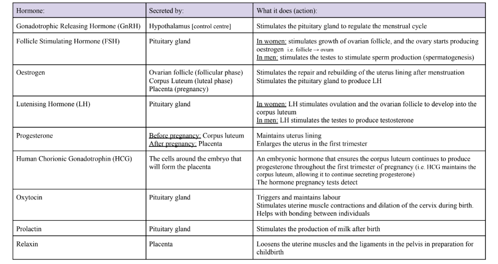
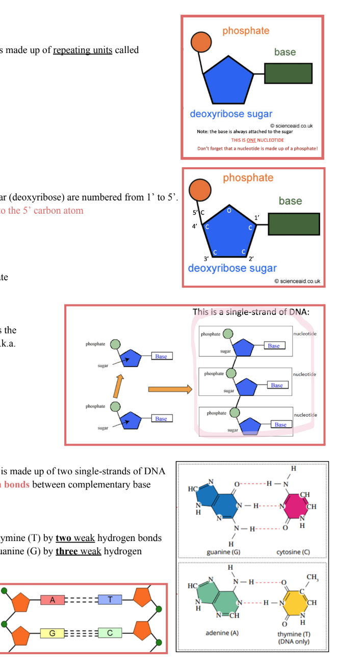
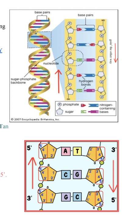
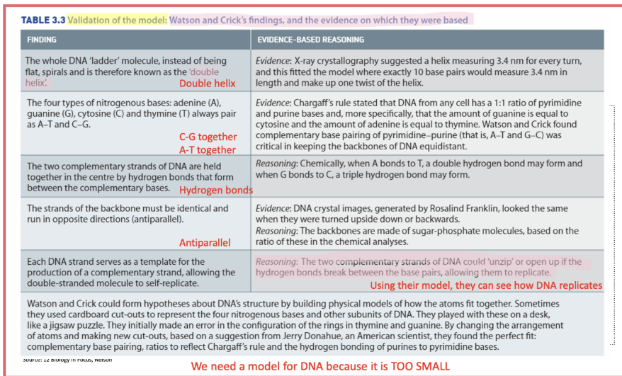
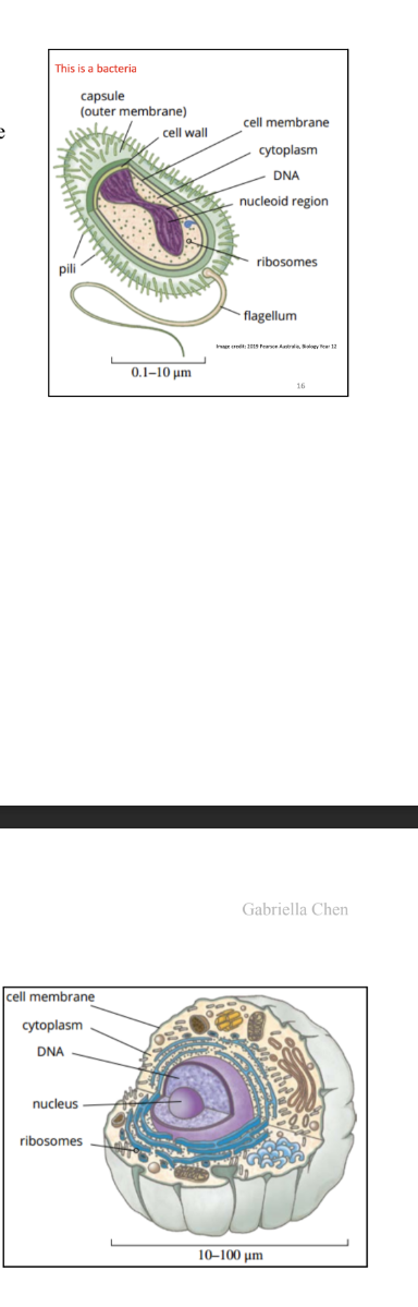
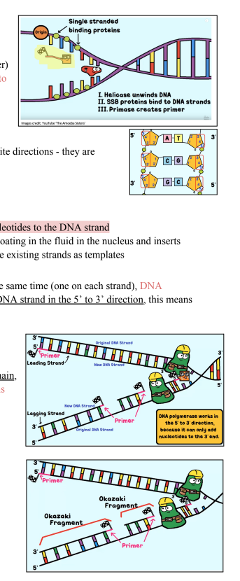
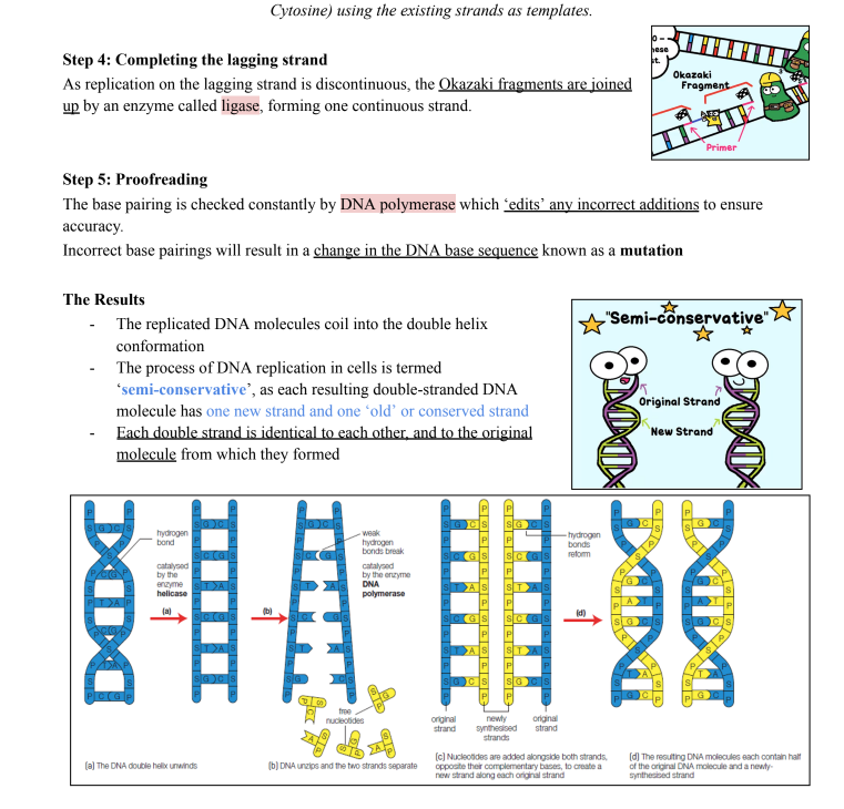
![<p>DNA replication using the Watson and Crick DNA model, including nucleotide composition,</p><p>pairing and bonding</p><p>● Mitosis and Meiosis</p><p>Cell Replication/Division: the process by which cells replicate their genetic material and divide to form new</p><p>cells.</p><p>Cell Division</p><p>- In UNICELLULAR organisms, cell division is by asexual binary fission: where one organism becomes</p><p>two; no gametes (sex cells) are involved</p><p>- In MULTICELLULAR organisms, cell division by mitosis leads to the formation of two new identical</p><p>cells that contribute to the growth of an organism. This occurs in the somatic (body) cells</p><p>Mitosis - makes body cells [<a target="_blank" rel="noopener noreferrer nofollow" href="https://youtu.be/L61Gp_d7evo">https://youtu.be/L61Gp_d7evo</a>]</p><p>Mitosis: a type of cell division where one parent cell divides once to produce two identical daughter cells for</p><p>asexual reproduction, growth, and replacement of old or damaged cells.</p><p>Mitosis plays an important role in:</p><p>● Growth of multicellular organisms</p><p>● Repair of damaged tissue and replacement of worn-out cells</p><p>● Asexual reproduction e.g. growing plants from cutting and cloning</p><p>● Genetic stability - mitosis ensures the precise and equal distribution of chromosomes</p><p>to each daughter nucleus, so that all resulting cells have the same chromosome</p><p>number and genetic information as each other</p><p>○ i.e. A cell will not function properly without a full set of correct genes</p><p>○ e.g. Down Syndrome (a.k.a. Trisomy 21) is caused when abnormal cell division</p><p>results in the 21st pair having an extra chromosome (3 chromosomes)</p><p>Cell Cycle</p><p>- The cell cycle: the life cycle of a cell, involving growth, replication of DNA and</p><p>division of cells to produce two identical daughter cells.</p><p>Has three main phases:</p><p>1. Interphase (cell spends most of its time in this phase)</p><p>2. Mitosis</p><p>3. Cytokinesis</p><p></p>](https://knowt-user-attachments.s3.amazonaws.com/6f0bdc39-d91b-45e7-8d13-aee9e313b347.png)
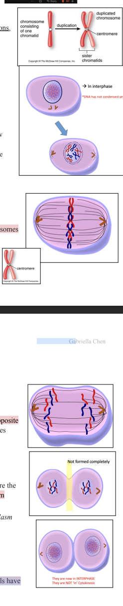

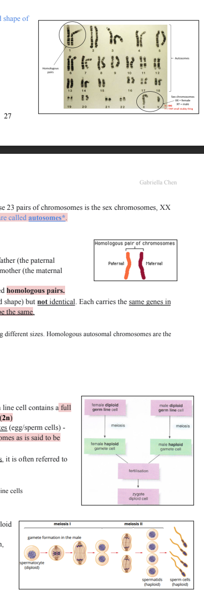
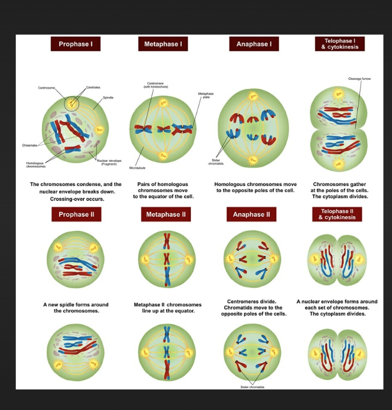
![<p>Proteins are the most abundant organic molecule in cells. They are responsible for forming the basic structure*</p><p>of cells and for carrying out all the work in cells**, by controlling every chemical reaction***.</p><p>*e.g. structural proteins: keratin, collagen</p><p>**e.g. proteins that carry out work: haemoglobin</p><p>***e.g. proteins that control chemical reactions: enzymes</p><p>Chemical Structure of Proteins</p><p>Shape is important!</p><p>→ Amino acids are the building blocks of proteins. They are made up of carbon, hydrogen, oxygen, nitrogen</p><p>and sometimes sulfur.</p><p>→ Amino acids are joined together in a linear sequence by chemical bonds called peptide bonds, forming a</p><p>polypeptide (which can be up to 300 amino acids). [Can be quite long!]</p><p>→ One or more polypeptides can be folded into a particular shape, making a protein. The sequence of amino</p><p>acids (there are 20 different types) and the shape of the protein determines its function.</p><p></p>](https://knowt-user-attachments.s3.amazonaws.com/fc7f9c8b-4ad6-4059-94e8-cbb1cf362e3e.png)
![<p>Types of Proteins in Cells</p><p>Different types of tissues contain different proteins.</p><p>There are two groups:</p><p>1. Fibrous Proteins: Stringy/hairlike</p><p>Form structural components in cells and tissues</p><p>They’re long and soluble in water</p><p>e.g. Collagen, found in skin</p><p>2. Globular Proteins: Honey-like, globby</p><p>Usually spherical in shape and are compact and</p><p>soluble in water</p><p>They are often transport proteins</p><p>e.g. Haemoglobin which transports oxygen around</p><p>the body</p><p>Function of Proteins</p><p>There are many different types of proteins in organisms. Each protein has a different function, and each plays a</p><p>vital role in the regulation*, functioning and maintenance of both individual cells and entire organisms. The</p><p>specific structure** of each protein enables it to carry out its own function.</p><p>Proteins can be classified according to their function [S.E.C.T.S]:</p><p>● Structural proteins for support and movement</p><p>● Enzymes which control biochemical reactions</p><p>● Proteins for cell communication, cell signalling and biological recognition</p><p>● Proteins for transport and storage</p><p>● Sensory proteins which respond to stimuli</p><p>*e.g. DNA Polymerase: regulates DNA to make sure it is the correct sequence</p><p>** Amino acid sequence</p><p>Structural Proteins</p><p>● Often fibrous</p><p>● Found in connective tissues such as skin, bone and ligaments</p><p>● Provide support by forming the structural components of cells and organs, e.g. collagen in</p><p>skin or assist in contractile functions such as muscle, e.g. myosin (protein that causes</p><p>muscles to contract)</p><p>Enzymes</p><p>Enzymes are protein molecules involved in all biochemical aspects</p><p>of cellular metabolism.</p><p>e.g. anabolic enzymes such as DNA Polymerase which catalyses the</p><p>formation of hydrogen bonds</p><p></p>](https://knowt-user-attachments.s3.amazonaws.com/9dac7402-d14b-42aa-9511-bebf2742edd7.png)
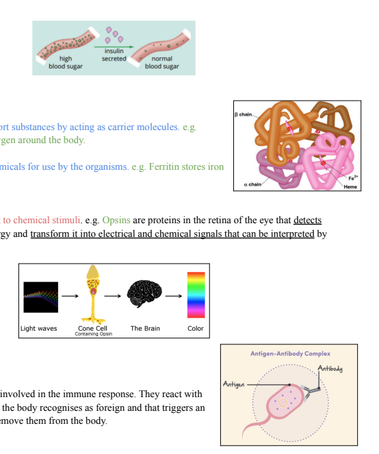
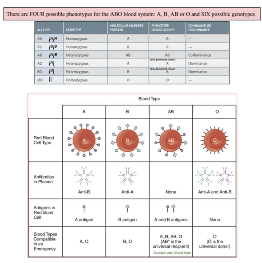
![<p>Multiple Alleles and Multiple Genes</p><p>Phenotypic trait that has multiple alleles ≠ Phenotypic traits that are coded by multiple genes</p><p>Linked Genes</p><p>Although many traits are inherited in accordance to the law of independent</p><p>assortment, it is not always the case. The exception occurs when two or</p><p>more genes are located on a single chromosome and are inherited</p><p>together. This is known as linkage.</p><p>Human Karyotype: (Left: Female, Right: Male)</p><p>Sex Determination</p><p>Sex Determination: the way in which sex</p><p>chromosomes separate during meiosis and recombine</p><p>during fertilisation to determine whether the offspring is</p><p>male or female.</p><p>During meiosis, sex chromosomes segregate (like other</p><p>homologous chromosomes) where only one of each</p><p>chromosome pair passes into a gamete.</p><p>A zygote that inherits an X chromosome from both the</p><p>mother and father will be female (XX). A zygote that</p><p>inherits an X chromosome from the mother and a Y</p><p>chromosome from the father will be a male (XY).</p><p>Depends on male for the sex of the of spring</p><p>Genes and Sex Linkage</p><p>Genes carried on sex chromosomes (X and Y) are called sex-linked genes.</p><p>X-chromosomes are larger than the Y and carry more genes.</p><p>Sex-linked genes in males and females tend to differ in their inheritance</p><p>patterns, since males lack one X chromosome and therefore have only one</p><p>allele (rather than a pair of alleles as present in females). Since males only</p><p>have one allele, whether it is dominant or recessive, males will express this</p><p>trait in their phenotype.</p><p><span data-name="sparkles" data-type="emoji">✨</span>SPECIAL NOTATION<span data-name="sparkles" data-type="emoji">✨</span>: SEX LINKED SYMBOLS</p><p>When a sex-linked gene is being inherited, the alleles of that gene, as well as the type of chromosome on which</p><p>it is carried (X or Y) must be shown.</p><p>e.g. Haemophillia is a recessive, sex-linked disorder.</p><p>NOTE: Only X chromosome expresses the trait, Y</p><p>does not have an allele [and even if Y did carry it, you</p><p>would ‘see’ it as an autosomal trait as you can’t differentiate</p><p>between sex-linked and autosomal here]</p>](https://knowt-user-attachments.s3.amazonaws.com/54fd337e-fbfb-43ce-899a-7e0dcbe03667.png)
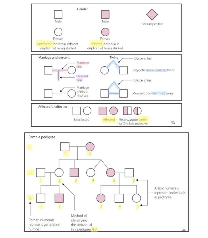
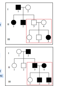
![<p>Sex-linked Inheritance</p><p>When the inheritance pattern is sex-linked, that means the trait affects males and females differently. This is</p><p>because females have two X chromosomes (XX) and males only have one (XY).</p><p>To determine whether a trait is sex-linked or autosomal, check these inheritance patterns:</p><p>● Differences in the incidence of the trait between males and females</p><p>● The frequency of the trait across generations</p><p>X-Linked Recessive Inheritance [males more affected]</p><p>Inheritance of colour blindness is X-linked recessive.</p><p>So, males are affected more frequently than females</p><p>(males only have one X chromosome). No colour-blind</p><p>females are observed in this particular pedigree.</p><p>NOTE: If they do not show the partner (like II-2), they do not</p><p>have the trait i.e. they are not affected</p><p>Males are more affected as there’s a higher chance of a man</p><p>inheriting one faulty X chromosome compared to the chances</p><p>of a woman inheriting two faulty chromosomes. This is because if she has a dominant, normal allele, it can mask the effects</p><p>of the faulty recessive allele.</p><p>53</p><p>Gabriella Chen</p><p>X-Linked Dominant Inheritance</p><p>Traits that are X-Linked Dominant are rare and affect more females than males</p><p>Evidence of X-linked dominance is seen in a pedigree in which affected males have daughters who are ALL</p><p>affected and sons who are NOT affected. This is because daughters inherit their father’s only X Chromosome,</p><p>while sons inherit their father’s Y Chromosome.</p><p>/</p><p>Ruling out X Linked Recessive</p><p>In order for a trait to be X-Linked Recessive,</p><p>affected mothers must have affected sons.</p><p>II-8 has two sons, one affected and one</p><p>unaffected. For the trait to be X-linked recessive,</p><p>the mother would be X</p><p>hX</p><p>h</p><p>in order to be affected.</p><p>The mother contributes one of these</p><p>chromosomes to her offspring, so both sons</p><p>would have received an affected allele. But, one</p><p>son does not show the trait. Therefore the trait is</p><p>not X linked recessive.</p><p>Ruling out X Linked Dominant</p><p>In order for a trait to be X-linked dominant, every daughter of</p><p>an affected male must be affected.</p><p>Daughters receive an X-chromosome from their father</p><p>(reminder, the father only has one X-chromosome).</p><p>I-1 has the trait, but his daughter II-6 does not. Therefore, NOT</p><p>X-Linked Dominant - it is autosomal.</p><p>Y-Linked Recessive</p><p>Only males are affected and affected fathers pass the trait on to</p><p>all their sons.</p><p>In this pedigree, only males are affected and the trait is present</p><p>in the offspring of all affected males. Y-linked inheritance of</p><p>disorders is relatively rare.</p><p>NOTE: II-7 is not affected as he is not related. Also, look out for a lot of black squares</p><p></p>](https://knowt-user-attachments.s3.amazonaws.com/4781f96e-3462-45c8-97e4-50cfcfc8cbe6.png)
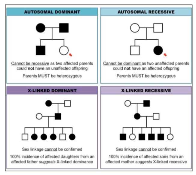
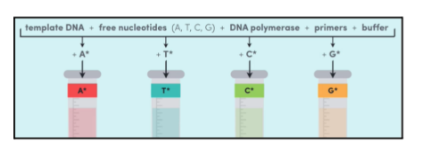
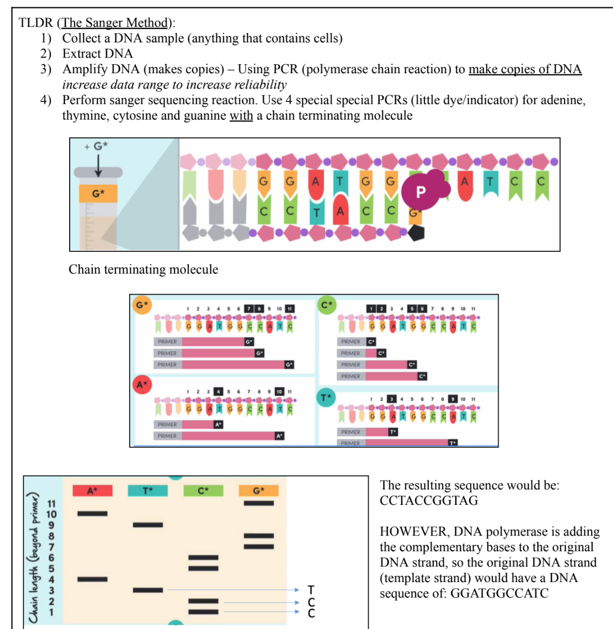
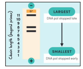
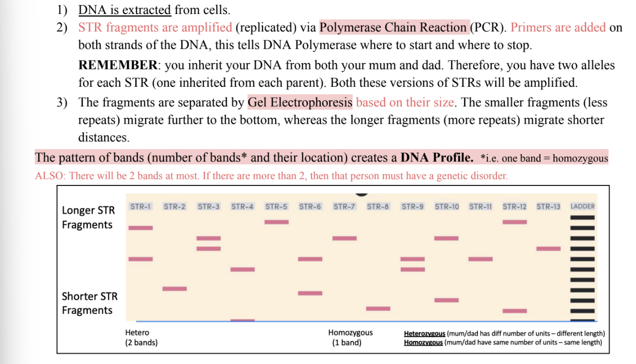
![<p>Since we receive half our DNA from our mother - it can be assumed that the</p><p>bands which match the mother (who is known) must have come from her</p><p>[blue].</p><p>Thus, all the remaining bands present in the child’s profile must come from</p><p>her dad [orange].</p><p>Therefore, ‘Male 1’ must be the father.</p><p>If there is ever an extra band that isn’t from mum/dad, it could either be due to a mutation, or</p><p>perhaps it’s someone related to the ‘father’ (e.g. the father’s cousin, brother).</p>](https://knowt-user-attachments.s3.amazonaws.com/070d1b7b-ddba-4a3c-a0e1-2c7065d3157a.png)